Clin Endosc.
2023 Mar;56(2):185-187. 10.5946/ce.2023.046.
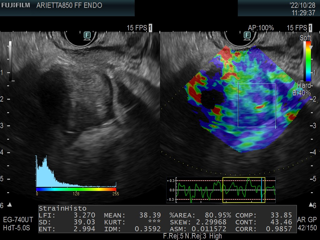
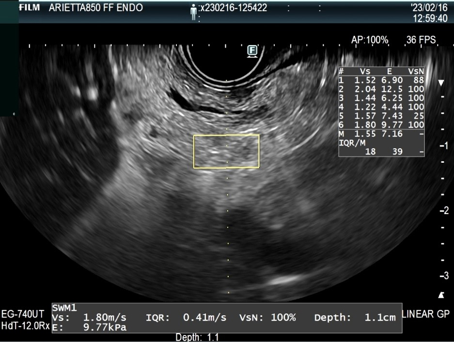
Standardization of endoscopic ultrasound shear wave elastography
- Affiliations
-
- 1Department of Gastroenterology and Hepatology, Health Research Institute of Santiago de Compostela, University Hospital of Santiago de Compostela, University of Santiago de Compostela, Santiago de Compostela, Spain
- KMID: 2540735
- DOI: http://doi.org/10.5946/ce.2023.046
Figure
Reference
-
1. Wani S, Wallace MB, Cohen J, et al. Quality indicators for EUS. Gastrointest Endosc. 2015; 81:67–80.2. Simons-Linares CR, Wander P, Vargo J, Chahal P. Endoscopic ultrasonography: an inside view. Cleve Clin J Med. 2020; 87:175–183.3. Iglesias-Garcia J, Lariño-Noia J, de la Iglesia-García D, Dominguez-Muñoz JE. Endoscopic ultrasonography: enhancing diagnostic accuracy. Best Pract Res Clin Gastroenterol. 2022; 60-61:101808.4. Iglesias-Garcia J, Lindkvist B, Lariño-Noia J, Abdulkader-Nallib I, Dominguez-Muñoz JE. Differential diagnosis of solid pancreatic masses: contrast-enhanced harmonic (CEH-EUS), quantitative-elastography (QE-EUS), or both? United European Gastroenterol J. 2017; 5:236–246.5. Iglesias-Garcia J, Lariño-Noia J, Nieto Bsn L, et al. Pancreatic elastography predicts endoscopic secretin-pancreatic function test result in patients with early changes of chronic pancreatitis: a prospective, cross-sectional, observational study. Am J Gastroenterol. 2022; 117:1264–1268.6. Giovannini M, Hookey LC, Bories E, Pesenti C, Monges G, Delpero JR. Endoscopic ultrasound elastography: the first step towards virtual biopsy? Preliminary results in 49 patients. Endoscopy. 2006; 38:344–348.7. Dietrich CF, Burmeister S, Hollerbach S, et al. Do we need elastography for EUS? Endosc Ultrasound. 2020; 9:284–290.8. Dietrich CF, Bibby E, Jenssen C, Saftoiu A, Iglesias-Garcia J, Havre RF. EUS elastography: How to do it? Endosc Ultrasound. 2018; 7:20–28.9. Ohno E, Hirooka Y, Kawashima H, Ishikawa T. Feasibility of EUS-guided shear-wave measurement: a preliminary clinical study. Endosc Ultrasound. 2019; 8:215–216.10. Wang TJ, Ryou M. Defining the optimal technique for endoscopic ultrasound shear wave elastography: a combined benchtop and animal model study with comparison to transabdominal shear wave elastography. Clin Endosc. 2023; 56:229–238.11. Yamashita Y, Yamazaki H, Shimokawa T, et al. Shear-wave versus strain elastography in endoscopic ultrasound for the diagnosis of chronic pancreatitis. Pancreatology. 2023; 23:35–41.12. Ohno E, Kawashima H, Ishikawa T, et al. Diagnostic performance of endoscopic ultrasonography-guided elastography for solid pancreatic lesions: shear-wave measurements versus strain elastography with histogram analysis. Dig Endosc. 2021; 33:629–638.
- Full Text Links
- Actions
-
Cited
- CITED
-
- Close
- Share
- Similar articles
-
- Diagnostic Performance of Quantitative Shear Wave Ultrasound Elastography for Thyroid Cancer
- Elastography of the Pancreas, Current View
- Ultrasound Elastography for Liver Disease with Focus on Hepatic Fibrosis
- Ultrasound elastography of the thyroid: principles and current status
- Future of breast elastography


