Direct Puncture of the Superficial Temporal Artery in Embolization of a Scalp Arteriovenous Fistula: A Case Report
- Walker GB
 1
1 - Wang AP2
- Hadwen J3
- Erdenebold UE1
- Bebedjian R1
- Sullivan P4
- Santos MP1,5
- Chenier C1
- Karwaski S1
- Caron K1
- Varga G1
- Lyon J1
- Lesiuk HJ1,2
- Heran N6
- Heran M7
- Quateen A1,5
- Drake BJ1,2
- Oliveira EPD1,5
- Kontolemos M5
- Fahed R1,3
- Affiliations
-
- 1Division of Interventional Neuroradiology, Department of Medical Imaging, The Ottawa Hospital, University of Ottawa, Ottawa, ON, Canada
- 2Division of Neurosurgery, Department of Surgery, The Ottawa Hospital, University of Ottawa, Ottawa, ON, Canada
- 3Division of Neurology, Department of Medicine, The Ottawa Hospital, University of Ottawa, Ottawa, ON, Canada
- 4Department of Anesthesiology, The Ottawa Hospital, University of Ottawa, Ottawa, ON, Canada
- 5Division of Radiology, Department of Medical Imaging, The Ottawa Hospital, University of Ottawa, Ottawa, ON, Canada
- 6Division of Neurosurgery, Department of Surgery, Royal Columbian Hospital, University of British Columbia, New Westminster, BC, Canada
- 7Division of Diagnostic and Therapeutic Neuroradiology, Department of Radiology, Vancouver General Hospital, University of British Columbia, Vancouver, BC, Canada
- KMID: 2539949
- DOI: http://doi.org/10.5469/neuroint.2022.00465
Abstract
- We describe a minimally invasive endovascular approach to treat an arteriovenous fistula of the scalp. We performed a direct puncture of the lesion through the patient’s scalp for liquid embolic agent injection along with external compression of the superficial temporal artery to perform a “manual pressure-cooker technique.” The combination of these minimally invasive techniques resulted in an excellent clinical and radiographic outcome.
Figure
-
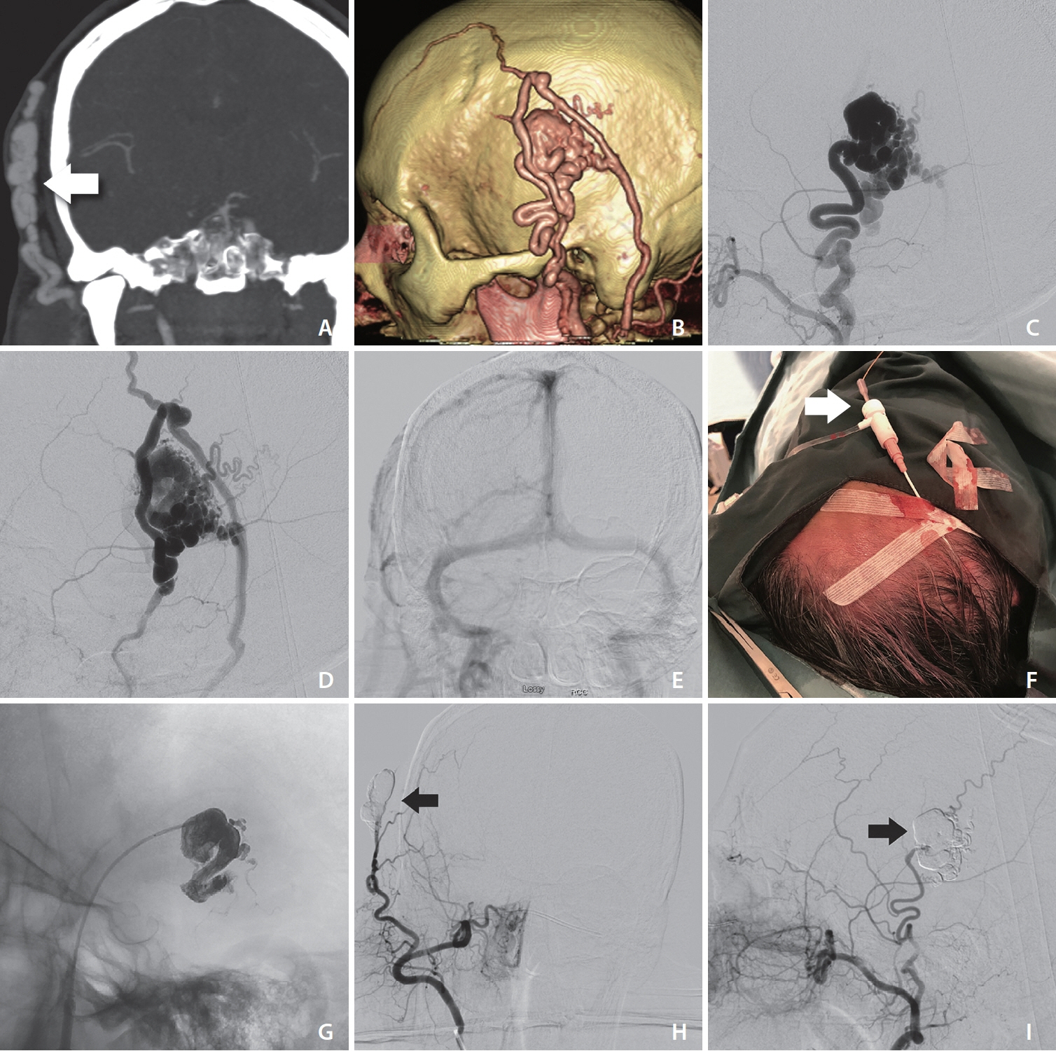
Fig. 1. Brain CTA in coronal view (A) shows enlarged vessels in the right scalp area (arrow), fed by the right superficial temporal artery as seen on 3D reconstruction (B). The angiogram (C, D) confirms the presence of an enlarged superficial temporal artery feeding an arteriovenous fistula with a large venous pouch and several enlarged veins, with eventual drainage into the superior sagittal sinus (E). (F) The Apollo catheter is inserted into the check flow valve using the tip of the Terumo wire housing as an introducer (arrow). The valve is attached to the Angiocath cannula from the thin wall entry needle, and together these act as a sheath directly placed inside the scalp lesion. Using this setup, we were able to inject liquid embolic agent directly into the nidus with no arterial reflux thanks to manual external compression of the feeding superficial temporal artery. (G) The patient’s symptoms fully resolved immediately after the procedure. (H, I) The 6-month follow-up angiogram confirmed the complete obliteration of the lesion (arrow).
Reference
-
1. Kojima D, Akamatsu Y, Fujimoto K, Oikawa K, Kashimura H, Kubo Y, et al. Utility of manual venous compression during transvenous Onyx injection for a scalp arteriovenous fistula: illustrative case. J Neurosurg Case Lessons. 2022; 4:CASE22317.
Article2. Gascón-Rubio MC, Merino-Bonilla JA, Guerra-Pérez H. Retroauricular arteriovenous fistula. Acta Otorrinolaringol Esp. 2012; 63:325–326.
Article3. Clarençon F, Shotar E, Pouvelle A, Mouyal S, Lenck S, Premat K, et al. Direct puncture of the superficial temporal artery for ethylene vinyl alcohol embolization of a type 3 arteriovenous fistula with a dual lumen balloon. J Neurointerv Surg. 2021; 13:493.
Article4. Gobin YP, Pasco A, Merland JJ, Aymard AA, Casasco A, Houdart E. Percutaneous puncture of the external carotid artery or its branches after surgical ligation. AJNR Am J Neuroradiol. 1994; 15:79–82.5. Oh JS, Yoon SM, Shim JJ, Bae HG. Transcranial direct middle meningeal artery puncture for the onyx embolization of dural arteriovenous fistula involving the superior sagittal sinus. J Korean Neurosurg Soc. 2015; 57:54–57.
Article6. Prashar A, Butt S, Shaida N. Introducing PHIL (precipitating hydrophobic injectable liquid) - a new embolic agent for the body interventional radiologist. Diagn Interv Radiol. 2020; 26:140–142.
Article7. Alawneh K, Al-Barbarawi M, Qawasmeh MA, Raffee LA, Al-Mistarehi AH. Successful use of neurovascular plug for embolization of scalp arteriovenous fistula: a novel technique. J Endovasc Ther. 2022; 29:827–834.
Article8. Koyanagi M, Mosimann PJ, Nordmeyer H, Heddier M, Krause J, Narata AP, et al. The transvenous retrograde pressure cooker technique for the curative embolization of high-grade brain arteriovenous malformations. J Neurointerv Surg. 2021; 13:637–641.
Article9. Sahu CD, Bhargava N. Intra-arterial onyx embolisation of sphenobasilar sinus fistula using pressure cooker technique: case report and review of the literature. Neuroradiol J. 2021; 34:131–134.
Article10. Zhang G, Zhu S, Wu P, Xu S, Shi H. The transvenous pressure cooker technique: a treatment for brain arteriovenous malformations. Interv Neuroradiol. 2017; 23:194–199.
Article11. Abud DG, de Castro-Afonso LH, Nakiri GS, Monsignore LM, Colli BO. Modified pressure cooker technique: an easier way to control onyx reflux. J Neuroradiol. 2016; 43:218–222.
Article12. Kamusella P, Scheer F, Lüdtke CW, Wiggermann P, Wissgott C, Andresen R. Interventional angiography: radiation protection for the examiner by using lead-free gloves. J Clin Diagn Res. 2017; 11:TC26–TC29.
Article
- Full Text Links
-
- Actions
-
Cited
- CITED
-
- Close
- Share
-
- Similar articles
-
- Surgical Treatment of a Post-Traumatic Arteriovenous Fistula of the Superficial Temporal Artery and Vein
- Post-Traumatic Arteriovenous Fistula of the Scalp
- A Case of Traumatic Arteriovenous Fistula of the Superficial Temporal Artery
- Embolization of a Bleeding Maxillary Arteriovenous Malformation via the Superficial Temporal Artery after External Carotid Artery Ligation
- Direct Puncture Embolization of Scalp Arteriovenous Malformation in a Patient with Severe Hemophilia A: A Case Report
