Delayed Cerebral Ischemia after Embolization in Ruptured Spinal Arteriovenous Fistula with Subarachnoid Hemorrhage : A Case Report
- Sani AF
 1,2,3
1,2,3 - Kurniawan D2,3
- Hamdan M2,3
- Swatan JP2,3
- Affiliations
-
- 1Doctoral Program of Medical Science, Faculty of Medicine, Universitas Airlangga, Surabaya, Indonesia
- 2Department of Neurology, Faculty of Medicine, Universitas Airlangga, Surabaya, Indonesia
- 3Department of Neurology, Dr. Soetomo General Hospital, Surabaya, Indonesia
- KMID: 2539882
- DOI: http://doi.org/10.3340/jkns.2022.0139
Abstract
- Delayed cerebral ischemia (DCI) remains a devastating complication in subarachnoid hemorrhage (SAH), however, there were no present reports that is associated with a ruptured spinal arteriovenous fistula (sAVF). We would like to present a rare case of DCI following embolization of a ruptured perimedullary sAVF. Initially, the patient clinical symptoms mimic a SAH caused by a ruptured intracranial aneurysm. Further evaluation revealed that the SAH was caused by a ruptured perimedullary sAVF and the patient’s condition improved following the embolization procedure. Three days later, the patient developed an acute left-sided facial and motor weakness, which persisted until the patient was discharged on the day-15 onset. A magnetic resonance imaging and angiography is performed 1.5 years after discharge and revealed no signs of cerebral infarction and hemorrhage. In this paper, we reported DCI after embolization in a ruptured sAVF with SAH, supported by evidence from the current literature. We would like to also stress the importance of complete spinal and cerebral vessel imaging to reveal the underlying abnormalities and determine the most appropriate intervention.
Keyword
Figure
-
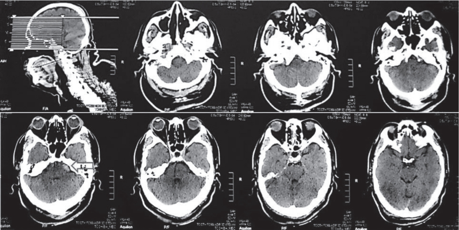
Fig. 1. An axial non-contrast head computed tomography-scan revealed subarachnoid hemorrhage in the left pontomedullary junction (arrow).
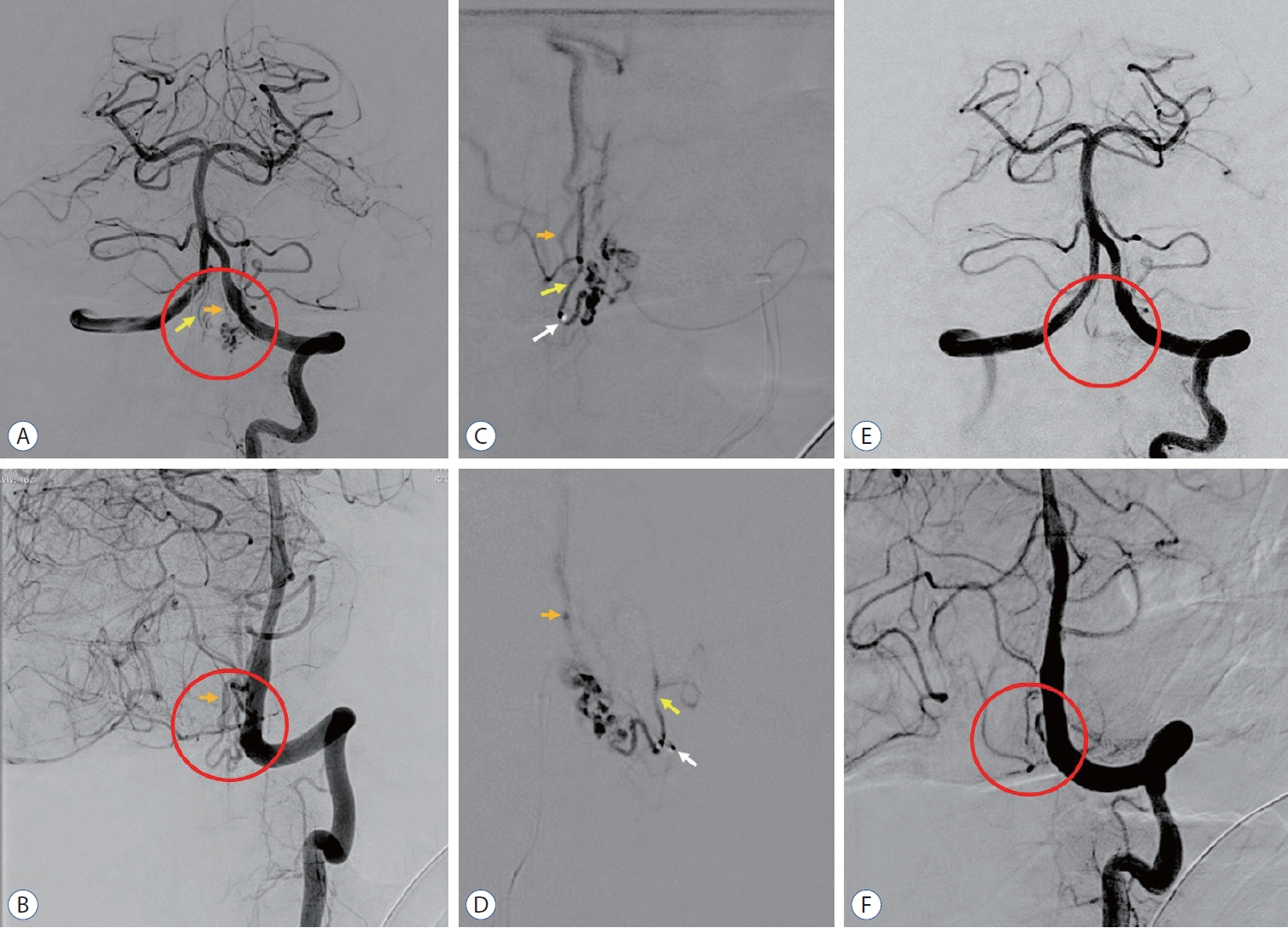
Fig. 2. A cerebral digital subtraction angiography in anteroposterior and oblique view following injection from left vertebral artery revealed a spinal arteriovenous fistula (sAVF) (red circle) with feeding artery from anterior spinal artery (yellow arrow) and drains to perimedullary vein (orange arrow) revealed the sAVF before embolization (A and B), during embolization procedure using N-buytl-2-cyanoacrylate (Histoacryl®; B-Braun, Melsungen, Germany) in a single super-selective injection (white arrow, microcatheter tip) of the arterial feeder (C and D), and complete obliteration after embolization procedure (E and F).
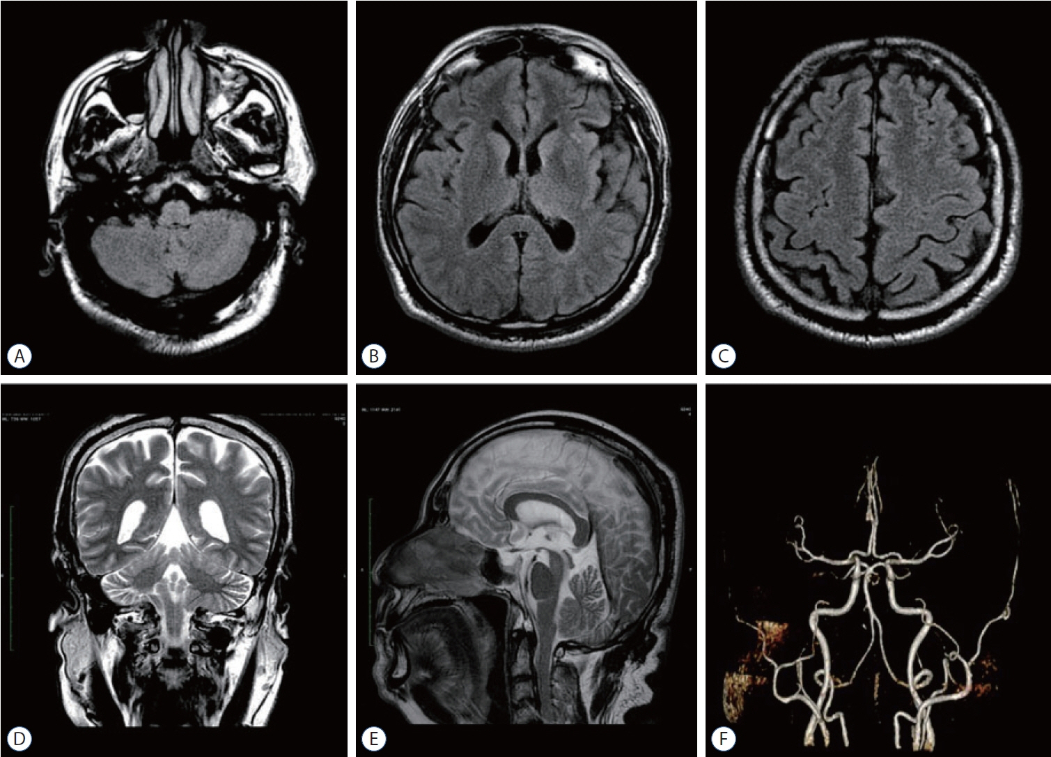
Fig. 3. Axial fluid-attenuated inversion recovery-sequence (A-C), coronal (D), and sagittal T2 (E) sequence of brain magnetic resonance imaging with contrast; and magnetic resonance angiography of cerebral blood vessels (F) conducted 1.5 years after the onset of delayed cerebral ischemia revealed no lesions or vascular abnormalities.
Reference
-
References
1. Ayata C, Lauritzen M. Spreading depression, spreading depolarizations, and the cerebral vasculature. Physiol Rev. 95:953–993. 2015.2. Dodd WS, Laurent D, Dumont AS, Hasan DM, Jabbour PM, Starke RM, et al. Pathophysiology of delayed cerebral ischemia after subarachnoid hemorrhage: a review. J Am Heart Assoc. 10:e021845. 2021.3. Francoeur CL, Mayer SA. Management of delayed cerebral ischemia after subarachnoid hemorrhage. Crit Care. 20:277. 2016.4. Goursaud S, Martinez de Lizarrondo S, Grolleau F, Chagnot A, Agin V, Maubert E, et al. Delayed cerebral ischemia after subarachnoid hemorrhage: is there a relevant experimental model? A systematic review of preclinical literature. Front Cardiovasc Med. 8:752769. 2021.5. Ikram A, Javaid MA, Ortega-Gutierrez S, Selim M, Kelangi S, Anwar SMH, et al. Delayed cerebral ischemia after subarachnoid hemorrhage. J Stroke Cerebrovasc Dis. 30:106064. 2021.6. Koch MJ, Stapleton CJ, Agarwalla PK, Torok C, Shin JH, Coumans JV, et al. Open and endovascular treatment of spinal dural arteriovenous fistulas: a 10-year experience. J Neurosurg Spine. 26:519–523. 2017.7. Krings T, Geibprasert S. Spinal dural arteriovenous fistulas. AJNR Am J Neuroradiol. 30:639–648. 2009.8. Murai S, Hiramatsu M, Suzuki E, Ishibashi R, Takai H, Miyazaki Y, et al. Trends in incidence of intracranial and spinal arteriovenous shunts: hospital-based surveillance in Okayama, Japan. Stroke. 52:1455–1459. 2021.9. Neifert SN, Chapman EK, Martini ML, Shuman WH, Schupper AJ, Oermann EK, et al. Aneurysmal subarachnoid hemorrhage: the last decade. Transl Stroke Res. 12:428–446. 2021.10. Qureshi AI, Luft AR, Sharma M, Guterman LR, Hopkins LN. Prevention and treatment of thromboembolic and ischemic complications associated with endovascular procedures: part II--clinical aspects and recommendations. Neurosurgery. 46:1360–1375. 2000.11. Sarrafzadeh AS, Vajkoczy P, Bijlenga P, Schaller K. Monitoring in neurointensive care - the challenge to detect delayed cerebral ischemia in high-grade aneurysmal SAH. Front Neurol. 5:134. 2014.12. Takai K. Spinal arteriovenous shunts: angioarchitecture and historical changes in classification. Neurol Med Chir (Tokyo). 57:356–365. 2017.13. van der Kleij LA, De Vis JB, Olivot JM, Calviere L, Cognard C, Zuithoff NP, et al. Magnetic resonance imaging and cerebral ischemia after aneurysmal subarachnoid hemorrhage: a systematic review and metaanalysis. Stroke. 48:239–245. 2017.14. Vergouwen MD, Vermeulen M, van Gijn J, Rinkel GJ, Wijdicks EF, Muizelaar JP, et al. Definition of delayed cerebral ischemia after aneurysmal subarachnoid hemorrhage as an outcome event in clinical trials and observational studies: proposal of a multidisciplinary research group. Stroke. 41:2391–2395. 2010.15. Zhao J, Esemen Y, Rane N, Nair R. Intracranial subarachnoid haemorrhage caused by cervical spinal dural arteriovenous fistulas: case report. Front Neurol. 12:685332. 2021.
- Full Text Links
-
- Actions
-
Cited
- CITED
-
- Close
- Share
-
- Similar articles
-
- Traumatic Intracerebral and Subarachnoid Hemorrhage Due to a Ruptured Pseudoaneurysm of Middle Meningeal Artery Accompanied by a Medial Sphenoid Wing Dural Arteriovenous Fistula
- Embolization Therapy for a Ruptured Spinal Artery Aneurysm Associated with Spinal Cord Arteriovenous Malformation and Presenting with Spontaneous Subarachnoid Hemorrhage
- A Rare Case of Subarachnoid Hemorrhage caused by Ruptured Venous Varix Due to Dural Arteriovenous Fistula at the Foramen Magnum Fed Solely by the Ascending Pharyngeal Artery
- Thoracic Spinal Arteriovenous Fistula with Subarachnoid Hemorrhage
- Delayed Pulmonary Edema after Coil Embolization in a Patient with Subarachnoid Hemorrhage: A Case Report
