Traumatic Intracerebral and Subarachnoid Hemorrhage Due to a Ruptured Pseudoaneurysm of Middle Meningeal Artery Accompanied by a Medial Sphenoid Wing Dural Arteriovenous Fistula
- Affiliations
-
- 1Department of Neurosurgery, Hallym University Kangdong Sacred Heart Hospital, Hallym University College of Medicine, Seoul, Korea. nsyjlee@gmail.com
- KMID: 2394554
- DOI: http://doi.org/10.13004/kjnt.2017.13.2.162
Abstract
- Traumatic pseudoaneurysms of middle meningeal artery (MMA) and medial sphenoid wing dural arteriovenous fistula (dAVF) are rare. These lesions usually result from traumatic brain injury, and associated with skull fracture. In this paper, the authors report a case of a patient with a ruptured traumatic pseudoaneurysm of MMA and medial sphenoid wing dAVF presented with an intracerebral hemorrhage in the left temporal region and subarachnoid hemorrhage. These lesions were completely obliterated by endovascular treatment, and the patient was recovered without any neurologic deficit. However, 18-day after the procedure, delayed neurologic deficits were developed due to cerebral vasospasm.
MeSH Terms
Figure
-
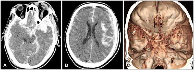
FIGURE 1 Initial radiological images after trauma. (A, B) Brain computed tomography (CT) showed intracerebral hemorrhage in the left temporal lobe with diffuse and large amount of subarachnoid hemorrhage and subdural hemorrhage at the left hemisphere. (C) Brain CT angiography revealed that abnormal vascular structure, considered as pseudoaneurysms, at the distal segment of the left lesser sphenoid ridge (arrow).
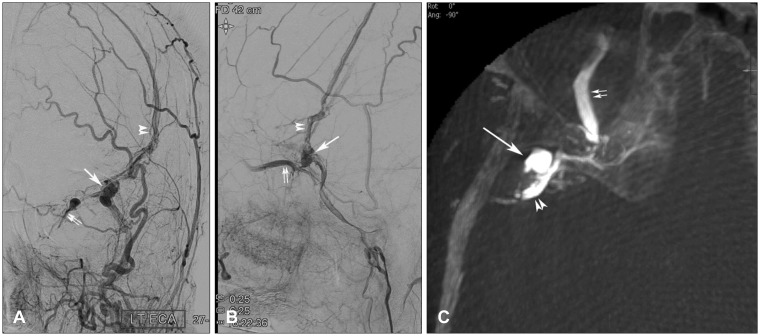
FIGURE 2 Left external carotid angiogram. (A) anteroposterior view, (B) lateral view showed a multilobulated pseudoaneurysm at the distal segment of the lesser sphenoid ridge (large arrow). Retrograde contrast filling through the left superior ophthalmic vein (double arrow) and middle meningeal artery-middle meningeal vein fistula (double arrow head) was also shown. (C) Cone-beam computed tomography image showed a multilobulated pseudoaneurysm at the distal segment of the lesser sphenoid ridge (large arrow) and the left superior ophthalmic vein (double arrow). Between these, complex vascular connections at the lesser sphenoid ridge resulting in medial sphenoid ridge dural arteriovenous fistula was shown.
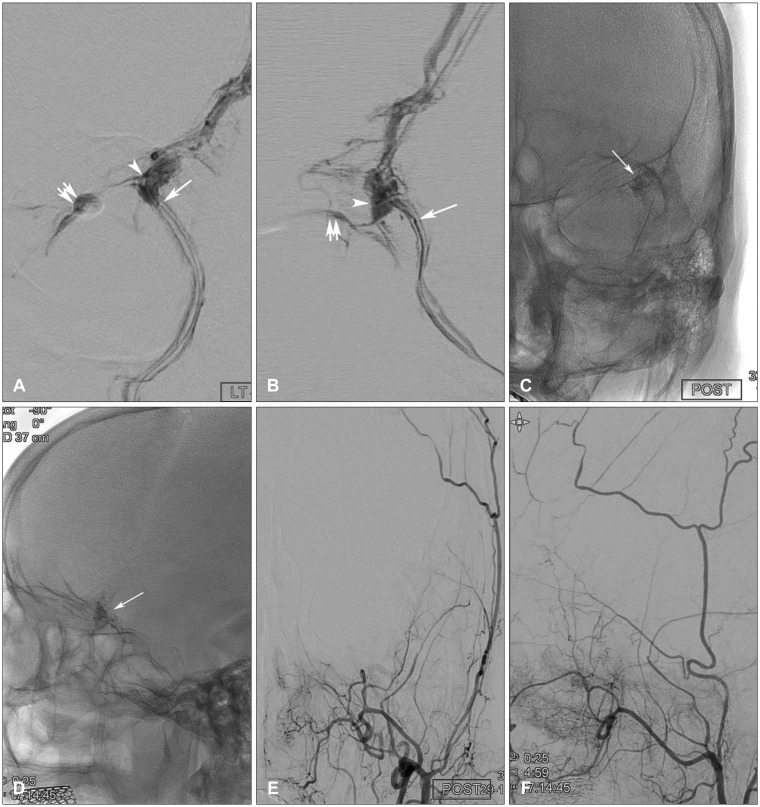
FIGURE 3 Superselective angiogram of the left middle meningeal artery (MMA). (A) anteroposterior view, (B) lateral view revealed an appropriate position of distal microcatheter tip (large arrow) showing a pseudoaneurysm (arrow head) and retrograde contrast filling through the left superior ophthalmic vein (double arrow). (C, D) N-butyl cyanoacrylate cast after embolization (large arrow). (E, F) Post-embolization external carotid angiogram showed complete occlusion of pseudoaneurysm, medial sphenoid ridge dural arteriovenous fistula and MMA-middle meningeal vein fistula.
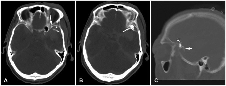
FIGURE 4 (A) Post-embolization computed tomography showed n-butyl cyanoacrylate cast at the dural vascular channel (arrow), (B) psneudoaneurysm (arrow), (C) proximal and distal segment of the left middle meningeal artery according to a pseudoaneurysm (arrow and arrow head, respectively).

FIGURE 5 Radiologic images 15 days after endovascular embolization with motor weakness and dysarthria. (A, B) Brain computed tomography (CT) showed multifocal low density lesions at the left middle cerebral artery (MCA) territory. (C) CT angiography revealed sparse vasculature of distal segment of the left MCA preserving M1 patency (large arrows). (D) Left internal carotid angiogram showed multiple luminal irregularities and narrowing at the distal segment of MCA (small arrows).
Reference
-
1. Bruneau M, Gustin T, Zekhnini K, Gilliard C. Traumatic false aneurysm of the middle meningeal artery causing an intracerebral hemorrhage: case report and literature review. Surg Neurol. 2002; 57:174–178. PMID: 12009543.2. Carney N, Totten AM, O'Reilly C, Ullman JS, Hawryluk GW, Bell MJ, et al. Guidelines for the management of severe traumatic brain injury, fourth edition. Neurosurgery. 2017; 80:6–15. PMID: 27654000.
Article3. Connolly ES Jr, Rabinstein AA, Carhuapoma JR, Derdeyn CP, Dion J, Higashida RT, et al. Guidelines for the management of aneurysmal subarachnoid hemorrhage: a guideline for healthcare professionals from the American Heart Association/american Stroke Association. Stroke. 2012; 43:1711–1737. PMID: 22556195.4. Lim DH, Kim TS, Joo SP, Kim SH. Intracerebral hematoma caused by ruptured traumatic pseudoaneurysm of the middle meningeal artery : a case report. J Korean Neurosurg Soc. 2007; 42:416–418. PMID: 19096582.
Article5. Oertel M, Boscardin WJ, Obrist WD, Glenn TC, McArthur DL, Gravori T, et al. Posttraumatic vasospasm: the epidemiology, severity, and time course of an underestimated phenomenon: a prospective study performed in 299 patients. J Neurosurg. 2005; 103:812–824. PMID: 16304984.
Article6. Paiva WS, de Andrade AF, Amorim RL, Figueiredo EG, Teixeira MJ. Traumatic pseudoaneurysm of the middle meningeal artery causing an intracerebral hemorrhage. Case Rep Med. 2010; 2010:219572. PMID: 20589087.
Article7. Salazar Flores J, Vaquero J, Garcia Sola R, Rossi E, Martinez R, Martinez P, et al. Traumatic false aneurysms of the middle meningeal artery. Neurosurgery. 1986; 18:200–203. PMID: 3960300.
Article8. San Millán Ruíz D, Fasel JH, Rüfenacht DA, Gailloud P. The sphenoparietal sinus of breschet: does it exist? An anatomic study. AJNR Am J Neuroradiol. 2004; 25:112–120. PMID: 14729539.9. Sandin JA 3rd, Salamat MS, Baskaya M, Dempsey RJ. Intracerebral hemorrhage caused by the rupture of a nontraumatic middle meningeal artery aneurysm. Case report and review of the literature. J Neurosurg. 1999; 90:951–954. PMID: 10223464.10. Shi ZS, Ziegler J, Feng L, Gonzalez NR, Tateshima S, Jahan R, et al. anatomic and treatment considerations. AJNR Am J Neuroradiol. 2013; 34:373–380. PMID: 22790245.
- Full Text Links
-
- Actions
-
Cited
- CITED
-
- Close
- Share
-
- Similar articles
-
- Intracerebral Hematoma Caused by Ruptured Traumatic Pseudoaneurysm of the Middle Meningeal Artery: A Case Report
- Angiographically Progressive Change of Traumatic Pseudoaneurysm Arising from the Middle Meningeal Artery
- Rupture of a Middle Meningeal Artery Pseudoaneurysm in Moyamoya Syndrome Related with Tuberculous Meningitis
- Subarachnoid Hemorrhage Due to a Ruptured Middle Cerebral Artery Bifurcation Aneurysm Superimposed by an Idiopathic Intracerebral Hematoma
- Borden Type I Sigmoid Sinus Dural Arteriovenous Fistula Presenting as Subarachnoid Hemorrhage from a Feeding Artery Aneurysm of the Anterior Inferior Cerebellar Artery: A Case Report
