A rare case of pure-type embryonal carcinoma in a 75-year-old woman mimicking epithelial ovarian carcinoma
- Affiliations
-
- 1Department of Obstetrics and Gynecology, Pusan National University Hospital, Busan, Korea
- 2Department of Radiology, Pusan National University Hospital, Busan, Korea
- 3Department of Pathology, Pusan National University Hospital, Busan, Korea
- 4Department of Obstetrics and Gynecology, Kosin University Gospel Hospital, Busan, Korea
- 5Biomedical Research Institute, Pusan National University School of Medicine, Busan, Korea
- KMID: 2538805
- DOI: http://doi.org/10.7180/kmj.22.026
Abstract
- Embryonal carcinoma, a very rare ovarian germ cell tumor, involves pure and mixed phenotypes. Pure-type embryonal carcinoma has never been reported in postmenopausal women. The current case was, thus, misdiagnosed as an epithelial ovarian carcinoma based on radiologic findings. Herein, we describe the case of ovarian embryonal carcinoma in a 75-year-old woman along with a literature review. Magnetic resonance imaging findings were suggestive of epithelial ovarian malignancy associated with endometrioma, including ureteral invasion. The patient underwent complete surgical staging, and a pathologic diagnosis of pure-type embryonal carcinoma was made. The patient’s postoperative course was uneventful, and adjuvant chemotherapy was administered. Embryonal carcinoma in the postmenopausal woman is a clinical challenge owing to the possibility of its misdiagnosis as epithelial ovarian carcinoma. To the best of our knowledge, this is the first report of pure-type ovarian embryonal carcinoma in a postmenopausal woman, with a description of the clinicopathologic characteristics and review of the relevant literature.
Figure
-
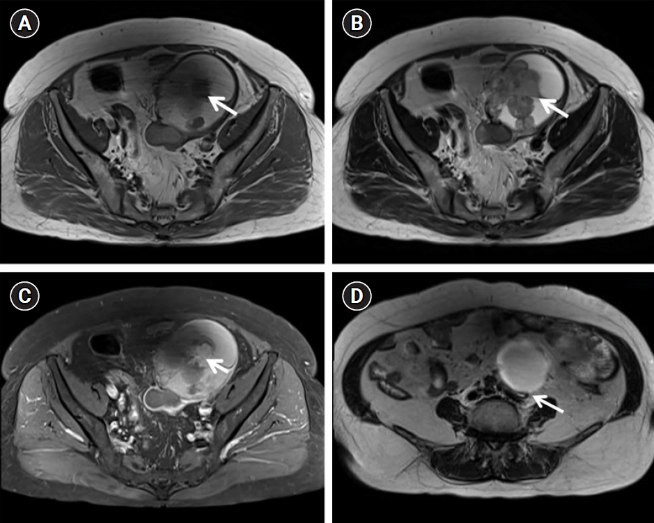
Fig. 1. Magnetic resonance images. Axial T1-weighted (A) and T2-weighted (B) images showing a septated hemorrhagic cystic mass with papillary projections (arrow) of the left ovary (ORADS 5). High T1 signal intensity with the T2 dark spot sign, suggestive of epithelial ovarian cancer associated with endometrioma (e.g., clear cell carcinoma or endometrioid carcinoma). (C) Contrast-enhanced axial T-weighted image shows enhancement (arrow) of the papillary projections in the left ovarian tumor. (D) Axial T2-weighted imaging revealed left hydronephrosis (arrow), showing abrupt narrowing at the ovarian mass level suggestive of ureteral invasion. ORADS, Ovarian-Adnexal Reporting & Data System.
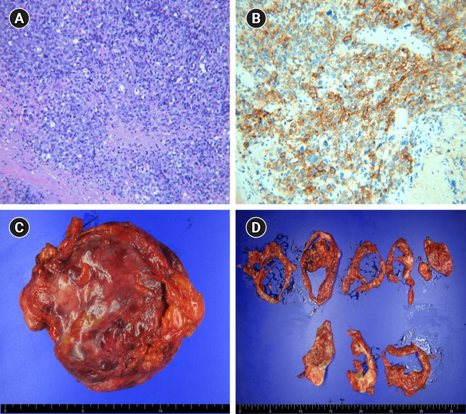
Fig. 2. Pathologic findings. (A) Tumor showing a predominantly solid pattern of highly anaplastic tumor cells and numerous mitotic figures (H&E, x200). (B) The tumor cells tested positive for CD30, a specific marker of embryonal carcinoma (x200). (C) Gross findings. A 12- to 15-cm cystic and solid mass was confined to the ovary with an intact capsule. (D) Cutting sections of the tumor.
Reference
-
References
1. Pectasides D, Pectasides E, Kassanos D. Germ cell tumors of the ovary. Cancer Treat Rev. 2008; 34:427–41.2. Euscher ED. Germ cell tumors of the female genital tract. Surg Pathol Clin. 2019; 12:621–49.3. Kammerer-Doak D, Baurick K, Black W, Barbo DM, Smith HO. Endodermal sinus tumor and embryonal carcinoma of the ovary in a 53-year-old woman. Gynecol Oncol. 1996; 63:133–7.4. Kurman RJ, Norris HJ. Embryonal carcinoma of the ovary: a clinicopathologic entity distinct from endodermal sinus tumor resembling embryonal carcinoma of the adult testis. Cancer. 1976; 38:2420–33.5. Hogg R, Friedlander M. Management of embryonal carcinoma of the ovary. CME J Gynecol Oncol. 2002; 7:234–7.6. Singh S, Gomathy E, Singh S, Kalyani R. Huge ovarian embryonal cell carcinoma in an adolescent girl: a case report. Indian J Obstet Gynecol Res. 2020; 7:133–5.7. Nasioudis D, Mastroyannis SA, Latif NA, Ko EM. Trends in the surgical management of malignant ovarian germcell tumors. Gynecol Oncol. 2020; 157:89–93.8. Turkmen O, Karalok A, Basaran D, Kimyon GC, Tasci T, Ureyen I, et al. Fertility-sparing surgery should be the standard treatment in patients with malignant ovarian germ cell tumors. J Adolesc Young Adult Oncol. 2017; 6:270–6.9. Smith HO, Berwick M, Verschraegen CF, Wiggins C, Lansing L, Muller CY, et al. Incidence and survival rates for female malignant germ cell tumors. Obstet Gynecol. 2006; 107:1075–85.10. Li J, Wu X. Current strategy for the treatment of ovarian germ cell tumors: role of extensive surgery. Curr Treat Options Oncol. 2016; 17:44.11. Brammer HM 3rd, Buck JL, Hayes WS, Sheth S, Tavassoli FA. From the archives of the AFIP. Malignant germ cell tumors of the ovary: radiologic-pathologic correlation. Radiographics. 1990; 10:715–24.
- Full Text Links
-
- Actions
-
Cited
- CITED
-
- Close
- Share
-
- Similar articles
-
- Ovarian Large Cell Neuroendocrine Carcinoma Associated with Endocervical-like Mucinous Borderline Tumor: A Case Report and Literature Review
- A Case of Testicular Embryonal Carcinoma
- A Case Report of Bilateral Squamous Cell Carcinoma Arising in Endometriosis of the Ovary
- Two Cases of Recovery of Ovarian Function and Spontaneous Pregnancy in Women Who Were Diagnosed as Premature Ovarian Failure
- Primary Epithelial Ovarian Carcinoma with Gastric Metastasis Mimic Gastrointestinal Stromal Tumor
