Small Cell Type Undifferentiated Carcinoma of Gall Bladder with Pas Positive Hyaline Globule Masquerading as Liver Mass: A Case Report and Literature Review
- Affiliations
-
- 1Department of Liver Transplantation & GI Surgery, Fortis Escort Liver and Digestive Institute, FEHI, New Delhi, India.
- 2Department of Gastroenterology and Hepatology, Fortis Escort Liver and Digestive Institute, FEHI, New Delhi, India.
- 3Department of Histopathology, SGT University Gurgaon, India.
- KMID: 2538687
- DOI: http://doi.org/10.4166/kjg.2022.100
Abstract
- An undifferentiated carcinoma (UC) of the gall bladder behaves aggressively and has a grave prognosis. Small cell type undifferentiated carcinoma of the gall bladder is a rare variant. This paper reports a case of UC of gall bladder with PAS-positive diastase-resistant eosinophilic hyaline globules present as liver mass (on imaging) in a male patient. The microscopic findings of the liver and gall bladder after a right tri-segmentectomy showed an un-differentiated malignant neoplasm composed of cells with round to oval nuclei, prominent nucleoli, and scanty neoplasm. No definite cell pattern was identified with these neoplastic cells. A section from the gall bladder revealed a tumor arising from the lining epithelium and infiltrating through the muscularis. This tumor was infiltrating the adherent liver tissue directly and forming a mass of undifferentiated malignant cells. The focal area within the tumor mass showed the presence of PAS-positive, diastase-resistant, eosinophilic hyaline globules within the neoplastic cells. The immunohistochemistry test was diffusely positive for perinuclear anti-neutrophil cytoplasmic antibodies and negative for chromogranin, vimentin, Desmin, alpha-fetoprotein, leukocyte common antigen, CD34, and bcl2. When the clinical and radiological data are inconclusive, careful analysis of the histological and immunophenotypic features is needed to make the final diagnosis of UC of the gall bladder. The biological behavior and prognosis of this tumor remain unclear because of its rarity. Further studies will be needed to understand the characteristics of this deadly tumor and to establish an effective therapy for it.
Keyword
Figure
-

Fig. 1 Contrast-enhanced CT scan. (A) Image showing a normal appearing gall bladder with a heterogeneous mass in segments 5 and 6 (arrow). (B) Image shows intensely enhancing huge vascular mass lesion occupying almost the entire right lobe of the liver.

Fig. 2 Gross finding of the resected specimen of gall bladder carcinoma. (A) Gross showing a large liver mass. (B) Gross cut surface showing whitish large liver mass. (C) Gross of the liver with gall bladder showing an infiltrative mass from the gall bladder directly infiltrating the liver.
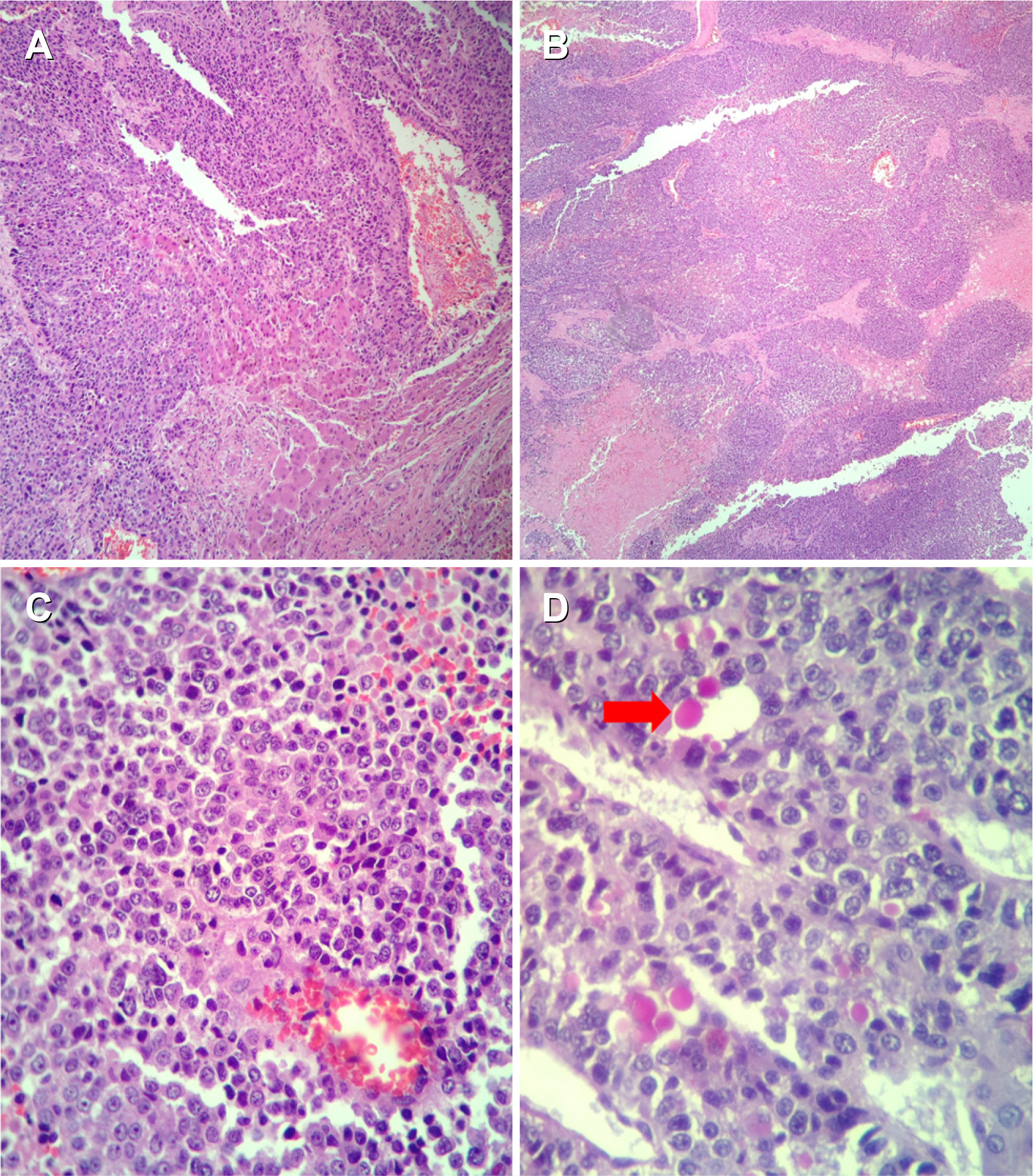
Fig. 3 Microscopic examination of the tumor mass. (A) Diffuse sheets of round tumor cells infiltrating hepatocytes (H&E, ×40). (B) Diffuse sheets of tumor cells (H&E, ×40). (C) Small round tumor cells (H&E, ×400). (D) PAS positive diastase resistant globules in the cytoplasm (arrow) (H&E, ×400). PAS, periodic acid-Schiff.
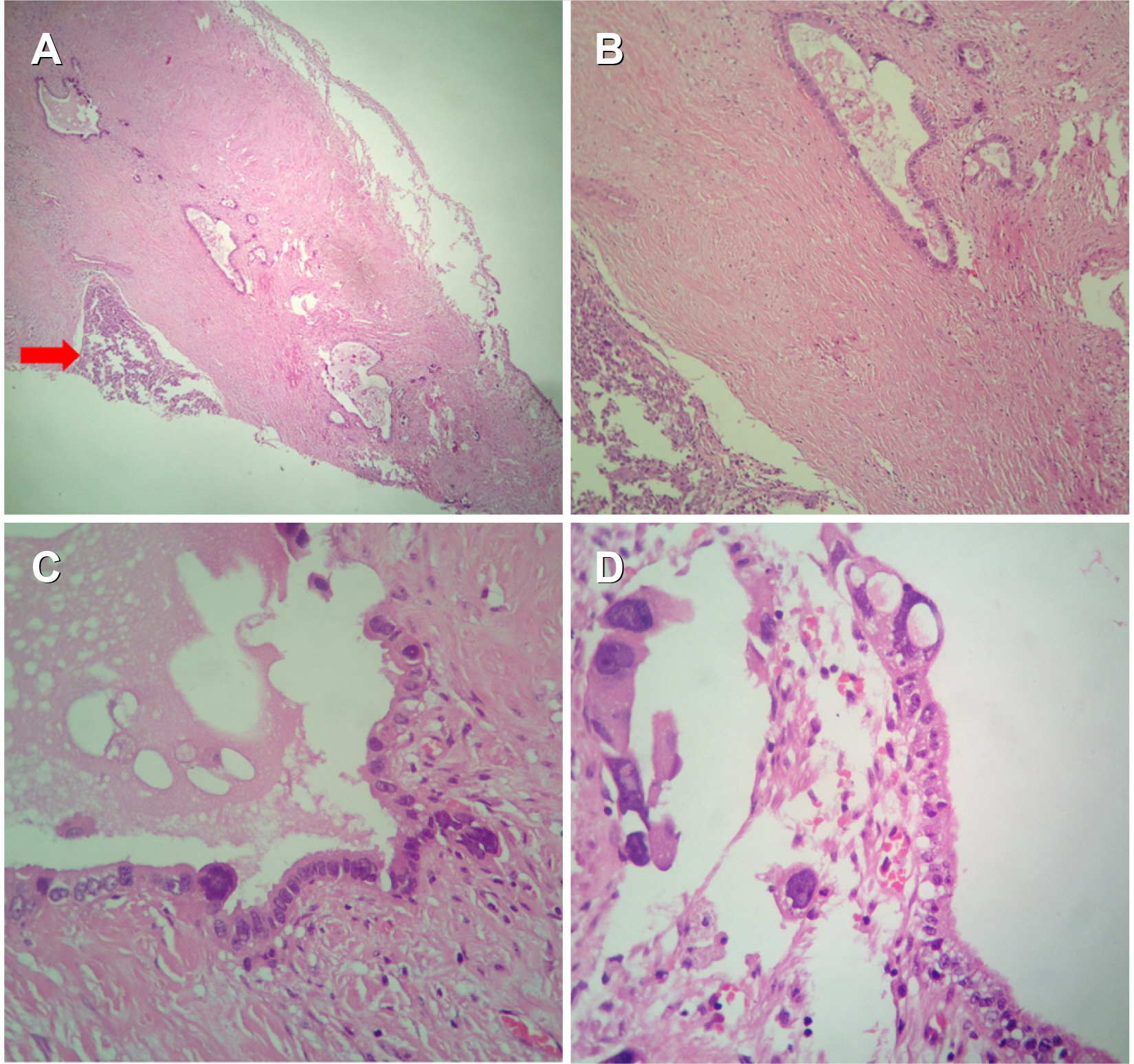
Fig. 4 Microscopic section from the gall bladder. (A) Tumor is seen arising from gall bladder (arrow) (H&E, ×40). (B) Malignant glands and cell nest (H&E, ×100). (C) Glands lined by malignant cells (H&E, ×400). (D) Tumor cells arising from lining epithelium (H&E, ×400).
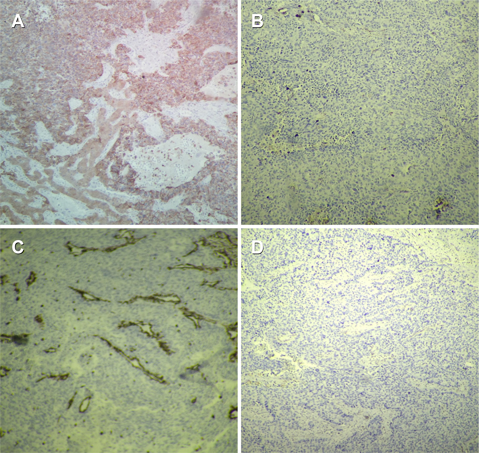
Fig. 5 Immuno-histochemistry. (A) IHC PanCK positive in tumor cells (H&E, ×100). (B) IHC LCA negative in tumor cells (H&E, ×100). (C) IHC vimentin negative in tumor cells (H&E, ×100). (D) IHC chromogranin negative in tumor cells (H&E, ×100). IHC, immunohistochemistry; LCA, leukocyte common antigen.
Reference
-
1. National Cancer Institute, tumour grade. [Internet]. Available from: https://www.cancer.gov/about-cancer/diagnosis-staging/prognosis/tumour-grade-fact-sheet. National Cancer Institute;cited 2014 Aug 18.2. Park HJ, Jang KT, Choi DW, Heo JS, Choi SH. 2014; Clinicopathologic analysis of undifferentiated carcinoma of the gallbladder. J Hepatobiliary Pancreat Sci. 21:58–63. DOI: 10.1002/jhbp.3. PMID: 23798378.
Article3. Guo KJ, Yamaguchi K, Enjoji M. 1988; Undifferentiated carcinoma of the gallbladder. A clinicopathologic, histochemical, and immunohistochemical study of 21 patients with a poor prognosis. Cancer. 61:1872–1879. DOI: 10.1002/1097-0142(19880501)61:9<1872::AID-CNCR2820610925>3.0.CO;2-Q. PMID: 2451557.
Article4. Manouras A, Genetzakis M, Lagoudianakis EE, et al. 2009; Undifferentiated giant cell type carcinoma of the gallbladder with sarcomatoid dedifferentiation: a case report and review of the literature. J Med Case Rep. 3:6496. DOI: 10.1186/1752-1947-3-6496. PMID: 19830109. PMCID: PMC2726484.
Article5. Kubota H, Kageoka M, Iwasaki H, et al. 2000; A patient with undifferentiated carcinoma of gallbladder presenting with hemobilia. J Gastroenterol. 35:63–68. DOI: 10.1007/PL00009979. PMID: 10632545.
Article6. Bosman FT. World Health Organization, International Agency for Research on Cancer. 2010. WHO classification of tumours of the digestive system. 4th ed. Lyon: International Agency for Research on Cancer.7. Nishihara K, Tsuneyoshi M. 1993; Undifferentiated spindle cell carcinoma of the gallbladder: a clinicopathologic, immunohistochemical, and flow cytometric study of 11 cases. Hum Pathol. 24:1298–1305. DOI: 10.1016/0046-8177(93)90263-G. PMID: 8276377.
Article8. Kubota K, Kakuta Y, Kawamura S, et al. 2006; Undifferentiated spindle-cell carcinoma of the gallbladder: an immunohistochemical study. J Hepatobiliary Pancreat Surg. 13:468–471. DOI: 10.1007/s00534-006-1100-x. PMID: 17013725.
Article9. Doval DC, Azam S, Mehta A, et al. 2014; A report of sarcomatoid carcinoma of the gallbladder treated with palliative deocetaxel and gemcitabine chemotherapy. J Gastrointest Cancer. 45 Suppl 1:270–274. DOI: 10.1007/s12029-014-9654-3. PMID: 25326734.
Article10. Akatsu T, Ueda M, Shimazu M, et al. 2005; Primary undifferentiated spindle-cell carcinoma of the gallbladder presenting as a liver tumor. J Gastroenterol. 40:993–998. DOI: 10.1007/s00535-005-1684-y. PMID: 16261437.
Article
- Full Text Links
-
- Actions
-
Cited
- CITED
-
- Close
- Share
-
- Similar articles
-
- A Case of Primary Undifferentiated Small Cell Carcinoma of the Urinary Bladder
- Metastatic Renal Cell Carcinoma of the Gallbladder
- Synchronous Triple Primary Cancers - Liver, Gallbladder and Pancreas
- Primary Undifferentiated Carcinoma of the Endometrium with Small Cell and Trophoblastic Differentiation
- Comprehensive Cytomorphologic Analysis of Pulmonary Adenoid Cystic Carcinoma: Comparison to Small Cell Carcinoma and Non-pulmonary Adenoid Cystic Carcinoma
