Ann Rehabil Med.
2022 Dec;46(6):303-311. 10.5535/arm.22110.
Correlation of Femoral Muscle Volume Using Three-Dimensional Modeling and Locomotor Function After Unilateral Trans-femoral Amputation
- Affiliations
-
- 1Department of Rehabilitation Medicine, Chungnam National University College of Medicine, Daejeon, Korea
- 2Department of Rehabilitation Medicine, Chungnam National University Sejong Hospital, Sejong, Korea
- KMID: 2537161
- DOI: http://doi.org/10.5535/arm.22110
Abstract
Objective
To evaluate the relationship between femoral muscle volume (FMV) and physiological outcomes after trans-femoral amputations (TFAs) affecting overall locomotor function in patients.
Methods
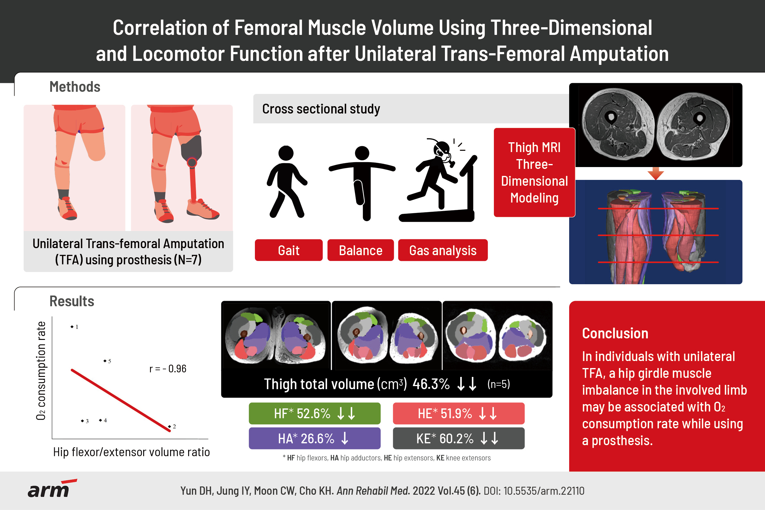
Seven individuals who underwent TFA and had been using a prosthesis participated in this cross-sectional study. Gait and balance were assessed using clinical tests, such as 10-m walk test, 6-minute walk test, Berg Balance Scale, and automatic balance system. Respiratory gas analysis was performed to check oxygen consumption rate. Five participants were evaluated for bilateral FMV by MR imaging and FMV was reconstructed using three-dimentional remodeling.
Results
In five participants, significant differences were found between the non-involved and involved sides in femur length, total FMV, and functional muscle volume (all p<0.01) in all groups except for the hip adductor volume. The %mean difference between the non-involved and involved sides was 30% for femur length, 52.55% for hip flexor volume, 26.55% for hip adductor volume, 51.86% for hip extensor volume, and 60.21% for knee extensor volume. The hip flexor volume to hip extensor volume ratio in the involved limb and oxygen consumption rate during comfortable gait were negatively correlated (r=-0.96, p=0.04).
Conclusion
In individuals who underwent unilateral TFA, hip girdle muscle imbalance in the involved limbs may be associated with oxygen consumption rate while using a prosthesis.
Keyword
Figure
-

Fig. 1. Three-dimensional modeling based on bilateral femoral magnetic resonance images. Hip flexor indicates shades of green; hip adductor, shades of purple; hip extensor, shades of red; knee extensor, shades of gray.

Fig. 2. Correlation of hip flexor/hip extensor volume ratio and oxygen consumption rate. Numbers in graph are same as ID in Table 1.
Reference
-
1. Ziegler-Graham K, MacKenzie EJ, Ephraim PL, Travison TG, Brookmeyer R. Estimating the prevalence of limb loss in the United States: 2005 to 2050. Arch Phys Med Rehabil. 2008; 89:422–9.2. Miller WC, Speechley M, Deathe B. The prevalence and risk factors of falling and fear of falling among lower extremity amputees. Arch Phys Med Rehabil. 2001; 82:1031–7.3. Langford J, Dillon MP, Granger CL, Barr C. Physical activity participation amongst individuals with lower limb amputation. Disabil Rehabil. 2019; 41:1063–70.4. Young AJ, Simon AM, Hargrove LJ. A training method for locomotion mode prediction using powered lower limb prostheses. IEEE Trans Neural Syst Rehabil Eng. 2014; 22:671–7.5. Isakov E, Burger H, Gregoric M, Marincek C. Stump length as related to atrophy and strength of the thigh muscles in trans-tibial amputees. Prosthet Orthot Int. 1996; 20:96–100.6. Genin JJ, Bastien GJ, Franck B, Detrembleur C, Willems PA. Effect of speed on the energy cost of walking in unilateral traumatic lower limb amputees. Eur J Appl Physiol. 2008; 103:655–63.7. Goktepe AS, Cakir B, Yilmaz B, Yazicioglu K. Energy expenditure of walking with prostheses: comparison of three amputation levels. Prosthet Orthot Int. 2010; 34:31–6.8. Waters RL, Mulroy S. The energy expenditure of normal and pathologic gait. Gait Posture. 1999; 9:207–31.9. Gottschalk FA, Stills M. The biomechanics of transfemoral amputation. Prosthet Orthot Int. 1994; 18:12–7.10. Thiele B, James U, Stalberg E. Neurophysiological studies on muscle function in the stump of above-knee amputees. Scand J Rehabil Med. 1973; 5:67–70.11. Rutkowska-Kucharska A, Kowal M, Winiarski S. Relationship between asymmetry of gait and muscle torque in patients after unilateral transfemoral amputation. Appl Bionics Biomech. 2018; 2018:5190816.12. Kowal M, Rutkowska-Kucharska A. Muscle torque of the hip joint flexors and extensors in physically active and inactive amputees. Biomed Hum Kinet. 2014; 6:63–8.13. Erskine RM, Jones DA, Maganaris CN, Degens H. In vivo specific tension of the human quadriceps femoris muscle. Eur J Appl Physiol. 2009; 106:827–38.14. Akagi R, Suzuki M, Kawaguchi E, Miyamoto N, Yamada Y, Ema R. Muscle size-strength relationship including ultrasonographic echo intensity and voluntary activation level of a muscle group. Arch Gerontol Geriatr. 2018; 75:185–90.15. Putz C, Block J, Gantz S, Heitzmann DW, Dreher T, Lehner B, et al. Structural changes in the thigh muscles following trans-femoral amputation. Eur J Orthop Surg Traumatol. 2017; 27:829–35.16. Leijendekkers RA, Marra MA, Ploegmakers MJ, Van Hinte G, Frolke JP, Van De Meent H, et al. Magnetic-resonance-imaging-based three-dimensional muscle reconstruction of hip abductor muscle volume in a person with a transfemoral bone-anchored prosthesis: a feasibility study. Physiother Theory Pract. 2019; 35:495–504.17. Jaegers SM, Arendzen JH, de Jongh HJ. Changes in hip muscles after above-knee amputation. Clin Orthop Relat Res. 1995; (319):276–84.18. Unver B, Baris RH, Yuksel E, Cekmece S, Kalkan S, Karatosun V. Reliability of 4-meter and 10-meter walk tests after lower extremity surgery. Disabil Rehabil. 2017; 39:2572–6.19. Major MJ, Fatone S, Roth EJ. Validity and reliability of the Berg Balance Scale for community-dwelling persons with lower-limb amputation. Arch Phys Med Rehabil. 2013; 94:2194–202.20. Donovan K, Lord SE, McNaughton HK, Weatherall M. Mobility beyond the clinic: the effect of environment on gait and its measurement in community-ambulant stroke survivors. Clin Rehabil. 2008; 22:556–63.21. Lusardi MM, Pellecchia GL, Schulman M. Functional performance in community living older adults. J Geriatr Phys Ther. 2003; 26:14–22.22. American College of Sports Medicine. ACSM’s resource manual for guidelines for exercise testing and prescription. 5th ed. Baltimore, MD: Lippincott Williams & Wilkins;2006.23. Downs S, Marquez J, Chiarelli P. Normative scores on the Berg Balance Scale decline after age 70 years in healthy community-dwelling people: a systematic review. J Physiother. 2014; 60:85–9.24. Batten HR, McPhail SM, Mandrusiak AM, Varghese PN, Kuys SS. Gait speed as an indicator of prosthetic walking potential following lower limb amputation. Prosthet Orthot Int. 2019; 43:196–203.25. Wentink EC, Prinsen EC, Rietman JS, Veltink PH. Comparison of muscle activity patterns of transfemoral amputees and control subjects during walking. J Neuroeng Rehabil. 2013; 10:87.26. Hong JH, Mun MS. Relationship between socket pressure and EMG of two muscles in trans-femoral stumps during gait. Prosthet Orthot Int. 2005; 29:59–72.
- Full Text Links
-
- Actions
-
Cited
- CITED
-
- Close
- Share
-
- Similar articles
-
- Femoral Neck Fracture in Bilateral Above Knee Amputee: A Case Report
- Relationship between Femoral Anteversion and Tibial Torsion in Intoeing Gait
- Studies on Femoral Neuralgia
- Relationship between Lateral Femoral Bowing and Varus Knee Deformity Based on Two-Dimensional Assessment of Side-to-Side Differences
- A Clinical Study of Slipped Capital Femoral epiphysis
