Endovascular Coiling of Fenestrated Vertebrobasilar Cerebral Aneurysms
- Affiliations
-
- 1Department of Vascular & Interventional Radiology, Loma Linda University Medical Center, Loma Linda, CA, USA
- 2Department of Vascular & Interventional Radiology, University of California–Irvine, Orange, CA, USA
- 3Department of Neurosurgery, Loma Linda University Medical Center, Loma Linda, CA, USA
- KMID: 2535956
- DOI: http://doi.org/10.5469/neuroint.2022.00360
Abstract
- Fenestrated vertebrobasilar junction aneurysms are uncommon vascular lesions. Surgical intervention remains extremely challenging due to the deep location and complex anatomy with adjacent cranial nerves and perforator vessels. Endovascular approach is safer and generally accepted as the primary treatment method. Optimal angiographic projections with three-dimensional reconstructions to guide microcatheter selection remain vital to successfully treating aneurysms with challenging fenestration anatomy. This report details the endovascular methods in two cases of fenestrated vertebrobasilar junction aneurysms with different coiling techniques.
Figure
-
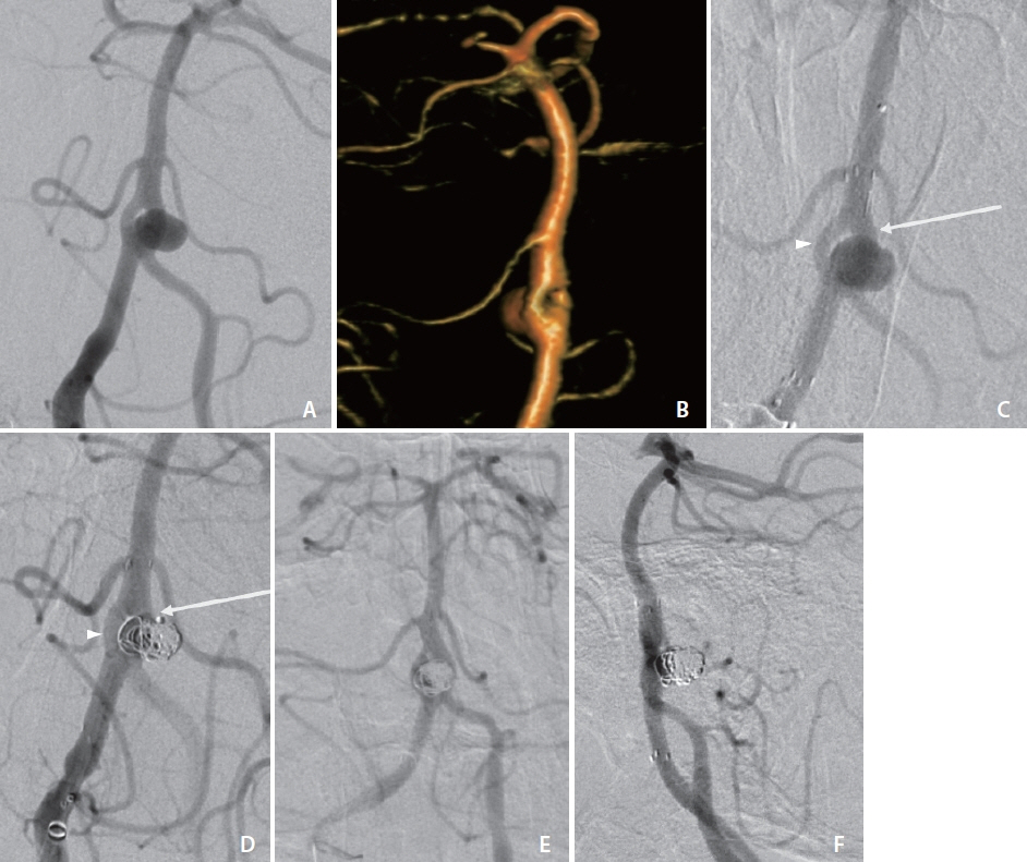
Fig. 1. Angiogram (A) and three-dimensional volume rendering (B) with obliqued view of case 1 demonstrating broad-based aneurysm arising from the vertebrobasilar artery proximal fenestration branchpoint with involvement of both fenestration branches. (C) Angiogram of case 1 following deployment of Neuroform Atlas stent through the dominant fenestration limb (arrow) across the aneurysm ostium. Additional microcatheter remains within non-dominant fenestration limb (arrowhead). (D) Coil deployment in case 1 via the dominant fenestration limb (arrow) through the Atlas stent interstices and (not shown) subsequently through the non-dominant fenestration limb (arrowhead). Thirteen-month surveillance cerebral angiogram in frontal (E) and lateral (F) views demonstrating no contrast opacification of the aneurysm and stable stent positioning.

Fig. 2. Angiogram (A) and three-dimensional (3D) volume rendering (B) of case 2 with obliqued view demonstrating narrow neck aneurysm arising from the vertebrobasilar artery proximal fenestration branchpoint, symmetric at bifurcation. Lateral view angiograms before (C) and following (D) primary coiling of case 2 with single microsystem. Eight-month surveillance cerebral angiograms in frontal (E) and lateral (F) views and 3D volume rendering (G) demonstrating contrast opacification of 1×2 mm neck remnant (arrows).
Reference
-
1. Kai Y, Hamada J, Morioka M, Yano S, Fujioka S, Kuratsu J. Endovascular treatment of ruptured aneurysms associated with fenestrated basilar artery. Two case reports. Neurol Med Chir (Tokyo). 2006; 46:244–247.
Article2. Campos J, Fox AJ, Viñuela F, Lylyk P, Ferguson GG, Drake CG, et al. Saccular aneurysms in basilar artery fenestration. AJNR Am J Neuroradiol. 1987; 8:233–236.3. Albanese E, Russo A, Ulm AJ. Fenestrated vertebrobasilar junction aneurysm: diagnostic and therapeutic considerations. J Neurosurg. 2009; 110:525–529.
Article4. Alqahtani SA, Felbaum DR, Tai A, Liu AH, Armonda RA. Endovascular treatment of large unruptured fusiform fenestrated vertebrobasilar junction aneurysm. Cureus. 2017; 9:e1219.
Article5. Vajpeyee A, Goyal G, Kant R, Mal N. Double microcatheter-assisted coiling of a basilar artery fenestration aneurysm. Neurointervention. 2013; 8:125–126.
Article6. Trivelato FP, Abud DG, Nakiri GS, de Castro Afonso LH, Ulhoa AC, Manzato LB, et al. Basilar artery fenestration aneurysms: endovascular treatment strategies based on 3D morphology. Clin Neuroradiol. 2016; 26:73–79.
Article7. Graves VB, Strother CM, Weir B, Duff TA. Vertebrobasilar junction aneurysms associated with fenestration: treatment with Guglielmi detachable coils. AJNR Am J Neuroradiol. 1996; 17:35–40.8. Miyamoto N, Ueno Y, Hira K, Kijima C, Nakajima S, Yamashiro K, et al. Characteristics of clinical symptoms, cerebral images and stroke etiology in vertebro-basilar artery fenestration-related infarction. Brain Sci. 2020; 10:243.
Article9. Tanaka M, Kikuchi Y, Ouchi T. Neuroradiological analysis of 23 cases of basilar artery fenestration based on 2280 cases of MR angiographies. Interv Neuroradiol. 2006; 12(Suppl 1):39–44.
Article10. Zhu DY, Fang YB, Wu YN, Li Q, Duan GL, Liu JM, et al. Treatment of fenestrated vertebrobasilar junction-related aneurysms with endovascular techniques. J Clin Neurosci. 2016; 28:112–116.
Article11. Peluso JP, van Rooij WJ, Sluzewski M, Beute GN. Aneurysms of the vertebrobasilar junction: incidence, clinical presentation, and outcome of endovascular treatment. AJNR Am J Neuroradiol. 2007; 28:1747–1751.
Article12. Lempert TE, Malek AM, Halbach VV, Phatouros CC, Meyers PM, Dowd CF, et al. Endovascular treatment of ruptured posterior circulation cerebral aneurysms. Clinical and angiographic outcomes. Stroke. 2000; 31:100–110.
Article13. Nelson PK, Sahlein D, Shapiro M, Becske T, Fitzsimmons BF, Huang P, et al. Recent steps toward a reconstructive endovascular solution for the orphaned, complex-neck aneurysm. Neurosurgery. 2006; 59(5 Suppl 3):S77-S92; discussion S3–S13.
Article14. Islak C, Kocer N, Kantarci F, Saatci I, Uzma O, Canbaz B. Endovascular management of basilar artery aneurysms associated with fenestrations. AJNR Am J Neuroradiol. 2002; 23:958–964.15. Aydin K, Arat A, Sencer S, Barburoglu M, Men S. Stent-assisted coiling of wide-neck intracranial aneurysms using low-profile LEO baby stents: initial and midterm results. AJNR Am J Neuroradiol. 2015; 36:1934–1941.
Article
- Full Text Links
-
- Actions
-
Cited
- CITED
-
- Close
- Share
-
- Similar articles
-
- Kissing Aneurysms at Fenestrated Proximal Basilar Artery: Double-barrel Stent-assisted Coiling Using Dual Closed-cell Stents
- Seven Intracranial Aneurysms in One Patient: Treatment and Review of Literature
- Application of Fenestrated Clip in the Intracranial Aneurysms: Report of Four Cases
- Endovascular treatment of intracranial aneurysms: Past and present
- Stent Application for the Treatment of Cerebral Aneurysms
