Stent Application for the Treatment of Cerebral Aneurysms
- Affiliations
-
- 1Interventional Neuroradiology, Department of Radiology, Yonsei University College of Medicine Severance Hospital, Seoul, Korea. bmoon21@hanmail.net
- KMID: 1783985
- DOI: http://doi.org/10.5469/neuroint.2011.6.2.53
Abstract
- Rapid and striking development in both the techniques and devices make it possible to treat most of cerebral aneurysms endovascularly. Stent has become one of the most important tools in treating difficult aneurysms not feasible for simple coiling. The physical features, the dimensions, and the functional characteristics of the stents show considerable differences. There are also several strategies and tips to treat difficult aneurysms by using stent and coiling. Nevertheless, they require much experience in clinical practice as well as knowledge of the stents to treat cerebral aneurysms safely and effectively. In this report, a brief review of properties of the currently available stents and strategies of their application is presented.
Figure
-
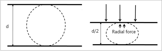
Fig. 1 Radial force measured by two plate method.

Fig. 2 Outward radial force measured by thin film method.

Fig. 3 Kinking or hugging

Fig. 4 Ovalization

Fig. 5 A. Two aneurysms at the basilar artery (BA) tip and BA-superior cerebellar artery (SCA).B. Several coil loops are deployed in the SCA aneurysm sac before stent deployment.C. After stent deployment, the extruded catheter tip is re-inserted into the sac over the pre-deployed coil loops.D. Complete coiling is performed after Y-stenting through the struts. Arrow head indicates coil mass in the BA-SCA aneurysm and white arrow indicates the catheter tip located in the BA aneurysm sac.

Fig. 6 Semi-jailing technique.A. A saccular aneurysm at the left middle cerebral artery (MCA) inferior division proximal portion, and a fusiform aneurysm of the inferior division just distal to the saccular aneurysm.B. A Solitaire stent is partially deployed after partial deployment of coil loops. A short arrow indicates distal markers of the Solitaire stent and a long arrow indicates the distal tip of a Prowler plus select catheter.C. Coiling is stably performed without catheter kickback owing to the partially deployed stent.D. After complete coil embolization, the stent is retrieved and is removed. In this case, the stent is used for the purpose of preventing anticipated catheter kickback due to the distal flaring of the parent artery. Balloon is also anticipated to be unstable due to a distal flaring of the artery. The stent is removed after complete coiling for the further treatment of a distal fusiform aneurysm of the parent artery by using flow diverter.
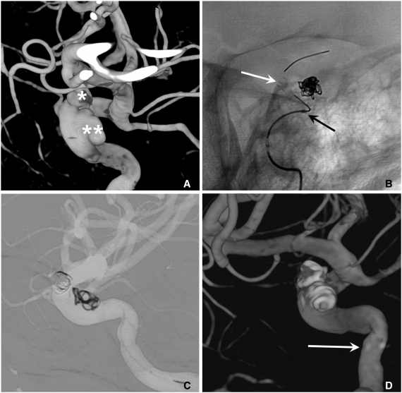
Fig. 7 A. Two aneurysms at the para-ophthalmic internal carotid artery (ICA) (*) and cavernous ICA (**).B. Coiling is performed after partial deployment of an Enterprise stent (white arrow: distal marker of stent, black arrow: proximal marker of stent).C. After completion of coil embolization of the para-ophthalmic aneurysm, the stent is partially re-sheathed. The catheter tip is repositioned into the cavernous aneurysm sac and coiling is performed after permanent placement of the stent covering both aneurysm necks.D. The 3D-reconstruction image after completion of coiling of both aneurysms. Arrow indicates proximal markers of the Enterprise stent.

Fig. 8 A. A wide necked right MCA aneurysm.B. Because catheterization of inferior division failed, coiling is performed with two catheters technique.C. The catheter is easily navigated into the inferior division after partial coiling of the aneurysm sac.D. Complete coil embolization is performed after a stent placement from M1 trunk to the inferior division.
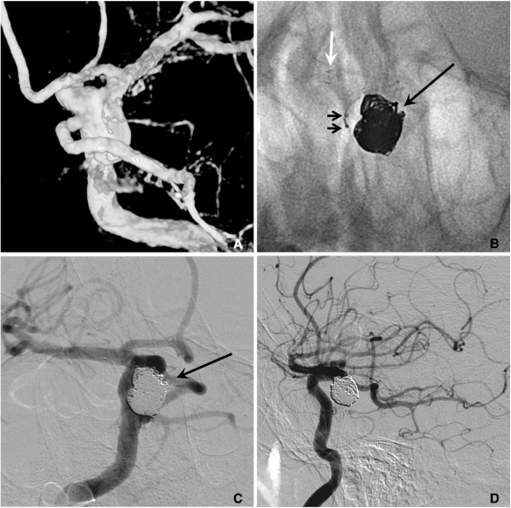
Fig. 9 A. A large aneurysm with a fetal type posterior cerebral artery (PCA) incorporated into the sac at the ICA posterior communicating artery (PComA) region.B. After a Neuroform stent placement, coiling is performed with two catheters technique for saving the origin of the fetal type PCA (white and long arrows: markers of stent, short arrows: distal markers of two microcatheters).C, D. The aneurysm sac is completely embolized with saving the origin (a long black arrow) of the fetal type PCA. A white arrow indicates distal markers of the deployed Neuroform stent and short arrows indicate proximal markers of two microcatheters.
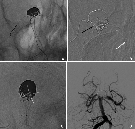
Fig. 10 A large aneurysm at the basilar artery tip.A. After Enterprise stent placement from BA to left PCA, the prowler plus select catheter is re-navigated to the left PCA over the stent loading wire left in-stent following the 1st stent deployment and a Hyperform balloon is placed at the right PCA through the stent struts.B. Coiling is completed by balloon- and stent-assisted technique. The black arrow indicates the Hyperform balloon and the white arrow indicates the distal tip of the Prowler plus select microcatheter.C. The 2nd Enterprise stent is placed using stent-within-stent technique for the purpose of saving the PCA lumen and promotion of flow diversion. Arrows indicate the patent left PCA.D. The 18-month follow-up angiogram reveals the stable occlusion state of the aneurysm sac and well preserved both PCAs.

Fig. 11 Horizontal stenting via circle of Willis (courtesy of Prf. Han MH in Seoul National University Hospital).A. A wide necked basilar tip aneurysm.B, C. Horizontal placement of an Enterprise stent (arrows) is performed from right PCA to left PCA via right PComA.D. The final control angiogram reveals complete occlusion of the aneurysm.
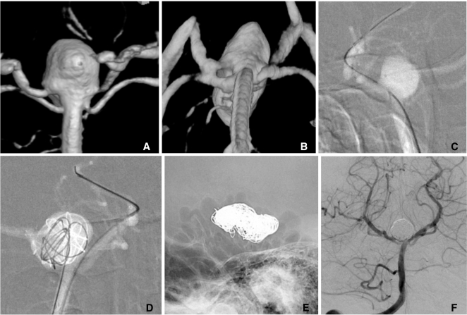
Fig. 12 Y-configuration stents through the struts.A, B. A large basilar tip aneurysm.C, D. Coil embolization is performed after placement of 2 stents with Y-configuration through the struts. A native lateral (E) and a control angiogram (F) show complete embolized state of the aneurysm and well preserved bilateral PCAs.

Fig. 13 Kissing 2 stents with Y-configuration (courtesy of Prf. Kim DI in Severance Hospital).A. A wide-necked aneurysm at the BA tip.B. Two Prowler plus microcatheters are navigated to both PCAs and 2 Enterprise stents are loaded in the catheters, respectively.C. Coiling is conducted after simultaneous deployment of 2 Enterprise stents from BA to both PCAs, respectively.D. The final control angiogram shows near complete occlusion of the aneurysm sac and well preserved both PCAs.

Fig. 14 Two stents with X-configuration (courtesy of Prf. Suh SH in Gangnam Severance Hospital).A, B. A large recurred aneurysm at the vertebrobasilar fenestration.C. Two Prowler plus select microcatheters (arrows) are navigated through the both arm of the fenestration, respectively, crossed at the aneurysm neck.D. After 2 stents are deployed in X-configuration at the aneurysm neck, complete coil embolization is performed.
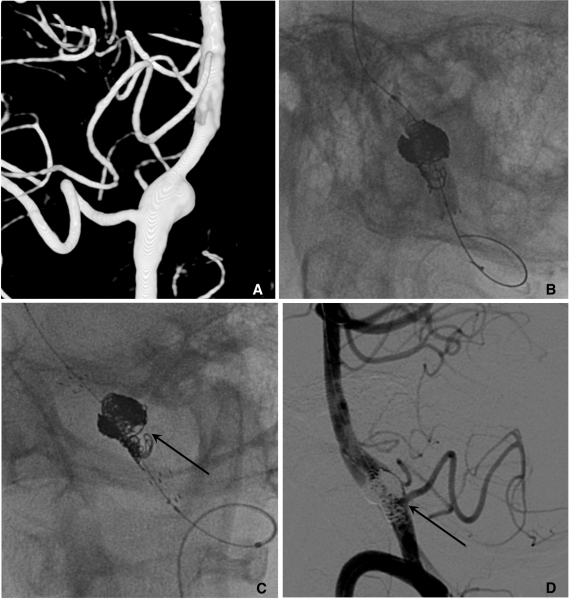
Fig. 15 Balloon-in-stent technique.A. A ruptured fusiform dissecting aneurysm of left vertebral artery (VA) with involvement of the segment bearing posterial inferior cerebellar artery (PICA) origin.B. After placement of a Neuroform stent, coiling is conducted with balloon-in-stent-technique.C. The 2nd Neuroform stent is (arrow) placed with stent-with-stent technique after completion of coiling.D. The final control angiogram reveals near complete occlusion of the dissecting aneurysm and well preserved VA and PICA. The black arrow indicates the origin of PICA.
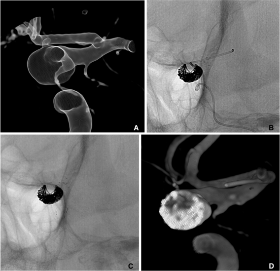
Fig. 16 Multiple overlapping Enterprise stents and coiling.A. The cross sectional image of 3D reconstruction shows an ultra-wide necked circumferential aneurysm of the ICA ophthalmic region.B. The 1st Enterprise stent is placed and the Prowler plus select microcatheter is re-navigated over the stent-loading wire left in-stent following the 1st stent placement. Then the stent-assisted coiling is performed.C. Finally, the 2nd Enterprise stent is placed with stent-within-stent technique.D. The cross sectional image of 3D reconstruction shows circumferential coil masses around the parent artery.
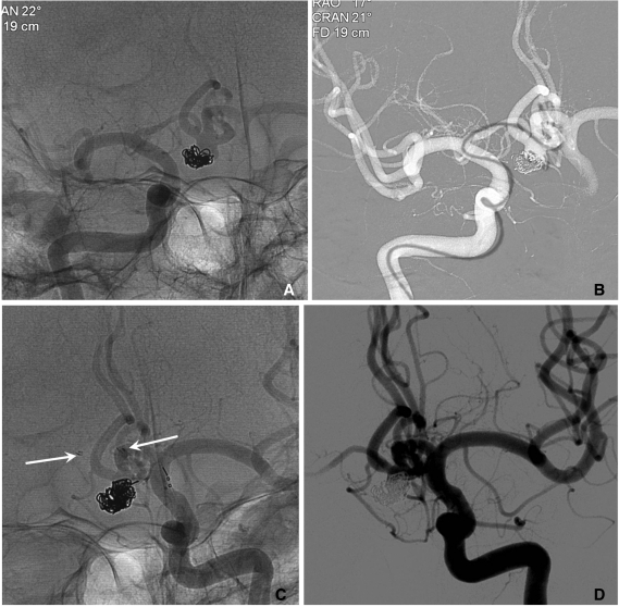
Fig. 17 A. A recurred AcomA aneurysm after coiling (courtesy of Prf. Yoon PH in Ilsan Hospital).B. A Prowler plus microcatheter is navigated to left ACA A2 portion across the aneurysm neck via right ACA A1.C. Coiling is performed after placement of an Enterprise stent (arrows) from right ACA A1 to left ACA A2 portion.D. The final control angiogram reveals near complete occlusion of the aneurysm sac.

Fig. 18 A, B. Images of 3D reconstruction and working projection show a wide necked aneurysm at the left VA-PICA origin.C. Stent-assisted coiling is performed. Arrows indicate proximal and distal markers of the Enterprise stent.D. The final control angiogram reveals complete occlusion of the aneurysm sac and widening of VA-PICA angle.
Cited by 10 articles
-
Initial Experience with the New Double-lumen Scepter Balloon Catheter for Treatment of Wide-necked Aneurysms
Myung Ho Rho, Byung Moon Kim, Sang Hyun Suh, Dong Joon Kim, Dong Ik Kim
Korean J Radiol. 2013;14(5):832-840. doi: 10.3348/kjr.2013.14.5.832.A Newly-Developed Flow Diverter (FloWise) for Internal Carotid Artery Aneurysm: Results of a Pilot Clinical Study
Byung Moon Kim, Keun Young Park, Jae Whan Lee, Joonho Chung, Dong Joon Kim, Dong Ik Kim
Korean J Radiol. 2019;20(3):505-512. doi: 10.3348/kjr.2018.0421.Clinical and Angiographic Outcomes of Aneurysms Treated with Two Self-expanding Stent-assisted Coiling Systems: A Comparison of Solitaire AB and Enterprise VRD Stents
Sung-Won Kim, Seng-Oun Sung, Kil-Sung Chae, Hwa-Seung Park, Sang-Hoon Lee
J Cerebrovasc Endovasc Neurosurg. 2015;17(3):149-156. doi: 10.7461/jcen.2015.17.3.149.Stent-Jack Technique for Ruptured Vertebral Artery Dissecting Aneurysm Involving the Origin of Posterior Inferior Cerebellar Artery
Toshitsugu Terakado, Yasunobu Nakai, Go Ikeda, Kazuaki Tsukada, Sho Hanai, Kazuki Akutagawa, Haruki Igarashi, Takahiro Konishi, Masanari Shiigai, Kazuya Uemura
Neurointervention. 2020;15(2):84-88. doi: 10.5469/neuroint.2019.00276.Pipeline Embolization Device for Large/Giant or Fusiform Aneurysms: An Initial Multi-Center Experience in Korea
Byung Moon Kim, Yong Sam Shin, Min Woo Baik, Deok Hee Lee, Pyoung Jeon, Seung Kug Baik, Tae Hong Lee, Dong-Hoon Kang, Sang-il Suh, Jun Soo Byun, Jin-Young Jung, Kihun Kwon, Dong Joon Kim, Keun Young Park, Bum-soo Kim, Jung Cheol Park, Seong Rim Kim, Young Woo Kim, Hoon Kim, Kyungil Jo, Chang Hyo Yoon, Young Soo Kim
Neurointervention. 2016;11(1):10-17. doi: 10.5469/neuroint.2016.11.1.10.Easy Advancement of a Large-Profile Microcatheter (Excelsior XT27™) by Parallel Use of Two Microguidewires For Stent Delivery
Yudhi Adrianto, Ku Hyun Yang, Hae-Won Koo, Wonhyoung Park, Jung Cheol Park, Deok Hee Lee
Neurointervention. 2016;11(1):24-29. doi: 10.5469/neuroint.2016.11.1.24.Comparison Between Balloon-Assisted and Stent-Assisted Technique for Treatment of Unruptured Internal Carotid Artery Aneurysms
Keun Young Park, Byung Moon Kim, Dong Joon Kim
Neurointervention. 2016;11(2):99-104. doi: 10.5469/neuroint.2016.11.2.99.Bench-top Comparison of Physical Properties of 4 Commercially-Available Self-Expanding Intracranial Stents
Su-hee Cho, Won-il Jo, Ye-eun Jo, Ku Hyun Yang, Jung Cheol Park, Deok Hee Lee
Neurointervention. 2017;12(1):31-39. doi: 10.5469/neuroint.2017.12.1.31.Semi-Jailing Technique Using a Neuroform3 Stent for Coiling of Wide-Necked Intracranial Aneurysms
Jun Kyeung Ko, Won Ho Cho, Seung Heon Cha, Chang Hwa Choi, Sang Weon Lee, Tae Hong Lee
J Korean Neurosurg Soc. 2017;60(2):146-154. doi: 10.3340/jkns.2016.0607.002.Delayed Rupture of an Anterior Communicating Artery Pseudoaneurysm Caused by Distal Occlusion Thrombectomy Using a Stent Retriever: A Case Report and Mechanism of Injury
Dong-Hyun Shim, Youngrok Do, Jin Kuk Do, Sung Won Youn
Neurointervention. 2022;17(2):121-125. doi: 10.5469/neuroint.2022.00101.
Reference
-
1. Higashida RT, Smith W, Gress D, Urwin R, Dowd CF, Balousek PA, et al. Intravascular stent and endovascular coil placement for a ruptured fusiform aneurysm of the basilar artery. Case report and review of the literature. J Neurosurg. 1997; 87:944–949. PMID: 9384409.2. Lylyk P, Ceratto R, Hurvitz D, Basso A. Treatment of a vertebral dissecting aneurysm with stents and coils: technical case report. Neurosurgery. 1998; 43:385–388. PMID: 9696097.
Article3. Lylyk P, Cohen JE, Ceratto R, Ferrario A, Miranda C. Combined endovascular treatment of dissecting vertebral artery aneurysms by using stents and coils. J Neurosurg. 2001; 94:427–432. PMID: 11235947.
Article4. Lylyk P, Cohen JE, Ferrario A, Ceratto R, Miranda C. Partially clipped intracranial aneurysm obliterated with combined stent and coil implantation. J Endovasc Ther. 2002; 9:160–164. PMID: 12010094.
Article5. Mohammed MI, Sandhu JS, Wakhloo AK. Stent-assisted coil placement in a wide-necked persistent trigeminal artery aneurysm with jailing of the trigeminal artery: a case report. AJNR Am J Neuroradiol. 2002; 23:437–441. PMID: 11901014.6. Henkes H, Bose A, Felber S, Miloslavski E, Berg-Dammer E, Kühne D. Endovascular coil occlusion of intracranial aneurysms assisted by a novel self-expandable nitinol microstent (neuroform). Interv Neuroradiol. 2002; 8:107–119. PMID: 20594519.
Article7. Howington JU, Hanel RA, Harrigan MR, Levy EI, Guterman LR, Hopkins LN. The Neurofrom stent, the first microcatheter-delivered stent for use in the intracranial circulation. Neurosurgery. 2004; 54:2–5. PMID: 14683535.8. Fiorella D, Albuquerque FC, Han P, Mcdougall CG. Preliminary experience using the Neuroform stent for the treatment of cerebral aneurysms. Neurosurgery. 2004; 54:6–16. PMID: 14683536.
Article9. Krischek O, Miloslavski E, Fischer S, Shrivastava S, Henkes H. A comparison of functional and physical properties of self-expanding intracranial stents [Neuroform3, Wingspan, Solitaire, Leo(+), Enterprise]. Minim Invasive Neurosurg. 2011; 54:21–28. PMID: 21506064.
Article10. Spelle L, Piotin M, Mounayer C, Moret J. Saccular aneurysms: endovascular treatment-devices, techniques and strategies, management of complications, results. Neuroimaging Clin N Am. 2006; 16:413–451. PMID: 16935709.11. Biondi A, Janardhan V, Katz JM, Salvaggio K, Riina HA, Gobin YP. Neuroform stent-assisted coil embolization of wide-neck intracranial aneurysms: strategies in stent deployment and midterm follow-up. Neurosurgery. 2007; 61:460–468. PMID: 17881956.12. Hong B, Patel NV, Gounis MJ, DeLeo MJ 3rd, Linfante I, Wojak JC, et al. Semi-jailing technique for coil embolization of complex, wide-necked intracranial aneurysms. Neurosurgery. 2009; 65:1131–1138. PMID: 19934972.
Article13. Gao X, Liang G, Li Z, Qu H, Wei X. Stent-assisted coil embolization of wide-necked intracranial aneurysms using a semi-deployment technique: angiographic and clinical outcomes in 31 consecutive patients. Interv Neuroradiol. 2010; 16:385–393. PMID: 21162768.
Article14. Thorell WE, Chow MM, Woo HH, Masaryk TJ, Rasmussen PA. Y-configured dual intracranial stent-assisted coil embolization for the treatment of wide-necked basilar tip aneurysms. Neurosurgery. 2005; 56:1035–1040. PMID: 15854251.15. Rohde S, Bendszus M, Hartmann M, Hähnel S. Treatment of a wide-necked aneurysm of the anterior cerebral artery using two Enterprise stents in "Y"-configuration stenting technique and coil embolization: a technical note. Neuroradiology. 2010; 52:231–235. PMID: 19844699.
Article16. Lozen A, Manjila S, Rhiew R, Fessler R. Y-stent-assisted coil embolization for the management of unruptured cerebral aneurysms: report of six cases. Acta Neurochir (Wien). 2009; 151:1663–1672. PMID: 19618104.
Article17. Spiotta AM, Gupta R, Fiorella D, Gonugunta V, Lobo B, Rasmussen PA, et al. Mid-term results of endovascular coiling of wide-necked aneurysms using double stents in "Y-configuration". Neurosurgery. 2011; 3. [Epub ahead of print].18. Yang TH, Wong HF, Yang MS, Ou Ch, Ho TL. "Waffle cone" technique for intra/extra-aneurysmal stent placement for the treatment of complex and wide-necked bifurcation aneurysm. Interv Neuroradiol. 2008; 14(Suppl 2):49–52. PMID: 20557801.
Article19. Sychra V, Klisch J, Werner M, Dettenborn C, Petrovitch A, Strasilla C, et al. Waffle-cone technique with Solitaire AB remodeling device: endovascular treatment of highly selected complex cerebral aneurysms. Neuroradiology. 2010; 9. [Epub ahead of print].
Article20. Kelly ME, Turner R, Gonugunta V, Woo HH, Rasmussen PA, Masaryk TJ, et al. Stent reconstruction of wide-necked aneurysms across the circle of Willis. Neurosurgery. 2007; 61(5 Suppl 2):249–254. PMID: 18091239.
Article21. Siddiqui MA, Bhattacharya J, Lindsay KW, Jenkins S. Horizontal stent-assisted coil embolization of wide-necked intracranial aneurysms with the Enterprise stent-a case series with early angiographic follow-up. Neuroradiology. 2009; 51:411–418. PMID: 19277620.22. de Paula Lucas C, Piotin M, Spelle L, Moret J. Stent-jack technique in stent-assisted coiling of wide-neck aneurysms. Neurosurgery. 2008; 62(5 Supple 2):ONS414–ONS416. PMID: 18596523.23. Menendez JY, Harrigan MR. X-configuration stent-assisted coiling. World Neurosurg. 2010; 74:143–144. PMID: 21300004.
Article24. Lubicz B. Linear stent-assisted coiling: another way to treat very wide-necked intracranial aneurysms. Neuroradiology. 2011; 53:457–459. PMID: 21088964.
Article25. Fiorella D, Albuquerque FC, Masaryk TJ, Rasmussen PA, McDougall CG. Balloon-in-stent technique for the constructive endovascular treatment of "ultra-wide necked" circumferential aneurysms. Neurosurgery. 2005; 57:1218–1227. PMID: 16331170.
Article26. Park SI, Kim BM, Kim DI, Shin YS, Suh SH, Chung EC, et al. Clinical and angiographic follow-up of stent-only therapy for acute intracranial vertebrobasilar artery dissecting aneurysms. AJNR Am J Neuroradiol. 2009; 30:1351–1356. PMID: 19342544.27. Heller RS, Malek AM. Delivery technique plays an important role in determining vessel wall apposition of the Enterprise self-expanding intracranial stent. J Neurointervent Surg. 2011; 7. [Epub].
Article28. Kim BM, Park SI, Kim DJ, Kim DI, Suh SH, Kwon TH, et al. Endovascular coil embolization of aneurysms with a branch incorporated into the sac. AJNR Am J Neuroradiol. 2010; 31:145–151. PMID: 19749218.
Article29. Kwon OK, Kim SH, Kwon BJ, Kang HS, Kim JH, Oh CW, et al. Endovascular treatment of wide-necked aneurysms by using two microcatheters: techniques and outcomes in 25 patients. AJNR Am J Neuroradiol. 2005; 26:894–890. PMID: 15814940.30. Fiorella D, Woo HH, Albuquerque FC, Nelson PK. Definitive reconstruction of circumferential, fusiform intracranial aneurysms with the pipeline embolization device. Neurosurgery. 2008; 62:1115–1120. PMID: 18580809.
Article31. Fiorella D, Kelly ME, Albuquerque FC, Nelson PK. Curative reconstruction of a giant midbasilar trunk aneurysm with the pipeline embolization device. Neurosurgery. 2009; 64:212–217. PMID: 19057425.
Article32. Turowski B, Macht S, Kulcsár Z, Hänggi D, Stummer W. Early fatal hemorrhage after endovascular cerebral aneurysm treatment with a flow diverter (SILK-Stent): do we need rethink our concepts? Neuroradiology. 2011; 53:37–41. PMID: 20339842.33. Kulcsár Z, Houdart E, Bonafé A, Parker G, Millar J, Goddard AJ, et al. Intra-aneurysmal thrombosis as a possible cause of delayed aneurysm rupture after flow-diversion treatment. AJNR Am J Neuroradiol. 2011; 32:20–25. PMID: 21071538.
Article34. Lubicz B, Collignon L, Raphaeli G, Pruvo JP, Bruneau M, De Witte O, et al. Flow-diverter stent for the endovascular treatment of intracranial aneurysms: a prospective study in 29 patients with 34 aneurysms. Stroke. 2010; 41:2247–2253. PMID: 20798369.35. Suh SH, Kim BM, Park SI, Kim DI, Shin YS, Kim EJ, et al. Stent-assisted coil embolization followed by a stent-within-a-stent technique for ruptured dissecting aneurysms of the intracranial vertebrobasilar artery. J Neurosurg. 2009; 111:48–52. PMID: 19326976.
Article36. Lee BH, Kim BM, Park MS, Park SI, Chung EC, Suh SH, et al. Reconstructive endovascular treatment of ruptured blood blister-like aneurysms of the internal carotid artery. J Neurosurg. 2009; 110:431–436. PMID: 19046039.
Article37. Fiorella D, Albuquerque FC, Woo H, Rasmussen PA, Masaryk TJ, McDougall CG. Neuroform in-stent stenosis: incidence, natural history, and treatment strategy. Neurosurgery. 2006; 59:34–42. PMID: 16823298.38. Kim DJ, Suh SH, Kim BM, Kim DI, Huh SK, Lee JW. Hemorrhagic complications related to the stent-remodeled coil embolization of intracranial aneurysms. Neurosurgery. 2010; 67:73–78. PMID: 20559093.
Article39. Kim BM, Kim DI, Park SI, Kim DJ, Suh SH, Won YS. Coil embolization of unruptured middle cerebral artery aneurysms. Neurosurgery. 2011; 68:346–353. PMID: 21135721.
Article40. Rasian A, Oztaskin M, Thompson E, Dogan A, Petersen B, Nesbit G, et al. Neuroform stent-assisted embolization of incidental anterior communicating aneurysms: long-term clinical and angiographic follow-up. Neurosurgery. 2011; 2. [Epub ahead of print].41. Huang Q, Xu Y, Hong B, Zhao R, Zhao W, Liu J. Stent-assisted embolization of wide-neck anterior communicating artery aneurysms: review of 21 consecutive cases. AJNR Am J Neuroradiol. 2009; 30:1502–1506. PMID: 19461055.
Article42. Vendrell JF, Costalat V, Brunel H, Riquelme C, Bonafe A. Stent-assisted coiling of complex middle cerebral artery aneurysms: initial and midterm results. AJNR Am J Neuroradiol. 2011; 32:259–263. PMID: 20966055.
Article43. Yang P, Liu J, Huang Q, Zhao W, Hong B, Xu Y, Zhao R. Endovascular treatment of wide-neck middle cerebral artery aneurysms with stents: a review of 16 cases. AJNR Am J Neuroradiol. 2010; 31:940–946. PMID: 20044506.
Article44. Turk AS, Niemann DB, Ahmed A, Aagaard-Kienitz B. Use of self-expanding stents in distal small cerebral vessels. AJNR Am J Neuroradiol. 2007; 28:533–536. PMID: 17353331.45. Tähtinen OI, Vanninen RL, Manninen HI, Rautio R, Haapanen A, Niskakangas T, et al. Wide-necked intracranial aneurysms: treatment with stent-assisted coil embolization during acute (<72 hours) subarachnoid hemorrhage-experience in 61 consecutive patients. Radiology. 2009; 253:199–208. PMID: 19710006.46. Lodi YM, Latorre JG, El-Zammar Z, Swarnkar A, Deshaies E, Fessler RD. Stent assisted coiling of the ruptured wide necked intracranial aneurysm. J Neurointervent Surg. 2011; 7. [Epub].
Article
- Full Text Links
-
- Actions
-
Cited
- CITED
-
- Close
- Share
-
- Similar articles
-
- Application of the Woven EndoBridge Device in the Treatment of Multiple Aneurysms of the Distal Posterior Cerebral Artery: A Case Report
- In-Stent Stenosis of Stent Assisted Endovascular Treatment on Intracranial Complex Aneurysms
- Stent-assisted Coil Embolization of Cerebral Aneurysms: Review Article
- Preliminary Results of Y-Stent-Assisted Coil Embolization of Wide-Necked Intracranial Aneurysms: 8 Consecutive Patients
- Double-Balloon-Assisted Coiling for Wide-Necked Posterior Communicating Artery Aneurysms with a Fetal-Type Variant of the Posterior Cerebral Artery: A Case Series
