Meningioma Originating From Choroid Plexus of Foramen of Luschka: A Rare Case Report and Tip of Differential Diagnosis
- Affiliations
-
- 1Departments of Neurosurgery, Chonnam National University Research Institute of Medical Science, Chonnam National University Hwasun Hospital, Chonnam National University Medical School, Hwasun, Korea
- 2Departments of Pathology, Chonnam National University Research Institute of Medical Science, Chonnam National University Hwasun Hospital, Chonnam National University Medical School, Hwasun, Korea
- KMID: 2534725
- DOI: http://doi.org/10.14791/btrt.2022.0025
Abstract
- Meningiomas are the most common benign brain tumors, and most of them originate from the dura mater. However, in some cases, they can originate from the choroid plexus, and they are rarely found in the posterior cranial fossa. A 63-year-old female patient presented with dizziness and swallowing difficulty and was found to have a homogeneously enhancing mass in the right posterior cranial fossa. Mass removal was performed through retrosigmoid suboccipital craniotomy, and the mass was confirmed to originate from the choroid plexus. The pathological diagnosis was meningothelial meningioma. The patient had temporary swallowing difficulty but recovered without any neurological sequelae. We report a rare case of a lower cerebellopontine angle meningioma without dural attachment originating from the choroid plexus of the foramen of Luschka.
Keyword
Figure
-
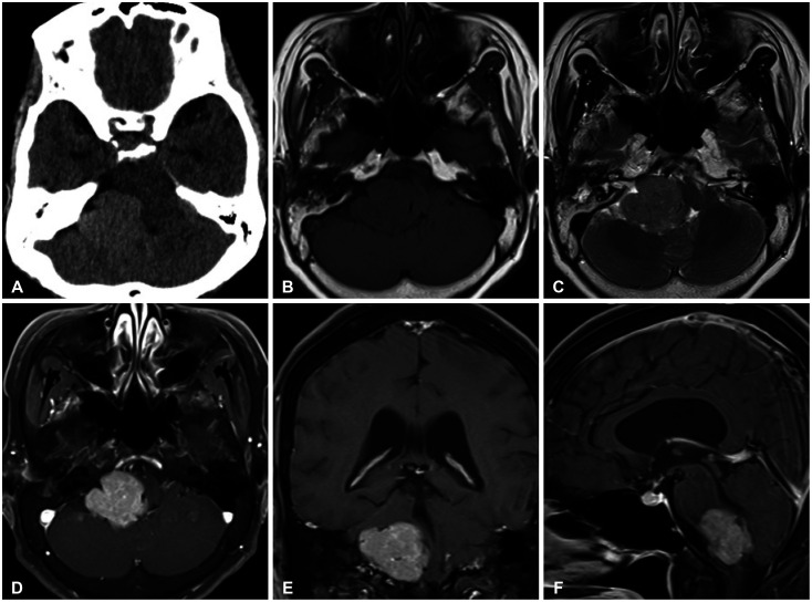
Fig. 1 Preoperative brain imaging. A: Initial brain CT revealing a hyperdense mass in the right posterior cranial fossa (right cerebellopontine angle and cerebellomedullary cistern). B and C: Iso-/hypointense signal mass in T1-weighted and iso-/hyperintense signal mass in T2-weighted images. D-F: Brain MRI with gadolinium enhancement revealing a homogeneously enhancing mass and mass effect on the 4th ventricle without definitive hydrocephalus.
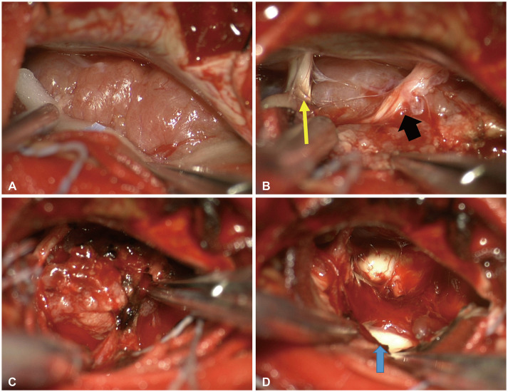
Fig. 2 Intraoperative findings. A: Mass of a large lower cerebellopontine angle region without dural attachment. B: 7th & 8th cranial nerves (yellow arrow) located in the cranial part and lower cranial nerves (9th & 10th) identified above the mass (black arrow). C: Mass originating from the choroid plexus of the foramen of Luschka. D: 4th ventricle lateral part (blue arrow) after mass removal and coagulation of the origin site.
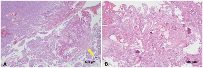
Fig. 3 Histopathologic features. A: Meningothelial pattern of the tumor with moderate hypercellularity after hematoxylin-eosin (H&E) staining. The choroid plexus was observed near the tumor (yellow arrow). B: Normal choroid plexus invaded by tumor cells.

Fig. 4 CT image of the choroid plexus-originated meningioma. A: Choroid plexus on the contralateral side (red arrow). B: The choroid plexus was mixed with the mass on the tumor side (red arrow).
Reference
-
1. Marosi C, Hassler M, Roessler K, Reni M, Sant M, Mazza E, et al. Meningioma. Crit Rev Oncol Hematol. 2008; 67:153–171. PMID: 18342535.
Article2. Ekşi MŞ, Canbolat Ç, Akbaş A, Özmen BB, Akpınar E, Usseli Mİ, et al. Elderly patients with intracranial meningioma: surgical considerations in 228 patients with a comprehensive analysis of the literature. World Neurosurg. 2019; 132:e350–e365. PMID: 31476477.
Article3. Hosainey SAM, Bouget D, Reinertsen I, Sagberg LM, Torp SH, Jakola AS, et al. Are there predilection sites for intracranial meningioma? A population-based atlas. Neurosurg Rev. 2022; 45:1543–1552. PMID: 34674099.
Article4. Papic V, Lasica N, Jelaca B, Vuckovic N, Kozic D, Djilvesi D, et al. Primary intraparenchymal meningiomas: a case report and a systematic review. World Neurosurg. 2021; 153:52–62. PMID: 34242832.
Article5. Bhatoe HS, Singh P, Dutta V. Intraventricular meningiomas: a clinicopathological study and review. Neurosurg Focus. 2006; 20:E9.6. Pereira BJA, de Almeida AN, Paiva WS, de Aguiar PHP, Teixeira MJ, Marie SKN. Natural history of intraventricular meningiomas: systematic review. Neurosurg Rev. 2020; 43:513–523. PMID: 30112665.
Article7. Leśniewski K, Kunert P, Matyja E, Czernicki T, Wójtowicz K, Wojciechowski J, et al. Trigone ventricular meningiomas - clinical characteristics, histopathology and results of surgical treatment. Neurol Neurochir Pol. 2019; 53:34–42. PMID: 30628049.
Article8. McDermott MW. Intraventricular meningiomas. Neurosurg Clin N Am. 2003; 14:559–569. PMID: 15024801.
Article9. Zhang J, Chi LY, Meng B, Li F, Zhu SG. Meningioma without dural attachment: case report, classification, and review of the literature. Surg Neurol. 2007; 67:535–539. PMID: 17445628.
Article10. Abraham J, Chandy J. Meningiomas of the posterior fossa without dural attachment: a case report. J Neurosurg. 1963; 20:177–179. PMID: 14192092.11. Shibuya M, Koketsu N, Osuka K. A meningioma of the lateral cerebellomedullary cistern without dural attachment. J Clin Neurosci. 1999; 6:50–52. PMID: 18639124.
Article12. Gonzalez-Quarante LH, Ruiz-Juretschke F, Iza-Vallejo B, Scola-Pliego E, Poletti D, Sola Vendrell E. Expanded endoscopic transclival approach for resection of a chordoid meningioma without dural attachment (MWODA) located in the prepontine cistern. World Neurosurg. 2016; 91:675.e5–675.e10.
Article13. Ishigaki D, Arai H, Sasoh M, Ogasawara K, Uesugi N, Sugai T, et al. Meningioma in the posterior fossa without dural attachment. Neurol Med Chir (Tokyo). 2007; 47:364–366. PMID: 17721053.
- Full Text Links
-
- Actions
-
Cited
- CITED
-
- Close
- Share
-
- Similar articles
-
- MR Findings of Choroid Plexus Papilloma: Case Report
- Choroid Plexus Tumor Located in Extra-Ventricular Area : A Case of Pigmented Choroid Plexus Carcinoma: A Report of a Case
- A Choroid Plexus Papilloma in Cerebellopontine Angle: Case Report
- Melanotic Choroid Plexus Carcinoma of the Posterior Fossa
- A Case of Choroid Plexus Carcinoma with Rapid Dissemination after Gross Total Resection
