Anat Cell Biol.
2022 Sep;55(3):376-379. 10.5115/acb.22.048.
A rare additional lobe of cerebellum, projecting from its superior surface
- Affiliations
-
- 1Division of Anatomy, Department of Basic Medical Sciences, Manipal Academy of Higher Education, Manipal, India
- 2Department of Anatomy, Kasturba Medical College, Manipal Academy of Higher Education, Manipal, India
- KMID: 2533364
- DOI: http://doi.org/10.5115/acb.22.048
Abstract
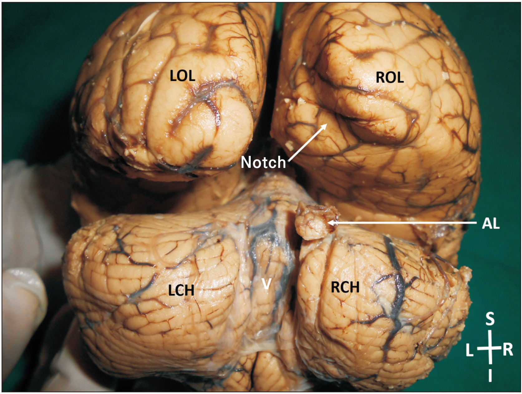
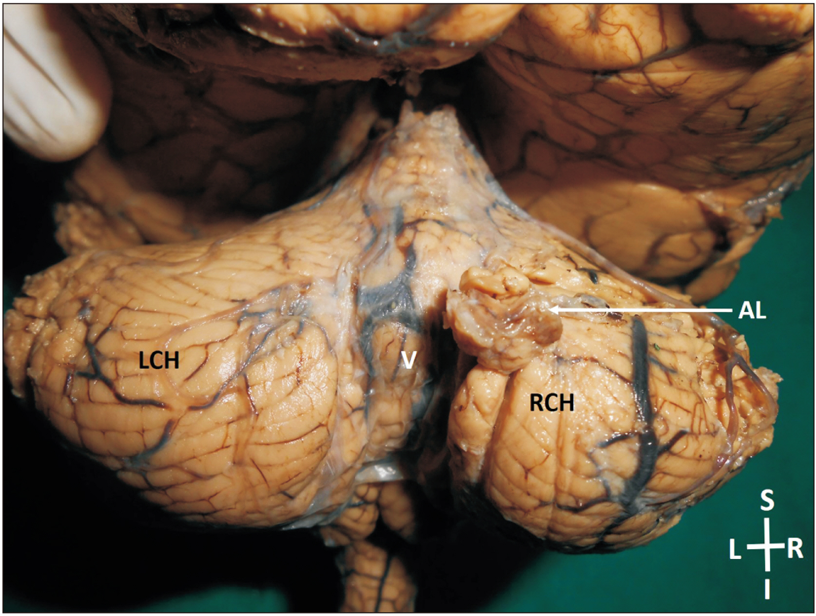
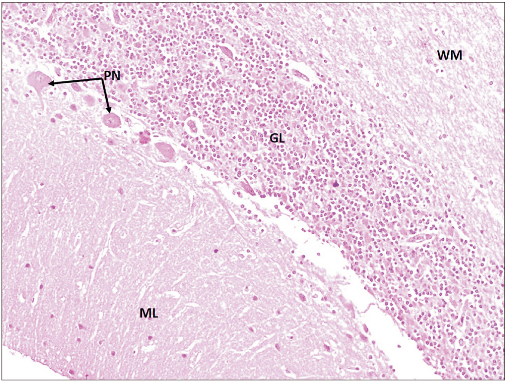
- Human cerebellum plays a vital role in motor coordination, regulation of muscle tone and maintaining the equilibrium of the body. It seldom shows anatomical/morphological variations. Herein, we report the presence of a small additional lobe projecting out on the superior surface of the right cerebellar hemisphere in the para-vermal area in an adult male cadaver. There was a notch on the tentorial surface of the occipital lobe of the right cerebral hemisphere, corresponding to the additional lobe of cerebellum. The additional lobe was histologically normal, with no evidence of any tumour cells. Knowledge of this variation is of importance to radiologists, neuroanatomists and neurosurgeons.
Keyword
Figure
Reference
-
References
1. Roostaei T, Nazeri A, Sahraian MA, Minagar A. 2014; The human cerebellum: a review of physiologic neuroanatomy. Neurol Clin. 32:859–69. DOI: 10.1016/j.ncl.2014.07.013. PMID: 25439284.2. Van Essen DC, Donahue CJ, Glasser MF. 2018; Development and evolution of cerebral and cerebellar cortex. Brain Behav Evol. 91:158–69. DOI: 10.1159/000489943. PMID: 30099464. PMCID: PMC6097530.
Article3. Witter L, De Zeeuw CI. 2015; Regional functionality of the cerebellum. Curr Opin Neurobiol. 33:150–5. DOI: 10.1016/j.conb.2015.03.017. PMID: 25884963.
Article4. Hawkes R. 2018; The ferdinando rossi memorial lecture: zones and stripes-pattern formation in the cerebellum. Cerebellum. 17:12–6. DOI: 10.1007/s12311-017-0887-0. PMID: 28965328.
Article5. Standring S. 2016. Gray's anatomy: the anatomical basis of clinical practice. 41st ed. Elsevier;Philadelphia:6. Ahmad T, Raybaud C, McNeely D, Khan N, Nadi M. 2019; Supernumerary cerebellar vermis: a unique cerebellar anomaly. Can J Neurol Sci. 46:255–7. DOI: 10.1017/cjn.2019.2.
Article7. Jackson JM, Sadove AM, Weaver DD, Edwards MK, Bull MJ. 1990; Unilateral duplication of the cerebellar hemisphere and internal, middle, and external ear: a clinical case study. Plast Reconstr Surg. 86:550–3. DOI: 10.1097/00006534-199009000-00028. PMID: 2385673.8. Hattapoğlu S, Hamidi C, Göya C, Çetinçakmak MG, Teke M, Ekici F. 2015; A surprising case: a supernumerary heterotopic hemicerebellum. Clin Neuroradiol. 25:431–4. DOI: 10.1007/s00062-015-0371-5. PMID: 25622771.
Article9. Agarwal S, Gathwala G. 2011; Third cerebellar hemisphere: an unusual new cerebellar anomaly. AJNR Am J Neuroradiol. 32:E73–4. DOI: 10.3174/ajnr.A2473. PMID: 21415147. PMCID: PMC7965887.
Article10. Barkovich AJ. 1995. Pediatric neuroimaging. 2nd ed. Raven Press;New York: p. 246–57. DOI: 10.1007/s00062-015-0371-5.11. Friede RL. 1989. Developmental neuropathology. 2nd ed. Springer-Verlag;Berlin: p. 361–71. DOI: 10.1007/978-3-642-73697-1_29.12. Haldipur P, Dang D, Millen KJ. 2018; Embryology. Handb Clin Neurol. 154:29–44. DOI: 10.1016/B978-0-444-63956-1.00002-3. PMID: 29903446. PMCID: PMC6231496.
Article13. Cho KH, Rodríguez-Vázquez JF, Kim JH, Abe H, Murakami G, Cho BH. 2011; Early fetal development of the human cerebellum. Surg Radiol Anat. 33:523–30. DOI: 10.1007/s00276-011-0796-8. PMID: 21380713.
Article14. Chung SH, Kim CT, Jung YH, Lee NS, Jeong YG. 2010; Early cerebellar granule cell migration in the mouse embryonic development. Anat Cell Biol. 43:86–95. DOI: 10.5115/acb.2010.43.1.86. PMID: 21190009. PMCID: PMC2998778.
Article
- Full Text Links
- Actions
-
Cited
- CITED
-
- Close
- Share
- Similar articles
-
- Ictal Hyperperfusion of Cerebellum and Basal Ganglia in Temporal Lobe Epilepsy: SPECT Subtraction
- Dysembryoplastic Neuroepithelial Tumor in the Cerebellum: Case Report
- Intracranial Cysticercosis: Report of 3 Cases
- Primary Occipital Malignant Melanoma
- Changes of Detrusor Function in Patients with Frontal Lobe Lesion


