J Korean Neurosurg Soc.
2022 Sep;65(5):751-757. 10.3340/jkns.2021.0071.
Profile and Outcome of Management of Brain Tumours in Kaduna Northwestern Nigeria
- Affiliations
-
- 1Department of Surgery, Barau Dikko Teaching Hospital, College of Medicine, Kaduna State University, Kaduna, Nigeria
- 2Department of Neurosurgery, Usmanu Danfodio University Teaching Hospital, Sokoto, Nigeria
- KMID: 2533037
- DOI: http://doi.org/10.3340/jkns.2021.0071
Abstract
Objective
: Tumours of the brain are a rare occurrence accounting for approximately 2% of all neoplasms in adults. Few studies have been done in Nigeria on the profile of brain tumours. The aim of this study is to determine the profile of brain tumours in general and determine the change in Kanofsky Performance Score (KPS) after treatment.
Methods
: This is a prospective hospital-based study in Kaduna. All consecutive patients over 18 years of age with diagnosis of brain tumours from January 2016 to December 2019 were included in the study. Demographic and clinical data was collected using a proforma during the study. Patients who received treatment were followed up for 12 months. The primary outcome data was the difference in the quality of life as measured by KPS at the point of first contact and at 1-month after treatment and at 12-month follow up. Data obtained was analysed with SPSS version 25.0 for Windows. Descriptive statistics was done to determine the profile. Paired t-test at 95% confidence interval was done to check for significant correlation between the mean KPS.
Results
: A total of 39 consecutive patients were included in the study. There was a slight male preponderance with a M : F of 1.17 : 1. Meningioma and metastasis were more common in females while gliomas and pituitary tumours were more common in males. The mean age of patients was 49.8 years and standard deviation of 11.8 years. Pituitary tumours were the most common tumours. The most common location of the tumour was frontal lobe followed by the pituitary gland. The mean duration of symptoms before neurosurgical consultation was 38 weeks. The most common presenting symptoms of patient with brain tumour was headache. The quality of life improve compare to the baseline in 81% of patient at discharge and at 1 year follow up. The overall mortality rate was 25.6%.
Conclusion
: The most common brain tumour in our study is pituitary tumour. Most patients present late. The most common presenting symptoms is headache. There is significant improvement in the KPS of patients following treatment. The overall mortality rate at 1-year post treatment is 25.6%.
Keyword
Figure
-
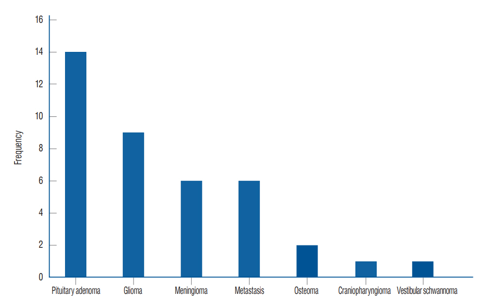
Fig. 1. Types of brain tumours.
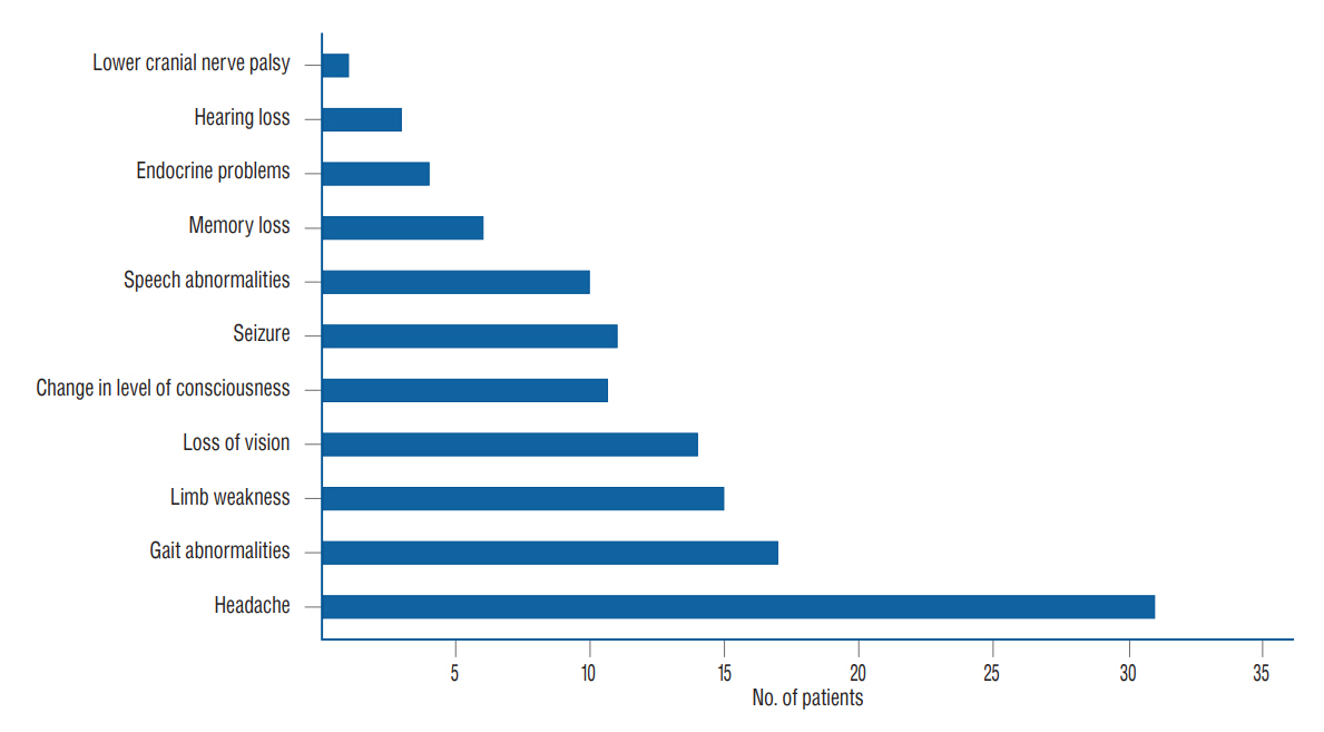
Fig. 2. Clinical features of brain tumors.
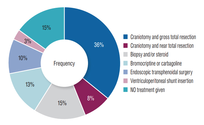
Fig. 3. Treatment of brain tumours.
Reference
-
References
1. Ali AM, Buji MA, Abubakar A. Patterns of computed tomographic findings in patients from Maiduguri, Nigeria, diagnosed with a brain tumor. Glioma. 2:153–156. 2019.2. Arora RS, Alston RD, Eden TO, Estlin EJ, Moran A, Birch JM. Age-incidence patterns of primary CNS tumors in children, adolescents, and adults in England. Neuro Oncol. 11:403–413. 2009.3. Chikani MC, Okpara S, Mathew M, Onuh A, Okwor V, Mezue W. Preliminary findings on metastatic brain tumors in Enugu, Southeast Nigeria. Niger J Med. 29:466–470. 2020.4. Das A, Chapman CA, Yap WM. Histological subtypes of symptomatic central nervous system tumours in Singapore. J Neurol Neurosurg Psychiatry. 68:372–374. 2000.5. Davis FG, McCarthy BJ. Current epidemiological trends and surveillance issues in brain tumors. Expert Rev Anticancer Ther. 1:395–401. 2001.6. DeAngelis LM. Brain tumors. N Engl J Med. 334:114–123. 2001.7. Dhar A, Bhat AR, Nizami FA, Kirmani AR, Zargar J, Ramzan AU, et al. Analysis of brain tumors in Kashmir Valley - a 10 year study. Bangladesh J Med Sci. 13:268–277. 2014.8. Ekpene U, Ametefe M, Akoto H, Bankah P, Totimeh T, Wepeba G, et al. Pattern of intracranial tumours in a tertiary hospital in Ghana. Ghana Med J. 52:79–83. 2018.9. Ferlay J, Soerjomataram I, Ervik M, Dikshit R, Eser S, Mathers C, et al. GLOBOCAN 2012 v1.0, Cancer Incidence and Mortality Worldwide: IARC CancerBase No. 11. Available at : https://publications.iarc.fr/Databases/Iarc-Cancerbases/GLOBOCAN-2012-Estimated-Cancer-Incidence-Mortality-And-Prevalence-Worldwide-In-2012-V1.0-2012.10. Ghanghoria S, Mehar R, Kulkarni C, Mittal M, Yadav A, Patidar H. Retrospective histological analysis of CNS tumors - a 5 year study. Int J Med Sci Public Health. 3:1205–1207. 2014.11. Grimes CE, Bowman KG, Dodgion CM, Lavy CB. Systematic review of barriers to surgical care in low-income and middle-income countries. World J Surg. 35:941–950. 2011.12. Hewedi I, Ibrahim R, Elserry T, Taha N, Mohamed H. Frequency of primary central nervous system tumors in a tertiary hospital, Cairo, Egypt. J Community Health Manag. 5:140–146. 2018.13. Idowu O, Akang E, Malomo A. Symptomatic primary intracranial neoplasms in Nigeria, West Africa. J Neurol Sci Turk. 24:212–218. 2017.14. Jalali R, Datta D. Prospective analysis of incidence of central nervous tumors presenting in a tertiary cancer hospital from India. J Neurooncol. 87:111–114. 2008.15. Jamal S, Moghal S, Mamoon N, Mushtaq S, Luqman M, Anwar M. The pattern of malignant tumours: tumour registry data analysis, AFIP, Rawalpindi, Pakistan (1992-2001). J Pak Med Assoc. 56:359–362. 2006.16. Jibrin P, Ibebuike K, Ado-Wanka AN. Histo-pathological pattern of intracranial tumours in the National Hospital, Abuja. Afr Health Sci. 18:281–286. 2018.17. Kleihues P, Cavenee W. Tumours of the nervous system: World Health Organization classification of tumours. France: IARC Press;2000.18. Kolo ES, Hassan I, Ibrahim M, Misbahu AH, Adamu A, Ismail NJ, et al. Early outcome of endoscopic trans-nasal trans-sphenoidal pituitary surgery in Kano, Nigeria. Niger J Basic Clin Sci. 17:145–50. 2020.19. Lee CH, Jung KW, Yoo H, Park S, Lee SH. Epidemiology of primary brain and central nervous system tumors in Korea. J Korean Neurosurg Soc. 48:145–152. 2010.20. Leece R, Xu J, Ostrom QT, Chen Y, Kruchko C, Barnholtz-Sloan JS. Global incidence of malignant brain and other central nervous system tumors by histology, 2003-2007. Neuro Oncol. 19:1553–1564. 2017.21. Louis DN. World Health Organization International Agency for Research on Cancer. 4th ed. Lyon: International Agency for Research on Cancer;2007. p. 309.22. Louis DN, Perry A, Reifenberger G, von Deimling A, Figarella-Branger D, Cavenee WK, et al. The 2016 World Health Organization classification of tumors of the central nervous system: a summary. Acta Neuropathol. 131:803–820. 2016.23. Mahmud MR, Idris MM. A glance at neurosurgery in Nigeria following the 3rd CAANS congress. AANS Neurosurgeon. 27:2018.24. Masoodi T, Gupta RK, Singh JP, Khajuria A. Pattern of central nervous system neoplasms: a study of 106 cases. JK Pract. 17:42–46. 2012.25. McKinney PA. Brain tumours: incidence, survival, and aetiology. J Neurol Neurosurg Psychiatry. 75 Suppl 2(Suppl 2):ii12–ii17. 2004.26. Mehta J, Bansal B, Mittal A, Mathur K, Vijay R. Histological analysis of primary brain tumors in a tertiary care hospital: a retrospective study of 5 years. Int J Med Res Prof. 3:14–18. 2017.27. Mondal S, Pradhan R, Pal S, Biswas B, Banerjee A, Bhattacharyya D. Clinicopathological pattern of brain tumors: a 3-year study in a tertiary care hospital in India. Clin Cancer Investig J. 5:437. 2016.28. NIH National Cancer Institute Surveillance, Epidemiology and End Result program. Cancer Stat Facts: Brain and Other Nervous System Cancer. Available at : https://seer.cancer.gov/statfacts/html/brain.html.29. Ohaegbulam SC, Saddeqi N, Ikerionwu S. Intracranial tumors in Enugu, Nigeria. Cancer. 46:2322–2324. 1980.30. Olasode BJ, Shokunbi MT, Aghadiuno PU. Intracranial neoplasms in Ibadan, Nigeria. East Afr Med J. 77:4–8. 2000.31. Ostrom QT, Gittleman H, Fulop J, Liu M, Blanda R, Kromer C, et al. CBTRUS statistical report: primary brain and central nervous system tumors diagnosed in the United States in 2008-2012. Neuro Oncol. 17 Suppl 4(Suppl 4):iv1–iv62. 2015.32. Salander P, Bergenheim AT, Hamberg K, Henriksson R. Pathways from symptoms to medical care: a descriptive study of symptom development and obstacles to early diagnosis in brain tumour patients. Fam Pract. 16:143–148. 1999.33. Soyemi SS, Oyewole OO. Spectrum of intracranial tumours in a tertiary health carefacility: our findings. Pan Afr Med J. 20:24. 2015.34. Stewart BW, Kleihues P. Tumours of the Nervous System. Lyon: IARC;2003. p. 265–269.35. Surawicz TS, McCarthy BJ, Kupelian V, Jukich PJ, Bruner JM, Davis FG. Descriptive epidemiology of primary brain and CNS tumors: results from the Central Brain Tumor Registry of the United States, 1990-1994. Neuro Oncol. 1:14–25. 1999.36. Tuly TI. Pattern of Brain Tumour among Admitted Patients in a Specialized Center of Dhaka City: A Cross Sectional Study. Available at : http://hdl.handle.net/10361/9327.37. Uche EO, Emejulu JKC, Okorie E, Onyia EE, Iloabachie I, Uche N, et al. Brain astrocytomas in South-East Nigeria: a 3-centre experience. J Clin Appl Neurosci. 1:37–43. 2015.38. World Health Organization. Cancer Incidence in Five Continents. Available at : http://ci5.iarc.fr.39. Yeole BB. Trends in the brain cancer incidence in India. Asian Pac J Cancer Prev. 9:267–270. 2008.
- Full Text Links
-
- Actions
-
Cited
- CITED
-
- Close
- Share
-
- Similar articles
-
- Andrographis paniculata protects against brain hippocampus and cerebellum from mercury chloride induced damage by attenuating oxidative stress
- Myositis ossificans of the platysma mimicking a malignancy: a case report with review of the literature
- A Case of Multiple Intraosseous Lipomas
- Three Cases of Primary Spinal Malignant Schwannomas: Case Reports
- Mitigating potential public health risks and challenges from hazardous materials contained in electronic waste items in a developing country setting
