Secondary Achalasia of Gastric Linitis Plastica
- Affiliations
-
- 1Department of Gastroenterology, Cha Bundang Medical Center, CHA University, School of Medicine, Seongnam, Korea
- 2Department of Gastroenterology, CHA Gangnam Medical Center, CHA University, School of Medicine, Seoul, Korea
- KMID: 2532685
- DOI: http://doi.org/10.4166/kjg.2022.101
Figure
-
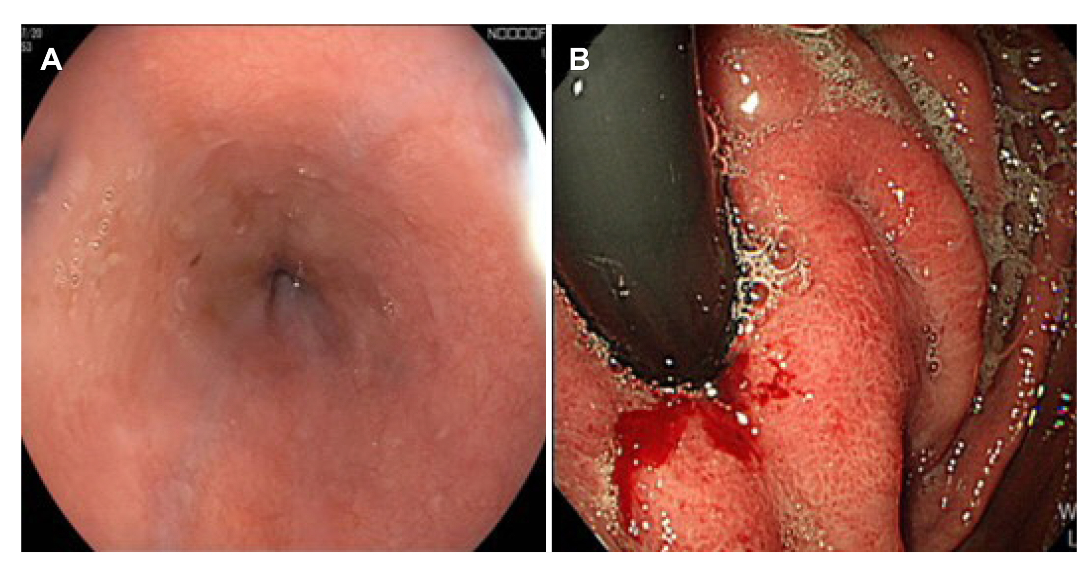
Fig. 1 Finding of esophagogastroduodenoscopy. (A) Endoscopy revealed stenosis in the esophagogastric junction. (B) The gastric rugal folds showed hypertrophy.
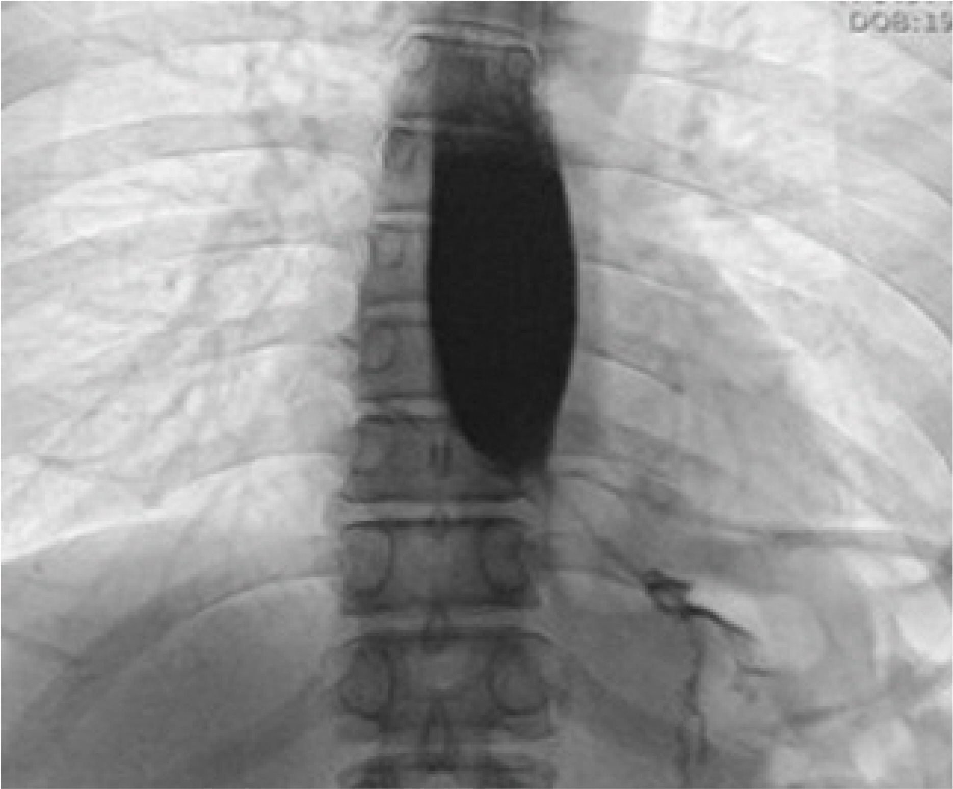
Fig. 2 The esophagography showed no passage of barium in the 5 minutes delayed phase.

Fig. 3 High-resolution manometry showed high integrated relaxation pressure (43 mmHg), and distal pressurization of the esophageal body.
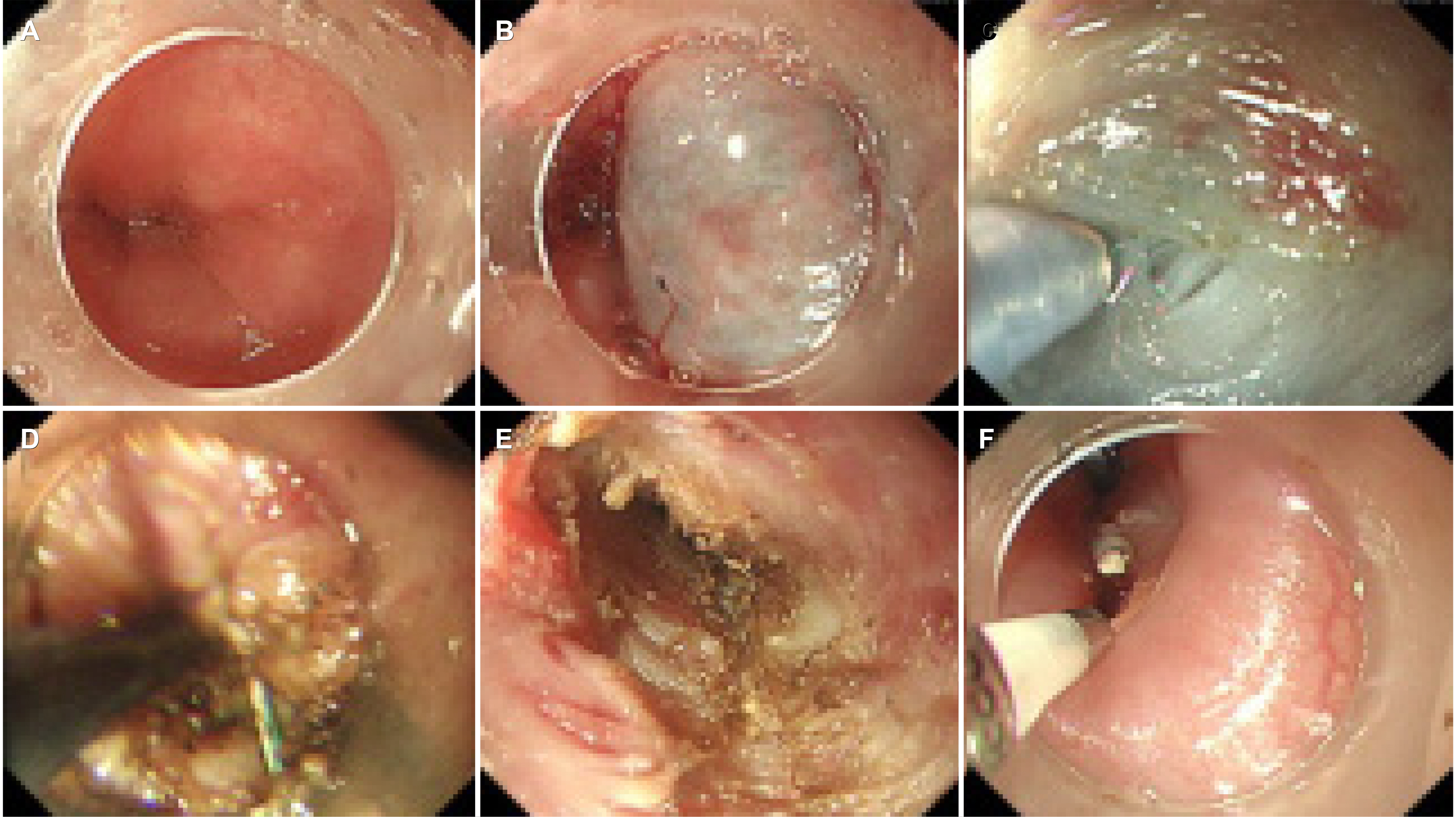
Fig. 4 Open peroral endoscopic myotomy (POEM). (A) Endoscopy reveals very tight esophagogastric junction (EGJ). (B) Submucosal injection was given 10 cm proximal to the EGJ. (C) Careful injection and dissection on the EGJ. (D) Open POEM around the stenotic part. (F) Selective myotomy was performed in the submucosal tunnel. (E) Entry site clipping.
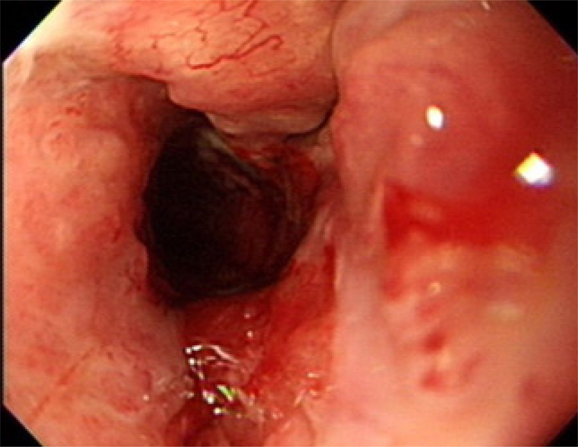
Fig. 5 The follow-up esophagogastroduodenoscopy revealed no stenosis on esophagogastric junction.

Fig. 6 (A) There was a ulceroinfiltrative lesion with irregular base and stomach was not fully distended with air-insufflation. (B) Histologic findings of the biopsy was adenocarcinoma, poorly differentiated (hematoxylin and eosin, ×12.5).
Reference
-
1. Liu W, Zeng XH, Yuan XL, et al. 2019; Open peroral endoscopic myotomy for the treatment of achalasia: a case series of 82 cases. Dis Esophagus. 32:1–7. DOI: 10.1093/dote/doz052. PMID: 31175357.2. Cohen S. 1979; Motor disorders of the esophagus. N Engl J Med. 301:184–192. DOI: 10.1056/NEJM197907263010404. PMID: 109758.3. Gregersen H, Lo KM. 2018; Pathophysiology and treatment of achalasia in a muscle mechanical perspective. Ann N Y Acad Sci. 1434:173–184. DOI: 10.1111/nyas.13711. PMID: 29756656.4. Cassella RR, Brown AL Jr, Sayre GP, Ellis FH Jr. 1964; Achalasia of the esophagus: pathologic and etiologic considerations. Ann Surg. 160:474–487. DOI: 10.1097/00000658-196409000-00010. PMID: 14206851. PMCID: PMC1408778.5. Kahrilas PJ, Kishk SM, Helm JF, Dodds WJ, Harig JM, Hogan WJ. 1987; Comparison of pseudoachalasia and achalasia. Am J Med. 82:439–446. DOI: 10.1016/0002-9343(87)90443-8. PMID: 3548347.6. Tucker HJ, Snape WJ Jr, Cohen S. 1978; Achalasia secondary to carcinoma: manometric and clinical features. Ann Intern Med. 89:315–318. DOI: 10.7326/0003-4819-89-3-315. PMID: 686541.7. Sandler RS, Bozymski EM, Orlando RC. 1982; Failure of clinical criteria to distinguish between primary achalasia and achalasia secondary to tumor. Dig Dis Sci. 27:209–213. DOI: 10.1007/BF01296916. PMID: 7075419.8. Kolodny M, Schrader ZR, Rubin W, Hochman R, Sleisenger MH. 1968; Esophageal achalasia probably due to gastric carcinoma. Ann Intern Med. 69:569–573. DOI: 10.7326/0003-4819-69-3-569. PMID: 5673174.9. Reynolds JC, Parkman HP. 1989; Achalasia. Gastroenterol Clin North Am. 18:223–255. DOI: 10.1016/S0889-8553(21)00676-2. PMID: 23452629. PMCID: PMC3618975.
- Full Text Links
-
- Actions
-
Cited
- CITED
-
- Close
- Share
-
- Similar articles
-
- Metastatic Gastric Linitis Plastica from Bladder Cancer Mimicking a Primary Gastric Carcinoma: a Case Report
- Primary Linitis Plastica of the Rectum: A Clinico-Pathologic Analysis of Five Cases with Special Reference to Comparison with Gastric Form
- Primary Linitis Plastica of the Colon with Mucinous Adenocarcinoma in Young Woman
- Gastric Outlet Obstruction Due to Gastric Amyloidosis Mimicking Malignancy in a Patient with Ankylosing Spondylitis
- Borrmann Type 4 Advanced Gastric Cancer: Focus on the Development of Scirrhous Gastric Cancer
