Korean Circ J.
2022 Aug;52(8):638-639. 10.4070/kcj.2022.0112.
Hemopericardium With Cardiac Tamponade After Percutaneous Vertebroplasty
- Affiliations
-
- 1Department of Cardiovascular Medicine, Regional Cardiocerebrovascular Center, Wonkwang University Hospital, Iksan, Korea
- KMID: 2532336
- DOI: http://doi.org/10.4070/kcj.2022.0112
Figure
-
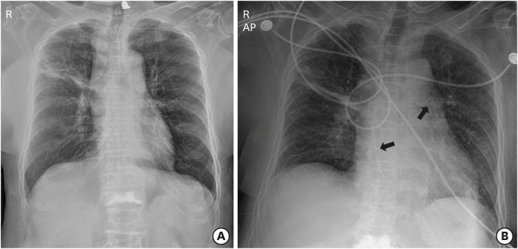
Figure 1 Chest radiography before (A) and after (B) percutaneous vertebroplasty. Linear radio-opaque materials (arrows) were observed within heart and left hilum.
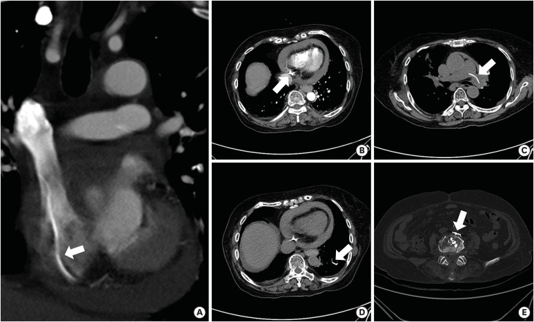
Figure 2 Computed tomography. With pericardial effusion, linear and sharp-edged embolic cements (arrows) were identified in superior vena cava/right atrium (A), pericardial space (B), left main (C)/subsegmental pulmonary arteries (D), and paravertebral vein (E).
Reference
- Full Text Links
-
- Actions
-
Cited
- CITED
-
- Close
- Share
-
- Similar articles
-
- A Case of Right Atrial Sarcoma Complicated with Hemopericardium and Cardiac Tamponade
- A Case of Cardiac Tamponade due to Penetration of the Right Ventricule by an Acupunture Needle
- Cardiac Tamponade Complicated by Acupuncture: Hemopericardium due to Shredded Coronary Artery Injury
- Cardiac Tamponade Associated with Acupuncture
- Fatal Hemothorax Following Percutaneous Vertebroplasty: A Case Report
