Conservative orthodontic treatment for severe pathologic migration following total glossectomy: A case report
- Affiliations
-
- 1Department of Orthodontics, Institute of Craniofacial Deformity, College of Dentistry, Yonsei University, Seoul, Korea
- 2Department of Advanced General Dentistry, College of Dentistry, Yonsei University, Seoul, Korea
- KMID: 2532153
- DOI: http://doi.org/10.4041/kjod21.215
Abstract
- Glossectomy combined with radiotherapy causes different levels of tongue function disorders and leads to severe malocclusion, with poor periodontal status in cancer survivors. Although affected patients require regular access to orthodontic care, special considerations are crucial for treatment planning. This case report describes the satisfactory orthodontic management for the correction of severe dental crowding in a 43-year-old female 6 years after treatment for tongue cancer with total glossectomy combined with radiotherapy, to envision the possibility of orthodontic care for oral cancer survivors. Extraction was performed to correct dental crowding and establish proper occlusion following alignment, after considering the possibility of osteoradionecrosis. Orthodontic mini-implants were used to provide skeletal anchorage required for closure of the extraction space and intrusion of the anterior teeth. The dental crowding was corrected, and Class I occlusal relationship was established after 36 months of treatment. The treatment outcome was sustained after 15 months of retention, and long-term follow-up was recommended.
Figure
-
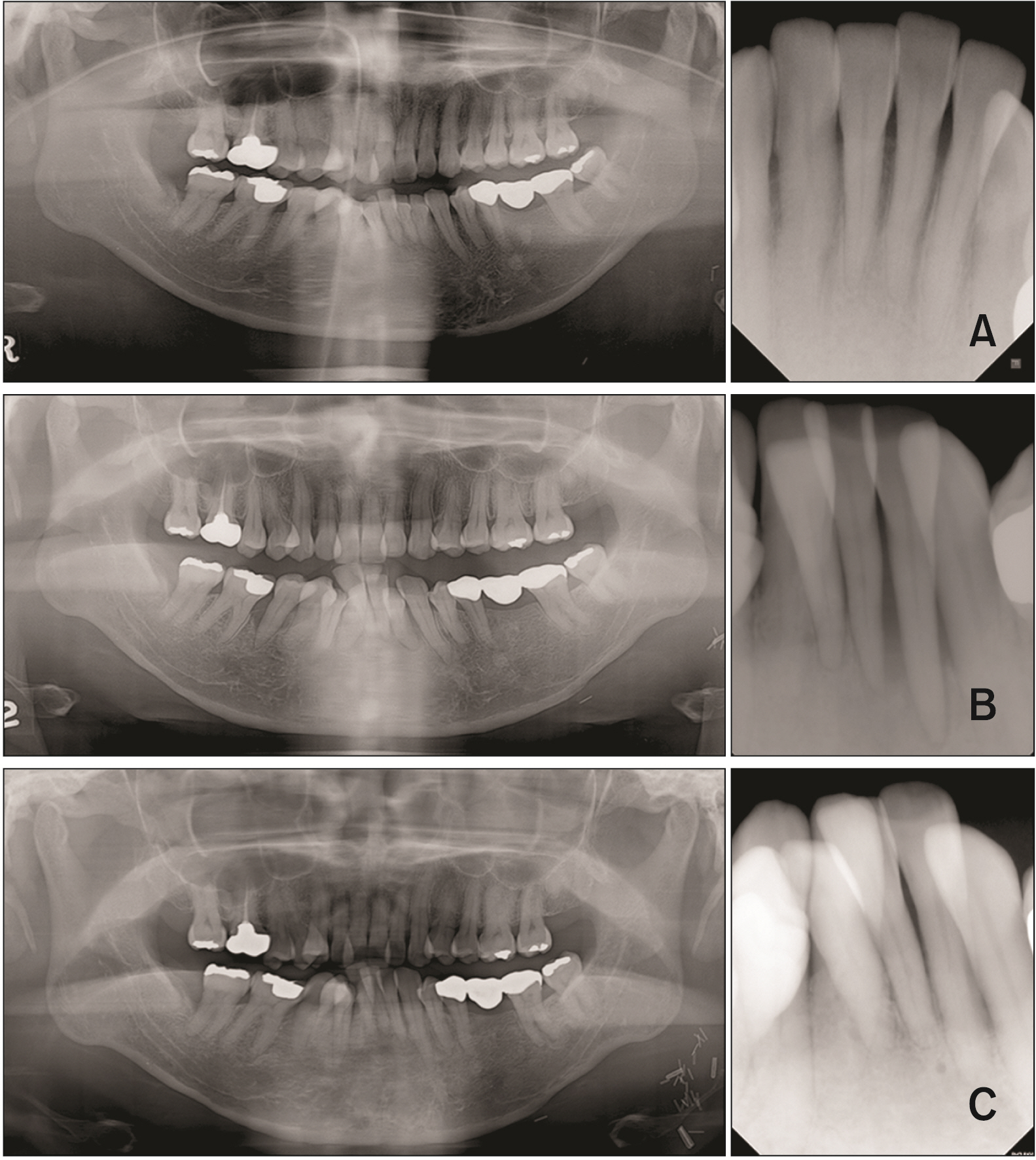
Figure 1 Serial panoramic and periapical radiographs showing the development of severe malocclusion over time. A, Before total glossectomy. B, Three years after tongue cancer treatment. C, Six years after tongue cancer treatment and pre-orthodontic treatment.
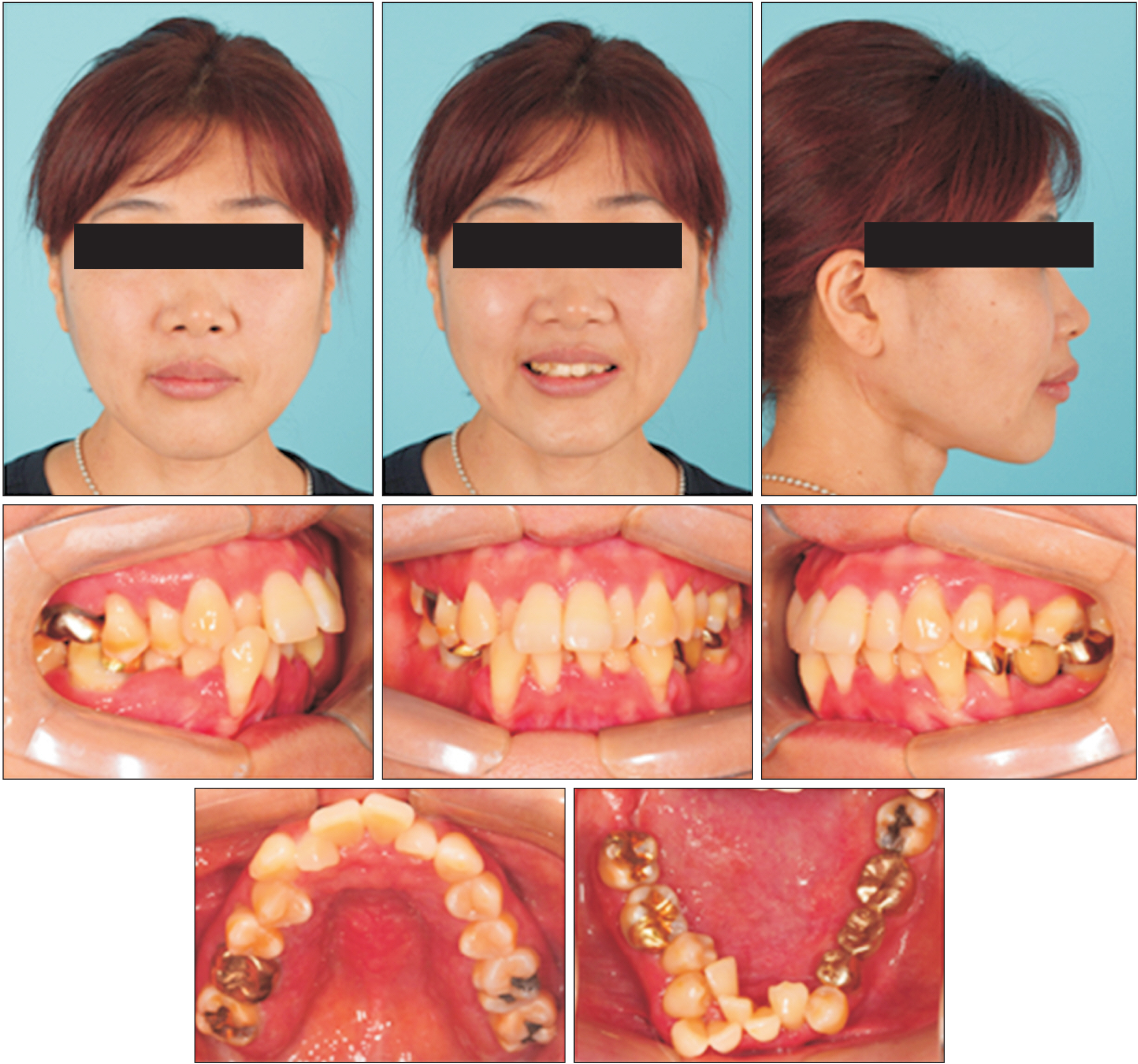
Figure 2 Pre-treatment lateral cephalometric radiograph and tracing.
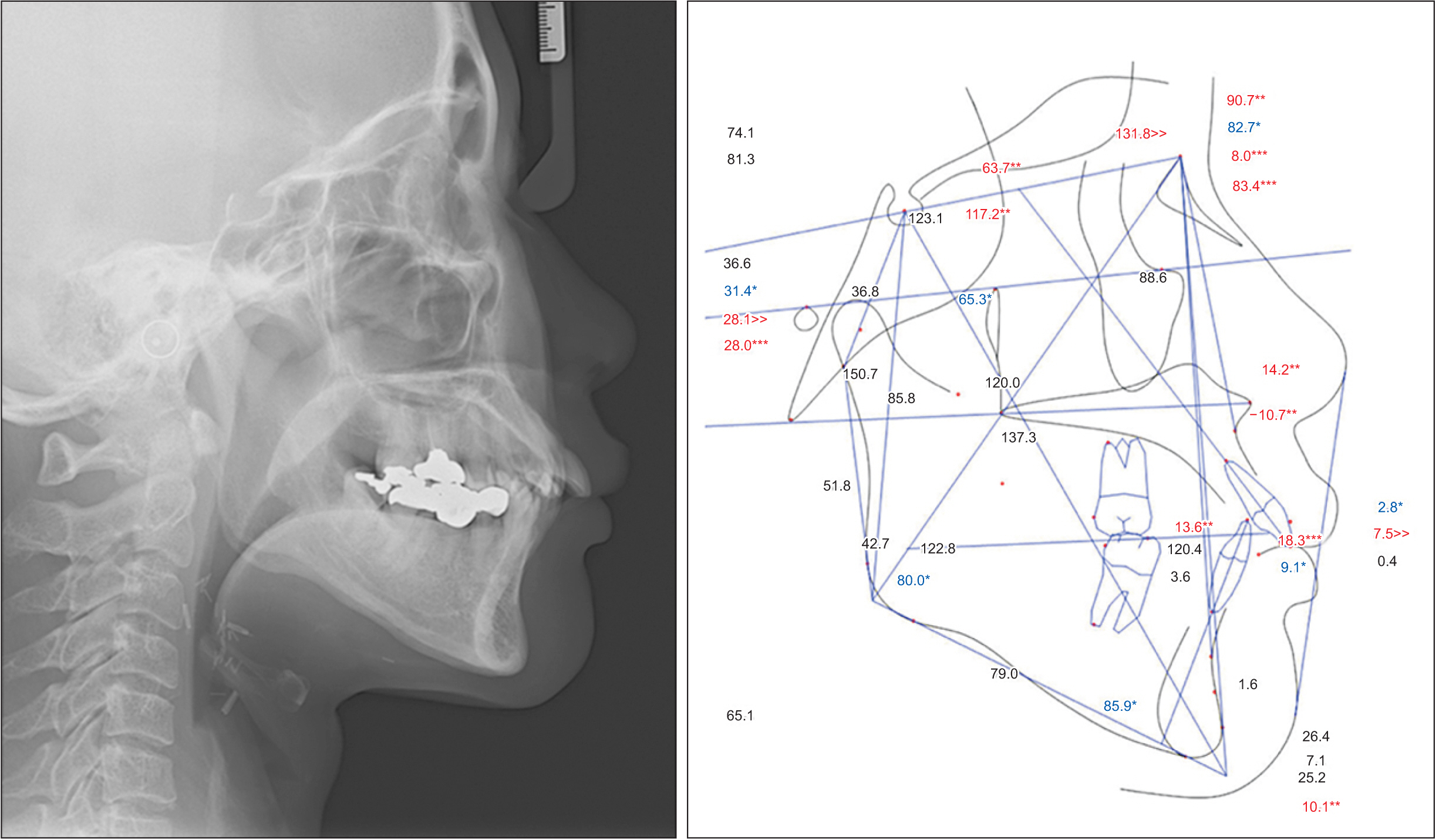
Figure 3 Pre-treatment facial and intraoral photographs.
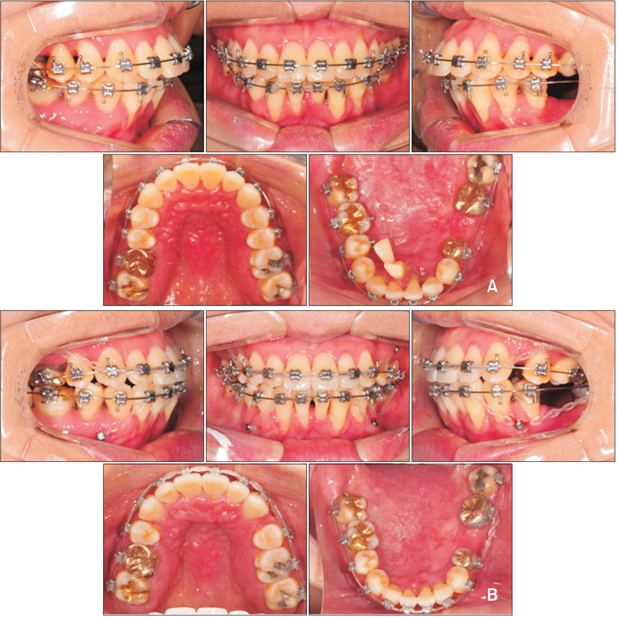
Figure 4 Intraoral photographs of treatment progress. A, Leveling and alignment. B, Closing extraction space and intruding maxillary and mandibular anterior teeth.

Figure 5 Periapical radiographs follow-up. A, Maxillary incisors. B, Mandibular incisors. T0, pre-treatment; T1, re-assessment after leveling and alignment; T2, T3, treatment progress with closing extraction space and intruding maxillary and mandibular anterior teeth; T4, post-treatment; T5, one year of retention.
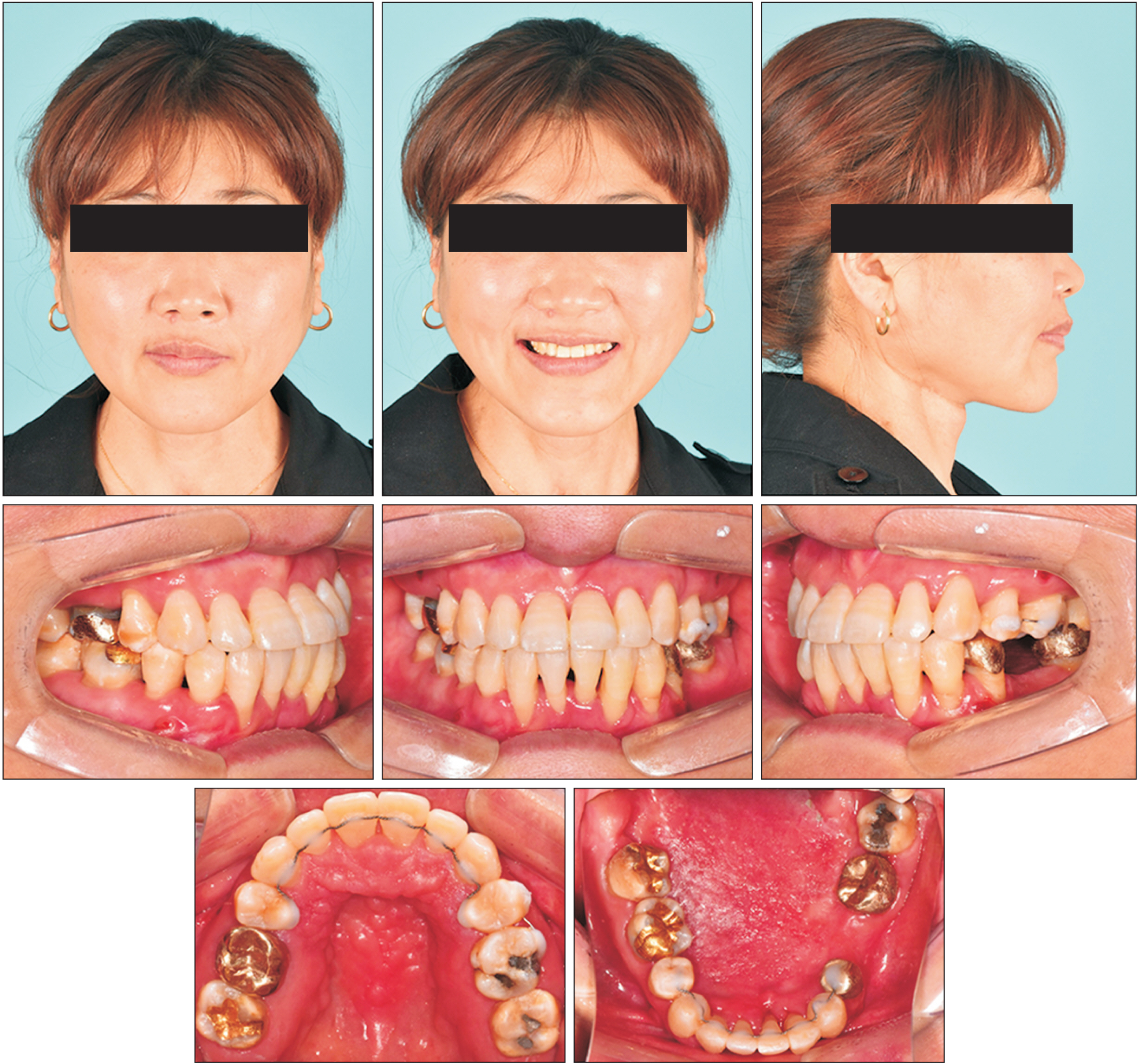
Figure 6 Post-treatment facial and intraoral photographs.

Figure 7 Post-treatment lateral cephalometric radiograph, tracing and panoramic radiograph.

Figure 8 Superimposition of pre- and post-treatment cephalometric tracings.

Figure 9 Intraoral photographs after 15 months of retention.

Figure 10 Panoramic radiograph after 1 year of retention.
Reference
-
1. Chitapanarux I, Lorvidhaya V, Sittitrai P, Pattarasakulchai T, Tharavichitkul E, Sriuthaisiriwong P, et al. 2006; Oral cavity cancers at a young age: analysis of patient, tumor and treatment characteristics in Chiang Mai University Hospital. Oral Oncol. 42:83–8. DOI: 10.1016/j.oraloncology.2005.06.015. PMID: 16249113. PMID: https://www.scopus.com/inward/record.uri?partnerID=HzOxMe3b&scp=29244492025&origin=inward.2. Patel SC, Carpenter WR, Tyree S, Couch ME, Weissler M, Hackman T, et al. 2011; Increasing incidence of oral tongue squamous cell carcinoma in young white women, age 18 to 44 years. J Clin Oncol. 29:1488–94. DOI: 10.1200/JCO.2010.31.7883. PMID: 21383286. PMID: https://www.scopus.com/inward/record.uri?partnerID=HzOxMe3b&scp=79955033400&origin=inward.3. Santos-Silva AR, Carvalho Andrade MA, Jorge J, Almeida OP, Vargas PA, Lopes MA. 2014; Tongue squamous cell carcinoma in young nonsmoking and nondrinking patients: 3 clinical cases of orthodontic interest. Am J Orthod Dentofacial Orthop. 145:103–7. DOI: 10.1016/j.ajodo.2012.09.026. PMID: 24373660. PMID: https://www.scopus.com/inward/record.uri?partnerID=HzOxMe3b&scp=84891585784&origin=inward.4. Mallet Y, Avalos N, Le Ridant AM, Gangloff P, Moriniere S, Rame JP, et al. 2009; Head and neck cancer in young people: a series of 52 SCCs of the oral tongue in patients aged 35 years or less. Acta Otolaryngol. 129:1503–8. DOI: 10.3109/00016480902798343. PMID: 19922105. PMID: https://www.scopus.com/inward/record.uri?partnerID=HzOxMe3b&scp=70450172632&origin=inward.5. Costa Bandeira AK, Azevedo EH, Vartanian JG, Nishimoto IN, Kowalski LP, Carrara-de Angelis E. 2008; Quality of life related to swallowing after tongue cancer treatment. Dysphagia. 23:183–92. DOI: 10.1007/s00455-007-9124-1. PMID: 17999111. PMID: https://www.scopus.com/inward/record.uri?partnerID=HzOxMe3b&scp=44549085327&origin=inward.6. Lin DT, Yarlagadda BB, Sethi RK, Feng AL, Shnayder Y, Ledgerwood LG, et al. 2015; Long-term functional outcomes of total glossectomy with or without total laryngectomy. JAMA Otolaryngol Head Neck Surg. 141:797–803. DOI: 10.1001/jamaoto.2015.1463. PMID: 26291031. PMID: https://www.scopus.com/inward/record.uri?partnerID=HzOxMe3b&scp=84942023954&origin=inward.7. Yu M, Gao X. 2019; Tongue pressure distribution of individual normal occlusions and exploration of related factors. J Oral Rehabil. 46:249–56. DOI: 10.1111/joor.12741. PMID: 30375017. PMCID: PMC7379747. PMID: https://www.scopus.com/inward/record.uri?partnerID=HzOxMe3b&scp=85056469618&origin=inward.8. Jawad H, Hodson NA, Nixon PJ. 2015; A review of dental treatment of head and neck cancer patients, before, during and after radiotherapy: part 1. Br Dent J. 218:65–8. DOI: 10.1038/sj.bdj.2015.28. PMID: 25613260. PMID: https://www.scopus.com/inward/record.uri?partnerID=HzOxMe3b&scp=84923113496&origin=inward.9. Jawad H, Hodson NA, Nixon PJ. 2015; A review of dental treatment of head and neck cancer patients, before, during and after radiotherapy: part 2. Br Dent J. 218:69–74. Erratum in: Br Dent J 2015;218: 290. DOI: 10.1038/sj.bdj.2015.29. PMID: 25613261. PMID: https://www.scopus.com/inward/record.uri?partnerID=HzOxMe3b&scp=84923097888&origin=inward.10. Mishra S. 2017; Orthodontic therapy for paediatric cancer survivors: a review. J Clin Diagn Res. 11:ZE01–4. DOI: 10.7860/JCDR/2017/23916.9404. PMID: 28511529. PMCID: PMC5427455. PMID: e2995bfc837a4ae49cab1677e82e1932. PMID: https://www.scopus.com/inward/record.uri?partnerID=HzOxMe3b&scp=85014366358&origin=inward.11. Burden D, Mullally B, Sandler J. 2001; Orthodontic treatment of patients with medical disorders. Eur J Orthod. 23:363–72. DOI: 10.1093/ejo/23.4.363. PMID: 11544786. PMID: https://www.scopus.com/inward/record.uri?partnerID=HzOxMe3b&scp=0035430697&origin=inward.12. Kurabeishi H, Tatsuo R, Makoto N, Kazunori F. 2018; Relationship between tongue pressure and maxillofacial morphology in Japanese children based on skeletal classification. J Oral Rehabil. 45:684–91. DOI: 10.1111/joor.12680. PMID: 29908035. PMID: https://www.scopus.com/inward/record.uri?partnerID=HzOxMe3b&scp=85050878287&origin=inward.13. Santin GC, Palma-Dibb RG, Romano FL, de Oliveira HF, Nelson Filho P, de Queiroz AM. 2015; Physical and adhesive properties of dental enamel after radiotherapy and bonding of metal and ceramic brackets. Am J Orthod Dentofacial Orthop. 148:283–92. DOI: 10.1016/j.ajodo.2015.03.025. PMID: 26232837. PMID: https://www.scopus.com/inward/record.uri?partnerID=HzOxMe3b&scp=84938388818&origin=inward.14. Martin P, Muller E, Paulus C. 2019; Alteration of facial growth after radiotherapy: orthodontic, surgical and prosthetic rehabilitation. J Stomatol Oral Maxillofac Surg. 120:369–72. DOI: 10.1016/j.jormas.2019.04.004. PMID: 30980947. PMID: https://www.scopus.com/inward/record.uri?partnerID=HzOxMe3b&scp=85065017297&origin=inward.15. Khoo SC, Nabil S, Fauzi AA, Yunus SSM, Ngeow WC, Ramli R. 2021; Predictors of osteoradionecrosis following irradiated tooth extraction. Radiat Oncol. 16:130. DOI: 10.1186/s13014-021-01851-0. PMID: 34261515. PMCID: PMC8278595. PMID: ebac24c028d84d94b99a1309dd0b973c. PMID: https://www.scopus.com/inward/record.uri?partnerID=HzOxMe3b&scp=85109853278&origin=inward.16. Yee JA, Türk T, Elekdağ-Türk S, Cheng LL, Darendeliler MA. 2009; Rate of tooth movement under heavy and light continuous orthodontic forces. Am J Orthod Dentofacial Orthop. 136:150.e1–9. discussion 150–1. DOI: 10.1016/j.ajodo.2008.06.027. PMID: 19651334. PMID: https://www.scopus.com/inward/record.uri?partnerID=HzOxMe3b&scp=67949094544&origin=inward.17. Burch JG, Bagci B, Sabulski D, Landrum C. 1992; Periodontal changes in furcations resulting from orthodontic uprighting of mandibular molars. Quintessence Int. 23:509–13. DOI: 10.1016/s0889-5406(08)80083-2. PMID: 1410254.18. Aarup-Kristensen SFM, Hansen CR, Johansen J. 2019; Osteoradionecrosis after radiotherapy for head and neck cancer: incidence, risk factors, and mandibular dose-volume effects. Int J Radiat Oncol Biol Phys. 105(Suppl 1):E400. DOI: 10.1016/j.ijrobp.2019.06.1585.19. Kuhnt T, Stang A, Wienke A, Vordermark D, Schweyen R, Hey J. 2016; Potential risk factors for jaw osteoradionecrosis after radiotherapy for head and neck cancer. Radiat Oncol. 11:101. DOI: 10.1186/s13014-016-0679-6. PMID: 27473433. PMCID: PMC4967325. PMID: https://www.scopus.com/inward/record.uri?partnerID=HzOxMe3b&scp=84979752489&origin=inward.20. Aarup-Kristensen S, Hansen CR, Forner L, Brink C, Eriksen JG, Johansen J. 2019; Osteoradionecrosis of the mandible after radiotherapy for head and neck cancer: risk factors and dose-volume correlations. Acta Oncol. 58:1373–7. DOI: 10.1080/0284186X.2019.1643037. PMID: 31364903. PMID: https://www.scopus.com/inward/record.uri?partnerID=HzOxMe3b&scp=85070302847&origin=inward.21. Epstein J, van der Meij E, McKenzie M, Wong F, Lepawsky M, Stevenson-Moore P. 1997; Postradiation osteonecrosis of the mandible: a long-term follow-up study. Oral Surg Oral Med Oral Pathol Oral Radiol Endod. 83:657–62. DOI: 10.1016/S1079-2104(97)90314-0. PMID: 9195618. PMID: https://www.scopus.com/inward/record.uri?partnerID=HzOxMe3b&scp=0031157940&origin=inward.22. Iqbal Z, Kyzas P. 2020; Analysis of the critical dose of radiation therapy in the incidence of Osteoradionecrosis in head and neck cancer patients: a case series. BDJ Open. 6:18. DOI: 10.1038/s41405-020-00044-3. PMCID: PMC7515872. PMID: 33042578. PMID: https://www.scopus.com/inward/record.uri?partnerID=HzOxMe3b&scp=85091442937&origin=inward.23. Irie MS, Mendes EM, Borges JS, Osuna LG, Rabelo GD, Soares PB. 2018; Periodontal therapy for patients before and after radiotherapy: a review of the literature and topics of interest for clinicians. Med Oral Patol Oral Cir Bucal. 23:e524–30. DOI: 10.4317/medoral.22474. PMID: 30148466. PMCID: PMC6167093. PMID: https://www.scopus.com/inward/record.uri?partnerID=HzOxMe3b&scp=85053079904&origin=inward.24. Upadhyay M, Yadav S, Nanda R. 2014; Biomechanics of incisor retraction with mini-implant anchorage. J Orthod. 41 Suppl 1:S15–23. DOI: 10.1179/1465313314Y.0000000114. PMID: 25138361. PMID: https://www.scopus.com/inward/record.uri?partnerID=HzOxMe3b&scp=84919455212&origin=inward.25. Harris EF. 2000; Root resorption during orthodontic therapy. Semin Orthod. 6:183–94. DOI: 10.1053/sodo.2000.8084. PMID: https://www.scopus.com/inward/record.uri?partnerID=HzOxMe3b&scp=0042949040&origin=inward.26. Ballard DJ, Jones AS, Petocz P, Darendeliler MA. 2009; Physical properties of root cementum: part 11. Continuous vs intermittent controlled orthodontic forces on root resorption. A microcomputed-tomography study. Am J Orthod Dentofacial Orthop. 136:8.e1–8. discussion 8–9. DOI: 10.1016/j.ajodo.2007.07.026. PMID: 19577132. PMID: https://www.scopus.com/inward/record.uri?partnerID=HzOxMe3b&scp=67649411604&origin=inward.27. Park HK, Sung EH, Cho YS, Mo SS, Chun YS, Lee KJ. 2011; 3-D FEA on the intrusion of mandibular anterior segment using orthodontic miniscrews. Korean J Orthod. 41:384–98. DOI: 10.4041/kjod.2011.41.6.384. PMID: https://www.scopus.com/inward/record.uri?partnerID=HzOxMe3b&scp=84855297271&origin=inward.28. González Del Castillo McGrath M, Araujo-Monsalvo VM, Murayama N, Martínez-Cruz M, Justus-Doczi R, Domínguez-Hernández VM, et al. 2018; Mandibular anterior intrusion using miniscrews for skeletal anchorage: a 3-dimensional finite element analysis. Am J Orthod Dentofacial Orthop. 154:469–76. DOI: 10.1016/j.ajodo.2018.01.009. PMID: 30268257. PMID: https://www.scopus.com/inward/record.uri?partnerID=HzOxMe3b&scp=85054341251&origin=inward.29. Feu D. 2020; Orthodontic treatment of periodontal patients: challenges and solutions, from planning to retention. Dental Press J Orthod. 25:79–116. DOI: 10.1590/2177-6709.25.6.079-116.sar. PMID: 33503129. PMCID: PMC7869805. PMID: https://www.scopus.com/inward/record.uri?partnerID=HzOxMe3b&scp=85100261656&origin=inward.30. Choi SH, Kim YH, Lee KJ, Hwang CJ. 2016; Effect of labiolingual inclination of a maxillary central incisor and surrounding alveolar bone loss on periodontal stress: a finite element analysis. Korean J Orthod. 46:155–62. DOI: 10.4041/kjod.2016.46.3.155. PMID: 27226961. PMCID: PMC4879318. PMID: https://www.scopus.com/inward/record.uri?partnerID=HzOxMe3b&scp=84969776657&origin=inward.
- Full Text Links
-
- Actions
-
Cited
- CITED
-
- Close
- Share
-
- Similar articles
-
- Simultaneous Glossectomy with Orthognathic Surgery for Mandibular Prognathism
- A case report on correction of Angle's Class III malocclusion with macroglossia
- Prosthetic rehabilitation for a glossectomy patient: a clinical report
- Central tongue reduction for macroglossia
- Intrusion of the extruded maxillary central incisor using skeletal anchorage system and unilateral segmental intrusion arch
