Clinical Manifestation and Radiologic Patterns of Spontaneous Cervicocephalic Dissection According to the Anatomic Location: A Single-Center Analysis in Korean Patients
- Affiliations
-
- 1Neurointervention Clinic, Department of Radiology, Asan Medical Center, University of Ulsan College of Medicine, Seoul, Korea
- 2Department of Neurology, Universitas Sebelas Maret Hospital, Sukoharjo, Indonesia
- 3Department of Neurointervention, GangNam St. Peter’s Hospital, Seoul, Korea
- KMID: 2531562
- DOI: http://doi.org/10.5469/neuroint.2022.00143
Abstract
- Purpose
Spontaneous cervicocephalic dissection (SCAD) is an important cause of stroke and shows various lesion locations and clinical features. The purpose of this study was to analyze the location of SCAD and its clinical and radiologic patterns in Korean patients.
Materials and Methods
Patients with SCAD who were evaluated between 2013 and 2018 at a tertiary center in Korea were reviewed. We classified and compared the morphological (aneurysm or steno-occlusion) and presenting (hemorrhage or infarction) patterns according to the lesion locations (anterior circulation [AC] vs. posterior circulation [PC]; intradural [ID] vs. extradural [ED]).
Results
A total of 166 patients were included in this study. The SCAD most commonly occurred in the PC-ID location (65.1%), followed by AC-ID (13.3%), AC-ED (13.3%), and PC-ED (8.4%). Aneurysm and steno-occlusion patterns were observed in 66.9% and 57.8% of the cases, respectively. The aneurysm pattern was significantly more common in the PC-ID location (78.7%) than in other locations. As for the presenting pattern, cerebral infarction was the most common pattern (39.8%), and intracranial hemorrhage was observed only in the ID location (7.2%).
Conclusion
In Korean patients, PC-ID, especially ID vertebral artery, was the most common location of SCAD, and most cases were accompanied by an aneurysm. It also suggested that these location trends differ by population or ethnicity.
Keyword
Figure
-

Fig. 1. Flow chart of the dissection analysis and comparison with other studies. CTA, computed tomography angiography; MRA, magnetic resonance angiography; MRI, magnetic resonance imaging; DSA, digital subtraction angiography; AC, anterior circulation; PC, posterior circulation; ID, intradural; ED extradural. *Number of patients undergoing each modality.
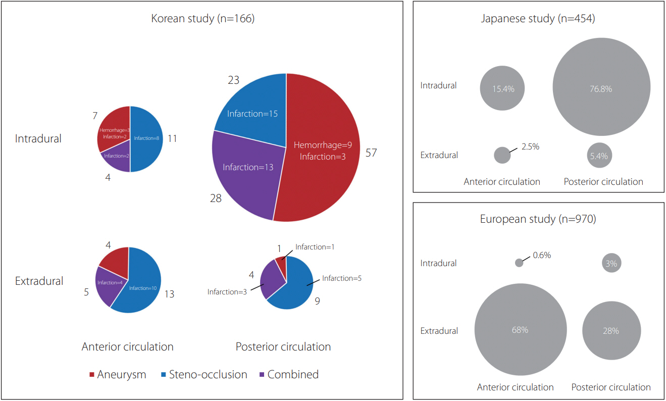
Fig. 2. Pie charts showing the differences of lesion locations among three studies according to the presenting and morphological patterns. The sizes of the circles reflect the number of patients in each group. The sector of the circle is marked with different colors according to the proportional patient number of each morphological pattern; the numbers of “aneurysm” and “steno-occlusion” do not include the numbers of “combined” cases. The pie charts of Japanese and European studies do not have colors because a detailed analysis of presenting patterns could not be done.
Reference
-
1. Schievink WI. Spontaneous dissection of the carotid and vertebral arteries. N Engl J Med. 2001; 344:898–906.
Article2. Kwak JH, Choi JW, Park HJ, Chae EY, Park ES, Lee DH, et al. Cerebral artery dissection: spectrum of clinical presentations related to angiographic findings. Neurointervention. 2011; 6:78–83.
Article3. Shin JH, Suh DC, Choi CG, Leei HK. Vertebral artery dissection: spectrum of imaging findings with emphasis on angiography and correlation with clinical presentation. Radiographics. 2000; 20:1687–1696.
Article4. Schievink WI, Mokri B, Piepgras DG. Spontaneous dissections of cervicocephalic arteries in childhood and adolescence. Neurology. 1994; 44:1607–1612.
Article5. Park MS, Cha J, Chung JW, Seo WK, Kim GM, Bang OY. Arterial dissection as a cause of intracranial stenosis in East Asians. J Am Coll Cardiol. 2017; 70:2205–2206.
Article6. Shin J, Chung JW, Park MS, Lee H, Cha J, Seo WK, et al. Outcomes after ischemic stroke caused by intracranial atherosclerosis vs dissection. Neurology. 2018; 91:e1751–e1759.
Article7. Kwon JY, Kim NY, Suh DC, Kang DW, Kwon SU, Kim JS. Intracranial and extracranial arterial dissection presenting with ischemic stroke: lesion location and stroke mechanism. J Neurol Sci. 2015; 358:371–376.
Article8. Debette S, Grond-Ginsbach C, Bodenant M, Kloss M, Engelter S, Metso T, Cervical Artery Dissection Ischemic Stroke Patients (CADISP) Group, et al. Differential features of carotid and vertebral artery dissections: the CADISP study. Neurology. 2011; 77:1174–1181.
Article9. Dziewas R, Konrad C, Dräger B, Evers S, Besselmann M, Lüdemann P, et al. Cervical artery dissection--clinical features, risk factors, therapy and outcome in 126 patients. J Neurol. 2003; 250:1179–1184.
Article10. Touzé E, Gauvrit JY, Moulin T, Meder JF, Bracard S, Mas JL; Multicenter Survey on Natural History of Cervical Artery Dissection. Risk of stroke and recurrent dissection after a cervical artery dissection: a multicenter study. Neurology. 2003; 61:1347–1351.
Article11. Arnold M, Bousser MG, Fahrni G, Fischer U, Georgiadis D, Gandjour J, et al. Vertebral artery dissection: presenting findings and predictors of outcome. Stroke. 2006; 37:2499–2503.12. Lee VH, Brown RD Jr, Mandrekar JN, Mokri B. Incidence and outcome of cervical artery dissection: a population-based study. Neurology. 2006; 67:1809–1812.
Article13. Huang YC, Chen YF, Wang YH, Tu YK, Jeng JS, Liu HM. Cervicocranial arterial dissection: experience of 73 patients in a single center. Surg Neurol. 2009; 72 Suppl 2:S20–7. discussion S27.
Article14. Tsukahara T, Minematsu K. Overview of spontaneous cervicocephalic arterial dissection in Japan. Acta Neurochir Suppl. 2010; 107:35–40.
Article15. Metso TM, Metso AJ, Helenius J, Haapaniemi E, Salonen O, Porras M, et al. Prognosis and safety of anticoagulation in intracranial artery dissections in adults. Stroke. 2007; 38:1837–1842.
Article16. Debette S, Compter A, Labeyrie MA, Uyttenboogaart M, Metso TM, Majersik JJ, et al. Epidemiology, pathophysiology, diagnosis, and management of intracranial artery dissection. Lancet Neurol. 2015; 14:640–654.
Article17. Debette S, Grond-Ginsbach C, Bodenant M, Kloss M, Engelter S, Metso T, et al. Differential features of carotid and vertebral artery dissections: The cadisp study. Neurology. 2011; 77:1174–1181.
Article18. Song Y, Lee D, Suh DC, Kim JG, Kim JK, Han M, et al. Cigarette smoking preferentially affects intracranial vessels in young males: a propensity-score matching analysis. Neurointervention. 2019; 14:43–52.
Article19. von Babo M, De Marchis GM, Sarikaya H, Stapf C, Buffon F, Fischer U, et al. Differences and similarities between spontaneous dissections of the internal carotid artery and the vertebral artery. Stroke. 2013; 44:1537–1542.
Article20. Kim BM, Kim SH, Kim DI, Shin YS, Suh SH, Kim DJ, et al. Outcomes and prognostic factors of intracranial unruptured vertebrobasilar artery dissection. Neurology. 2011; 76:1735–1741.
Article21. Shin DH, Hong JM, Lee JS, Nasim R, Sohn SI, Kim SJ, et al. Comparison of potential risks between intracranial and extracranial vertebral artery dissections. Eur Neurol. 2014; 71:305–312.
Article22. Kim DJ. Intracranial stenting; the current landscape. Neurointervention. 2021; 16:2–5.
Article23. Lee JS, Hwang YH, Sohn SI. Factors contributing to an efficacious endovascular treatment for acute ischemic stroke in Asian population. Neurointervention. 2021; 16:91–110.
Article24. Bang OY. Intracranial atherosclerosis: current understanding and perspectives. J Stroke. 2014; 16:27–35.
Article25. Kim JS, Bonovich D. Research on intracranial atherosclerosis from the east and west: why are the results different? J Stroke. 2014; 16:105–113.
Article26. Song Y, Kwon B, Al-Abdulwahhab AH, Nam YK, Ahn Y, Jeong SY, et al. Rare neurovascular diseases in Korea: classification and related genetic variants. Korean J Radiol. 2021; 22:1379–1396.
Article27. Ulbricht D, Diederich NJ, Hermanns-Lê T, Metz RJ, Macian F, Piérard GE. Cervical artery dissection: an atypical presentation with Ehlers-Danlos-like collagen pathology? Neurology. 2004; 63:1708–1710.
Article28. Grond-Ginsbach C, Debette S. The association of connective tissue disorders with cervical artery dissections. Curr Mol Med. 2009; 9:210–214.
Article29. Longoni M, Grond-Ginsbach C, Grau AJ, Genius J, Debette S, Schwaninger M, et al. The ICAM-1 E469K gene polymorphism is a risk factor for spontaneous cervical artery dissection. Neurology. 2006; 66:1273–1275.
Article30. Kim JS, Lee HB, Kwon HS. RNF213 polymorphism in intracranial artery dissection. J Stroke. 2018; 20:404–406.
Article31. Pezzini A, Del Zotto E, Archetti S, Negrini R, Bani P, Albertini A, et al. Plasma homocysteine concentration, C677T MTHFR genotype, and 844ins68bp CBS genotype in young adults with spontaneous cervical artery dissection and atherothrombotic stroke. Stroke. 2002; 33:664–669.
Article32. Kim BJ, Yang E, Kim NY, Kim MJ, Kang DW, Kwon SU, et al. Vascular tortuosity may be associated with cervical artery dissection. Stroke. 2016; 47:2548–2552.
Article33. Lee RM. Morphology of cerebral arteries. Pharmacol Ther. 1995; 66:149–173.
Article34. Wu Y, Chen H, Xing S, Tan S, Chen X, Tan Y, et al. Predisposing factors and radiological features in patients with internal carotid artery dissection or vertebral artery dissection. BMC Neurol. 2020; 20:445.
Article35. Choi MH, Hong JM, Lee JS, Shin DH, Choi HA, Lee K. Preferential location for arterial dissection presenting as golf-related stroke. AJNR Am J Neuroradiol. 2014; 35:323–326.
Article
- Full Text Links
-
- Actions
-
Cited
- CITED
-
- Close
- Share
-
- Similar articles
-
- Management of Dissecting Aneurysm of Cervicocephalic Carotid and Vertebral Artery
- Recurrent cervicocephalic artery dissection in a patient with Churg-Strauss syndrome treated with long-term corticosteroid therapy
- Spontaneous Coronary Artery Dissection in a female patient with fragile X syndrome
- Clinical Experiences of Unruptured Vertebral Artery Dissection
- Surgical Technique for the Functional Preservation of the Inferior Parathyroid Glands
