A rare case of primary plasma cell leukemia exhibiting the small-cell variant of plasma cells
- Affiliations
-
- 1Department of Laboratory Medicine, Soonchunhyang University Seoul Hospital, Seoul, Korea
- 2Department of Internal Medicine, Soonchunhyang University Seoul Hospital, Seoul, Korea
- KMID: 2530884
- DOI: http://doi.org/10.5045/br.2022.2022059
Figure
-
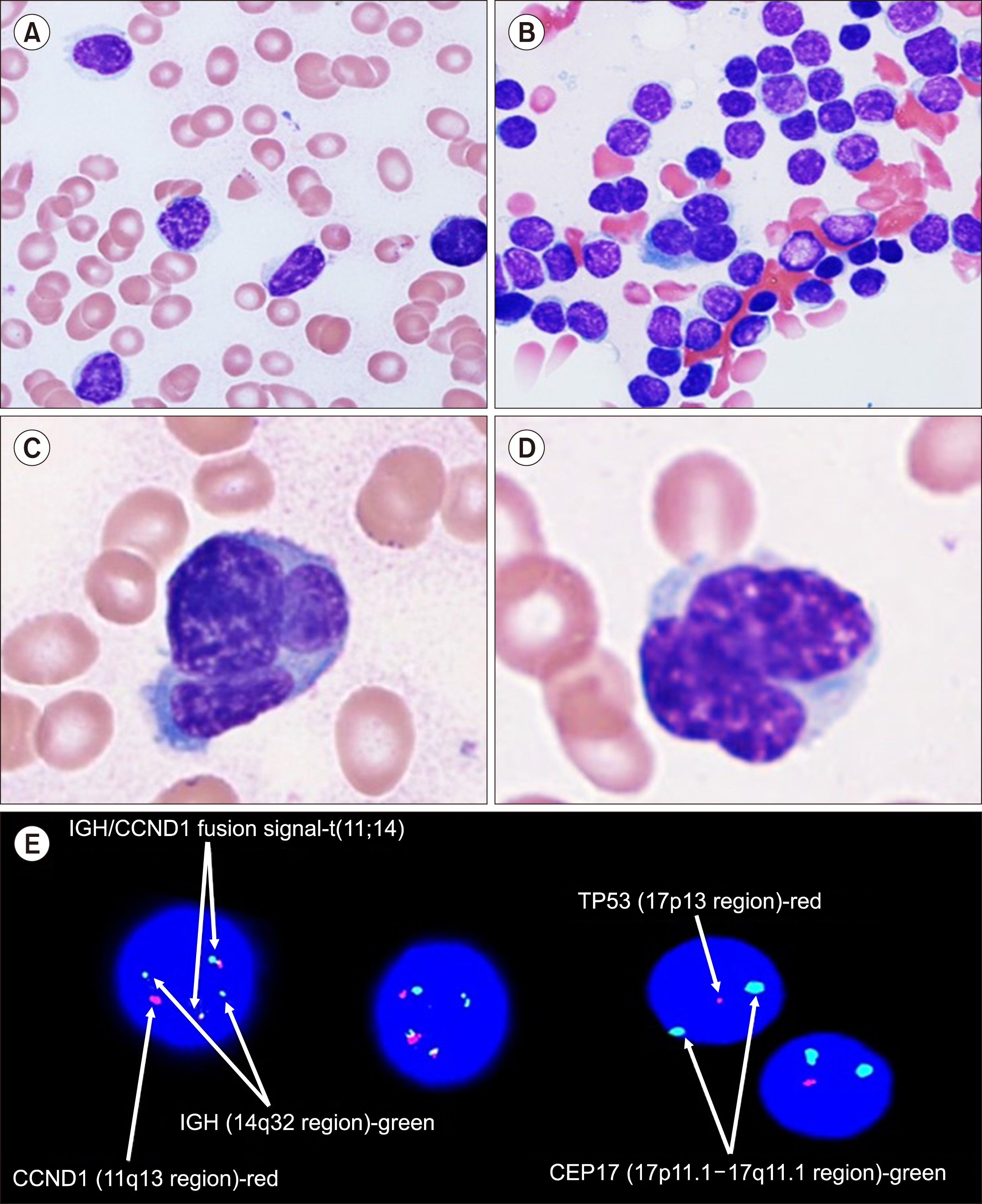
Fig. 1 Microscopic findings and FISH analysis of the small-cell type of plasma cell leukemia. Peripheral blood and bone marrow aspirate smears: small-cell type neoplastic plasma cells and atypical multinu-clear plasma cells (A), peripheral blood, Wright–Giemsa stain, ×400; (B), bone marrow, Wright–Giemsa stain, ×400; (C, D) bone marrow, Wright–Giemsa stain, ×1,000. (E) FISH analysis showing an IGH/ CCND1 rearrangement and p53 (17p) deletion (white arrow). Abbreviation: FISH, fluorescence in situ hybridization.
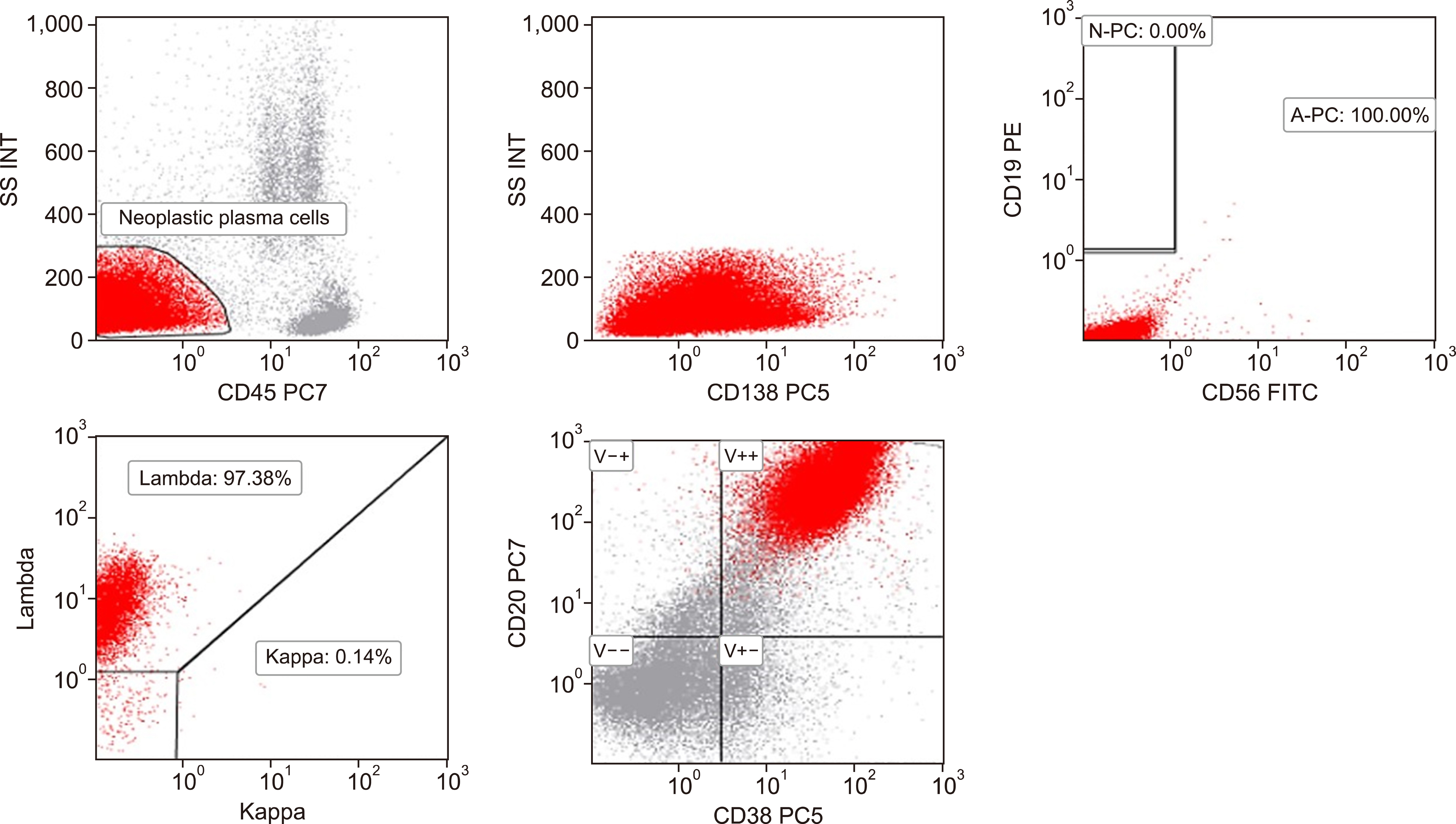
Fig. 2 Flow-cytometric immunophenotyping of the small-cell type of plasma cell leukemia. Clonal plasma cells in the bone marrow exhibited a CD19-/CD20+/CD38+/CD56-/CD138+ phenotype and cytoplasmic lambda light-chain restriction gated by CD45- and low side scatter.
Reference
-
1. Gonsalves WI, Rajkumar SV, Go RS, et al. 2014; Trends in survival of patients with primary plasma cell leukemia: a population-based analysis. Blood. 124:907–12. DOI: 10.1182/blood-2014-03-565051. PMID: 24957143. PMCID: PMC4126330.
Article2. Mina R, Joseph NS, Kaufman JL, et al. 2019; Survival outcomes of patients with primary plasma cell leukemia (pPCL) treated with novel agents. Cancer. 125:416–23. DOI: 10.1002/cncr.31718. PMID: 30332496.
Article3. Bartl R, Frisch B, Fateh-Moghadam A, Kettner G, Jaeger K, Sommerfeld W. 1987; Histologic classification and staging of multiple myeloma. A retrospective and prospective study of 674 cases. Am J Clin Pathol. 87:342–55. DOI: 10.1093/ajcp/87.3.342. PMID: 3825999.
Article4. Raja KR, Kovarova L, Hajek R. 2010; Review of phenotypic markers used in flow cytometric analysis of MGUS and MM, and applicability of flow cytometry in other plasma cell disorders. Br J Haematol. 149:334–51. DOI: 10.1111/j.1365-2141.2010.08121.x. PMID: 20201947.
Article5. Pellat-Deceunynck C, Barillé S, Jego G, et al. 1998; The absence of CD56 (NCAM) on malignant plasma cells is a hallmark of plasma cell leukemia and of a special subset of multiple myeloma. Leukemia. 12:1977–82. DOI: 10.1038/sj.leu.2401211. PMID: 9844928.
Article6. Robillard N, Avet-Loiseau H, Garand R, et al. 2003; CD20 is associated with a small mature plasma cell morphology and t(11;14) in multiple myeloma. Blood. 102:1070–1. DOI: 10.1182/blood-2002-11-3333. PMID: 12702507.
Article7. Heerema-McKenney A, Waldron J, Hughes S, et al. 2010; Clinical, immunophenotypic, and genetic characterization of small lymphocyte-like plasma cell myeloma: a potential mimic of mature B-cell lymphoma. Am J Clin Pathol. 133:265–70. DOI: 10.1309/AJCPUS3PRRT5ZXVS. PMID: 20093236. PMCID: PMC4433023.
Article8. Gounari E, Kaiafa G, Koletsa T, et al. 2018; CD5+ B lymphoproliferative disorder with subsequent development of plasma cell leukaemia: diagnostic and aetiologic reasoning. Cytometry B Clin Cytom. 94:688–94. DOI: 10.1002/cyto.b.21596. PMID: 29024518.
Article9. Loureiro AD, Gonçalves MV, Ikoma MR, et al. 2017; Plasma cell leukemia with t(11;14)(q13;q32) simulating lymphoplasmacytic lymphoma - a diagnostic challenge solved by flow cytometry. Rev Bras Hematol Hemoter. 39:66–9. DOI: 10.1016/j.bjhh.2016.10.001. PMID: 28270351. PMCID: PMC5339392.
Article10. Teriaky A, Hsia CC. 2011; Plasma cell leukemia mimicking chronic lymphocytic leukemia. Blood. 117:2991. DOI: 10.1182/blood-2010-02-269977. PMID: 21528511.
Article
- Full Text Links
-
- Actions
-
Cited
- CITED
-
- Close
- Share
-
- Similar articles
-
- A case of primary plasma cell leukemia exhibiting hemophagocytic plasma cells relapsed with multiple cutaneous plasmacytoma
- A case of primary plasma cell leukemia
- Plasma cell leukemia with rouleaux formation involving neoplastic cells and RBC
- A Case of Metastatic Ovarian Cancer Arising in Plasma Cell Leukemia
- Plasma cell leukemia
