Korean Circ J.
2022 Jun;52(6):481-482. 10.4070/kcj.2022.0060.
Recurrent Pericardial and Pleural Effusion due to Pericardial Pleomorphic Mesothelioma
- Affiliations
-
- 1Cardiac Surgery Department, Clinic University Hospital of Valladolid, Valladolid, Spain
- 2Thoracic Surgery Department, Clinic University Hospital of Valladolid, Valladolid, Spain
- KMID: 2530458
- DOI: http://doi.org/10.4070/kcj.2022.0060
Figure
-
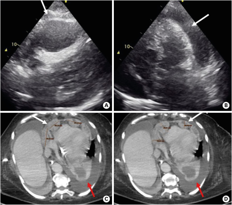
Figure 1 (A, B) Echocardiogram images: severe pericardial effusion with compressing the apex and the right ventricle (white arrows). (C, D) Computerized tomography images: nodular pericardium with diameter maximum of 38 mm (white arrows) and pericardial and pleural effusion (red arrows).
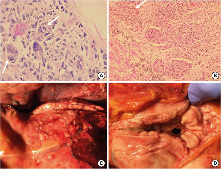
Figure 2 (A) Pathologic examination zoom ×40: pericardium with infiltration of pleomorphic cells is observed (white arrows). (B) Pathologic examination: in the upper right quadrant pericardium with infiltration of pleomorphic cells (white arrow) with progressive invasion of myocardial tissue in the lower left quadrant, in the central area there is lymphatic dissemination. (C, D) Autopsy showed invasion and thickening of the pericardium and myocardium.
- Full Text Links
-
- Actions
-
Cited
- CITED
-
- Close
- Share
-
- Similar articles
-
- CT findings of intrathoracic mesothelioma
- A case of massive pericardial effusion caused by acute recurrent pancreatitis with complication
- Pericardial mesothelioma in a dog with lymph node metastasis and chylothorax
- Disappearance of pericardial effusion by suspected pericardial-pleural fistulain a Miniature Schnauzer dog
- A "Vanishing", Tuberculous, Pericardial Effusion
