Juxtacortical chondromyxoid fibroma in the small bones: two cases with unusual location and a literature review
- Affiliations
-
- 1Department of Pathology, Kosin University Gospel Hospital, Kosin University College of Medicine, Busan, Korea
- 2Department of Orthopedic Surgery, Kosin University Gospel Hospital, Kosin University College of Medicine, Busan, Korea
- KMID: 2529807
- DOI: http://doi.org/10.4132/jptm.2021.12.15
Abstract
- Chondromyxoid fibroma is a rare bone tumor of cartilaginous origin, representing less than 1% of all bone tumors. It preferentially arises in the eccentric location of the metaphysis of a long tubular bone. Juxtacortical locations are reported infrequently in the long bones and even more rarely in short tubular bones, with only three cases documented. Here we present two new cases of juxtacortical chondromyxoid fibroma in the small bones. One was an intracortical osteolytic lesion of the metatarsal bone of the foot with degenerative atypia that histologically should be differentiated from chondrosarcoma. The other was a phalangeal mass protruding into the interphalangeal joint of the hand, which had been labeled mistakenly as a soft tissue mass preoperatively. These cases illustrated that chondromyxoid fibromas have various the manifestations and should be included in the differential diagnosis of an osteolytic lesion or an exophytic mass in the small bones.
Keyword
Figure
-
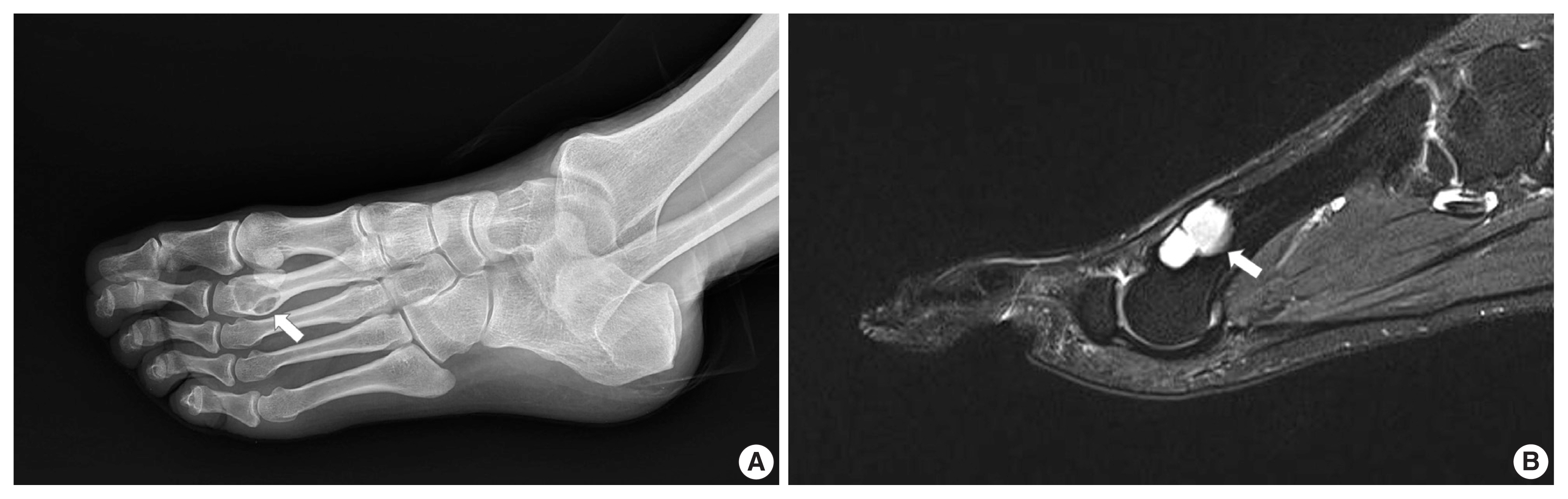
Fig. 1 Radiologic images of the case 1. (A) Oblique radiograph of the foot shows an intracortical lytic lesion (arrow) in the right second metatarsal neck. (B) Sagittal magnetic resonance image shows a hyperintense mass (arrow) with an intact shell of cortical bone.
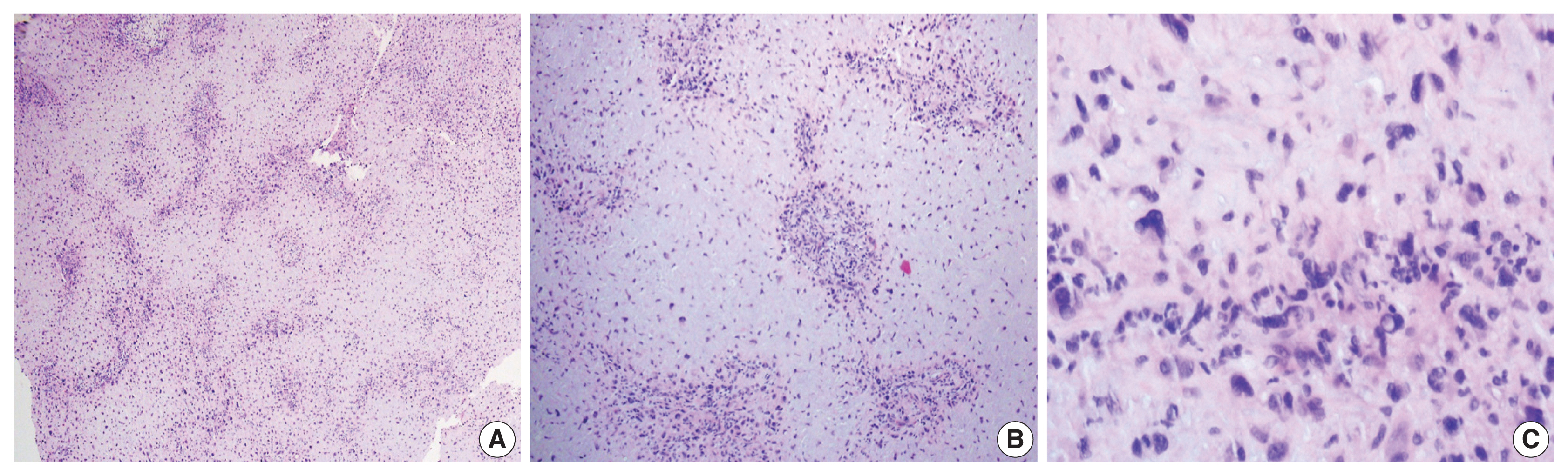
Fig. 2 Histologic findings of the case 1. (A) Low-power view of the tumor shows a prominent lobular patten. (B) The lobules consist of a hypocellular center with abundant myxoid matrix and condensation of tumor cells toward the periphery. (C) The tumor cells in the hypercellular periphery reveal degenerative features including bizarre nuclei, hyperchromasia, and vacuolization.

Fig. 3 Radiologic and histologic images of the case 2. (A) Lateral radiograph of the right index finger reveals a soft tissue mass (arrow) with calcification in the proximal interphalangeal joint. The lesion erodes the epiphysis of the mid phalanx. (B) Magnetic resonance image shows a protruding mass (arrow) with heterogeneous intensity. (C) Microphotographs show a characteristic lobular growth pattern. Spotty calcifications are scattered throughout the lesion. (D) Hypercellular area surrounding the hypocellular myxoid lobules occasionally contains multinucleated giant cells.
Reference
-
References
1. Unni KK, Inwards CY. Dahlin’s bone tumors. 6th ed. Philadelphia: Lippincott Williams and Wilkins;2010. p. 50–9.2. Baker AC, Rezeanu L, O’Laughlin S, Unni K, Klein MJ, Siegal GP. Juxtacortical chondromyxoid fibroma of bone: a unique variant: a case study of 20 patients. Am J Surg Pathol. 2007; 31:1662–8.3. Jaffe HL, Lichtenstein L. Chondromyxoid fibroma of bone; a distinctive benign tumor likely to be mistaken especially for chondrosarcoma. Arch Pathol (Chic). 1948; 45:541–51.4. Takenaga RK, Frassica FJ, McCarthy EF. Subperiosteal chondromyxoid fibroma: a report of two cases. Iowa Orthop J. 2007; 27:104–7.5. Estrada-Villasenor E, Cedillo ED, Martinez GR, Chavez RD. Periosteal chondromyxoid fibroma: a case study using imprint cytology. Diagn Cytopathol. 2005; 33:402–6.
Article6. Harrington KA, Hoda S, La Rocca Vieira R. Surface-type chondromyxoid fibroma in an elderly patient: a case report and literature review. Skeletal Radiol. 2019; 48:823–30.
Article7. Han JS, Shim E, Kim BH, Choi JW. An intracortical chondromyxoid fibroma in the diaphysis of the metatarsal. Skeletal Radiol. 2017; 46:1757–62.
Article8. Abdelwahab IF, Klein MJ. Surface chondromyxoid fibroma of the distal ulna: unusual tumor, site, and age. Skeletal Radiol. 2014; 43:243–6.
Article9. Slotcavage RL, Dickson BC, Ogilvie CM. Chondromyxoid fibroma involving the metacarpophalangeal joint. Orthopedics. 2009. 32: orthosupersite.com/view.asp?rID=38065 .
Article10. Wu CT, Inwards CY, O’Laughlin S, Rock MG, Beabout JW, Unni KK. Chondromyxoid fibroma of bone: a clinicopathologic review of 278 cases. Hum Pathol. 1998; 29:438–46.
Article11. Won KY, Lee J, Kim YW, Kim EJ, Kim SW, Park YK. Chondromyxoid fibroma of the ethmoid sinus complicated by a brain abscess: a case report and literature review. Korean J Pathol. 2010; 44:547–50.
Article
- Full Text Links
-
- Actions
-
Cited
- CITED
-
- Close
- Share
-
- Similar articles
-
- Chondromyxoid fibroma of iliac bone: Report of a Case
- Slipped Capital Femoral Epiphysis after Curettage of Juxtaphyseal Chondromyxoid Fibroma of the Femoral Neck
- A Rare Case of Epiphyseal Chondromyxoid Fibroma of the Proximal Tibia
- Chondromyxoid Fibroma Involving Distal Phalanx of the Big Toe Treated with Reconstructive Surgery: A Case Report
- Chondromyxoid fibroma of the femur: a case report with intra-cortical location
