Korean J Radiol.
2011 Dec;12(6):761-764. 10.3348/kjr.2011.12.6.761.
A Rare Case of Epiphyseal Chondromyxoid Fibroma of the Proximal Tibia
- Affiliations
-
- 1Department of Radiology, Eulji Hospital, Eulji University, Seoul 139-711, Korea. cys0128@eulji.ac.kr
- 2Department of Orthopedic Surgery, Eulji Hospital, Eulji University, Seoul 139-711, Korea.
- 3Department of Pathology, Eulji Hospital, Eulji University, Seoul 139-711, Korea.
- 4Department of Pathology, Kyung Hee University Hospital, Seoul 130-702, Korea.
- KMID: 1101934
- DOI: http://doi.org/10.3348/kjr.2011.12.6.761
Abstract
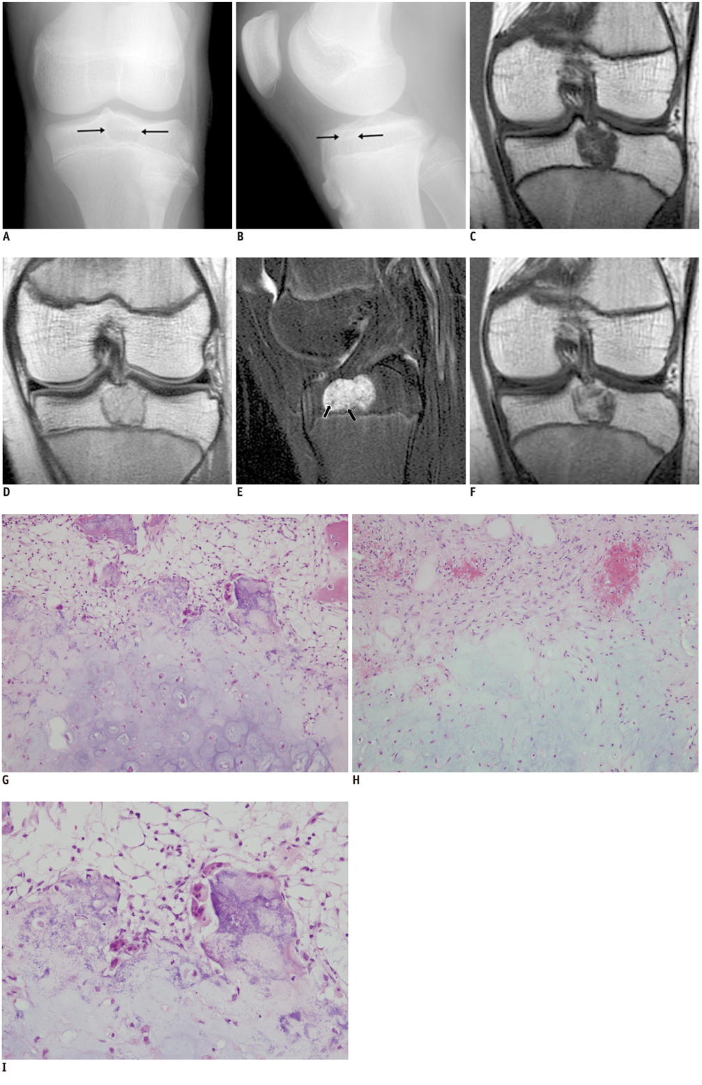
- Chondromyxoid fibroma is an uncommon benign cartilaginous tumor of the bone. It occurs most frequently in the metaphysis of long tubular bones, and an epiphyseal location is exceedingly rare. We present here an unusual case of a chondromyxoid fibroma that occurred in the epiphysis of the proximal tibia with an open growth plate. MR imaging findings of this tumor, which has, to the best of our knowledge, never been described in an epiphyseal location, makes the present case unique.
MeSH Terms
Figure
Reference
-
1. Giudici MA, Moser RP Jr, Kransdorf MJ. Cartilaginous bone tumors. Radiol Clin North Am. 1993. 31:237–259.2. Feldman F, Hecht HL, Johnston AD. Chondromyxoid fibroma of bone. Radiology. 1970. 94:249–260.3. Rahimi A, Beabout JW, Ivins JC, Dahlin DC. Chondromyxoid fibroma: a clinicopathologic study of 76 cases. Cancer. 1972. 30:726–736.4. Wilson AJ, Kyriakos M, Ackerman LV. Chondromyxoid fibroma: radiographic appearance in 38 cases and in a review of the literature. Radiology. 1991. 179:513–518.5. Wu CT, Inwards CY, O'Laughlin S, Rock MG, Beabout JW, Unni KK. Chondromyxoid fibroma of bone: a clinicopathologic review of 278 cases. Hum Pathol. 1998. 29:438–446.6. Gardner DJ, Azouz EM. Solitary lucent epiphyseal lesions in children. Skeletal Radiol. 1988. 17:497–504.7. Fotiadis E, Akritopoulos P, Samoladas E, Akritopoulou K, Kenanidis E. Chondromyxoid fibroma: a rare tumor with an unusual location. Arch Orthop Trauma Surg. 2008. 128:371–375.8. Jaffe HL, Lichtenstein L. Chondromyxoid fibroma of bone; a distinctive benign tumor likely to be mistaken especially for chondrosarcoma. Arch Pathol (Chic). 1948. 45:541–551.9. Soler R, Rodriguez E, Suarez I, Gayol A. Magnetic resonance imaging of chondromyxoid fibroma of the fibula. Eur J Radiol. 1994. 18:210–211.10. Adams MJ, Spencer GM, Totterman S, Hicks DG. Quiz: case report 776. Chondromyxoid fibroma of femur. Skeletal Radiol. 1993. 22:358–361.11. Budny AM, Ismail A, Osher L. Chondromyxoid fibroma. J Foot Ankle Surg. 2008. 47:153–159.12. Jee WH, Park YK, McCauley TR, Choi KH, Ryu KN, Suh JS, et al. Chondroblastoma: MR characteristics with pathologic correlation. J Comput Assist Tomogr. 1999. 23:721–726.
- Full Text Links
- Actions
-
Cited
- CITED
-
- Close
- Share
- Similar articles
-
- Slipped Capital Femoral Epiphysis after Curettage of Juxtaphyseal Chondromyxoid Fibroma of the Femoral Neck
- A Chondromyxoid Fibroma of the Fibula: A Case Report
- Chondromyxoid Fibroma Involving Distal Phalanx of the Big Toe Treated with Reconstructive Surgery: A Case Report
- Chondromyxoid fibroma of iliac bone: Report of a Case
- Juxtacortical chondromyxoid fibroma in the small bones: two cases with unusual location and a literature review


