Pharmaceutical Activation of Nrf2 Accelerates Diabetic Wound Healing by Exosomes from Bone Marrow Mesenchymal Stem Cells
- Affiliations
-
- 1Department of Burn Rectification, Affiliated Hospital of Nantong University, Nantong, China
- KMID: 2529788
- DOI: http://doi.org/10.15283/ijsc21067
Abstract
- Background and Objectives
Despite advances in wound treatments, chronic diabetic wounds remain a significant medi-cal challenge. Exosomes from mesenchymal stem cells (MSCs) and small molecule activators of nuclear factor erythroid 2–related factor 2 (Nrf2) have emerged as potential therapies for nonhealing diabetic wounds. This study aimed to evaluate the effects of exosomes from bone marrow-derived MSCs (BMSCs) alone, or in combination with a small molecule activator of Nrf2 on diabetic wound healing.
Methods and Results
BMSCs and endothelial progenitor cells (EPCs) were isolated from the femur and tibia bone marrow of Sprague-Dawley (SD) rats and culture-expanded. Exosomes were harvested from the BMSC culture supernatants through ultracentrifugation. The effects of the exosomes and Nrf2 knockdown, alone or in combination, on EPC tube formation were evaluated. Streptozotocin-induced diabetic rats bearing a fresh full-thickness round wound were treated with the exosomes alone, or in combination with a lentiviral shRNA targeting Nrf2 (Lenti-sh-Nrf2) or tert-butylhydroquinone (tBHQ), a small molecule activator of Nrf2. Two weeks later, wound closure, re-epithelization, collagen deposition, neovascularization, and local inflammation were evaluated. BMSC exosomes promoted while Nrf2 knockdown inhibited EPC tube formation. BMSC exosomes accelerated wound closure, re-epithelization, collagen deposition, and neovascularization, and reduced wound inflammation in diabetic rats. These regenerative and anti-inflammatory effects of the exosomes were inhibited by Lenti-sh-Nrf2 but enhanced by tBHQ administration.
Conclusions
BMSC exosomes in combination with a small molecule Nrf2 activator hold promise as a new therapeutic option for chronic diabetic wounds.
Keyword
Figure
-

Fig. 1 Identification of BMSCs and BMSC exosomes. (A) The detection of the surface markers CD105, CD90, and CD45 in isolated BMSCs with flow cytometry. (B) A TEM image of BMSC exosomes. Scale bar=100 nm. (C) The particle size distribution of BMSC exosomes determined with light scattering. (D) The detection of the exosome markers CD9, CD63, and TSG101 in BMSC exosomes with western blot analysis.

Fig. 2 The effects of BMSC exosomes and Nrf2 knockdown, alone or in combination, on EPC tube formation. (A) The detection of the surface markers CD31, CD34, and CD45 in isolated EPCs with flow cytometry. (B) EPCs were transfected with Lenti-sh-Nrf2 or Lenti-sh-NC for 48 hours. The Nrf2 protein levels were determined with western blot analysis. (C, D) EPCs stably expressing sh-Nrf2 or sh-NC were treated with BMSC exosomes (200 μg/ml) for 72 hours. (C) Tube formation was observed under a microscope. Scale bar=100 μm. (D) The relative tube length in each treatment group was determined. N=3, *p<0.05, **p< 0.01.

Fig. 3 The effects of BMSC exosomes alone, or in combination with knockdown or pharmaceutical activation of Nrf2 on wound closure in diabetic rats. STZ-induced diabetic rats received 100 μg/ml subcutaneous BMSC exosomes alone at the wound site, or in combination with 200 μl intravenous Lenti-sh-Nrf2 or Lenti-sh-NC, or 50 mg/kg intraperitoneal tBHQ immediately after the wounding and again a week after. Rats that received PBS were included as control. (A) The photo images of the wounds at day 0, 7, and 14 after the wounding. (B) The wound size at day 0, 7, and 14 after the wounding. N=5; *p<0.05, **p<0.01 vs. PBS control.

Fig. 4 The effects of BMSC exosomes alone, or in combination with knockdown or pharmaceutical activation of Nrf2 on wound tissue regeneration in diabetic rats. (A) The Nrf2 protein levels in the wound tissues at day 14 after the wounding were evaluated with western blot analysis. The STZ-induced diabetic rats were treated after skin wounding as described in Fig. 3. (A, B) Wound re-epithelization and collagen deposition were evaluated with H&E (B) and Masson (C) staining of the wound tissues at day 14 after the wounding. Scale bar=100 μm. N=5; *p<0.05, **p<0.01 vs. PBS control.
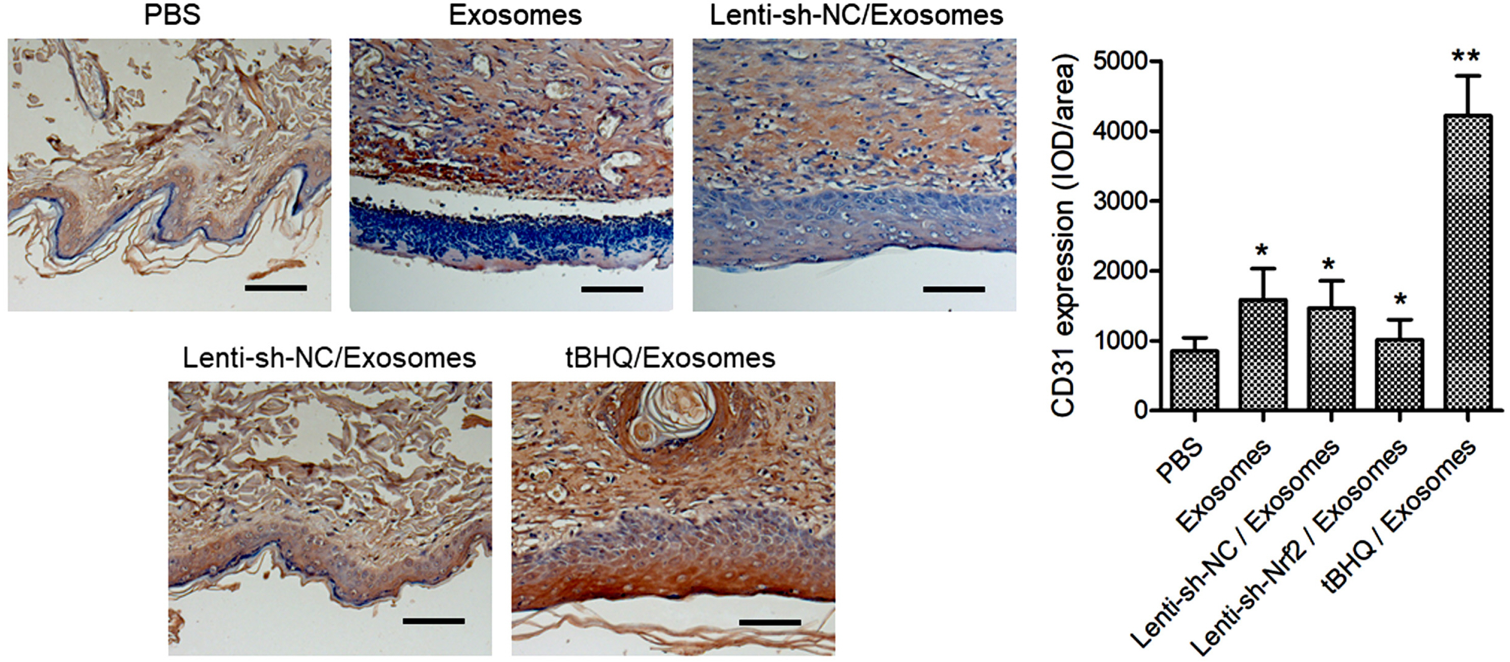
Fig. 5 The effects of BMSC exosomes alone, or in combination with knockdown or pharmaceutical activation of Nrf2 on wound neovascularization in diabetic rats. The STZ-induced diabetic rats were treated after skin wounding as described in Fig. 3. The CD31 expression in the wound tissues at day 14 after the wounding was detected with immunohistochemistry. Scale bar=100 μm. N=5; *p<0.05, **p<0.01 vs. PBS control.

Fig. 6 The effects of BMSC exosomes alone, or in combination with knockdown or pharmaceutical activation of Nrf2 on wound inflammation in diabetic rats. The STZ-induced diabetic rats were treated after skin wounding as described in Fig. 3. The levels of TNF-α, IL-1β, IL-4, and IL-10 in the wound tissues at day 14 after the wounding were determined with ELISA. N=5; *p<0.05, **p<0.01 vs. PBS control.
Reference
-
References
1. Nussbaum SR, Carter MJ, Fife CE, DaVanzo J, Haught R, Nusgart M, Cartwright D. 2018; An economic evaluation of the impact, cost, and medicare policy implications of chronic nonhealing wounds. Value Health. 21:27–32. DOI: 10.1016/j.jval.2017.07.007. PMID: 29304937.
Article2. Kosaric N, Kiwanuka H, Gurtner GC. 2019; Stem cell therapies for wound healing. Expert Opin Biol Ther. 19:575–585. DOI: 10.1080/14712598.2019.1596257. PMID: 30900481.
Article3. Hettich BF, Ben-Yehuda Greenwald M, Werner S, Leroux JC. 2020; Exosomes for wound healing: purification optimization and identification of bioactive components. Adv Sci (Weinh). 7:2002596. DOI: 10.1002/advs.202002596. PMID: 33304765. PMCID: PMC7709981.
Article4. Silva AM, Teixeira JH, Almeida MI, Gonçalves RM, Barbosa MA, Santos SG. 2017; Extracellular vesicles: immunomodulatory messengers in the context of tissue repair/regeneration. Eur J Pharm Sci. 98:86–95. DOI: 10.1016/j.ejps.2016.09.017. PMID: 27644894.
Article5. Hoang DH, Nguyen TD, Nguyen HP, Nguyen XH, Do PTX, Dang VD, Dam PTM, Bui HTH, Trinh MQ, Vu DM, Hoang NTM, Thanh LN, Than UTT. 2020; Differential wound healing capacity of mesenchymal stem cell-derived exosomes originated from bone marrow, adipose tissue and umbilical cord under serum- and xeno-free condition. Front Mol Biosci. 7:119. DOI: 10.3389/fmolb.2020.00119. PMID: 32671095. PMCID: PMC7327117. PMID: d8551c1647974a088970fa3aafd4b75b.
Article6. Ding J, Wang X, Chen B, Zhang J, Xu J. 2019; Exosomes derived from human bone marrow mesenchymal stem cells stimulated by deferoxamine accelerate cutaneous wound healing by promoting angiogenesis. Biomed Res Int. 2019:9742765. DOI: 10.1155/2019/9742765. PMID: 31192260. PMCID: PMC6525840.
Article7. Wu D, Kang L, Tian J, Wu Y, Liu J, Li Z, Wu X, Huang Y, Gao B, Wang H, Wu Z, Qiu G. 2020; Exosomes derived from bone mesenchymal stem cells with the stimulation of Fe3O4 nanoparticles and static magnetic field enhance wound healing through upregulated miR-21-5p. Int J Nanomedicine. 15:7979–7993. DOI: 10.2147/IJN.S275650. PMID: 33116513. PMCID: PMC7585514.8. Wang C, Liang C, Wang R, Yao X, Guo P, Yuan W, Liu Y, Song Y, Li Z, Xie X. 2019; The fabrication of a highly efficient self-healing hydrogel from natural biopolymers loaded with exosomes for the synergistic promotion of severe wound healing. Biomater Sci. 8:313–324. DOI: 10.1039/C9BM01207A. PMID: 31701966.
Article9. Wang C, Wang M, Xu T, Zhang X, Lin C, Gao W, Xu H, Lei B, Mao C. 2019; Engineering bioactive self-healing antibacterial exosomes hydrogel for promoting chronic diabetic wound healing and complete skin regeneration. Theranostics. 9:65–76. DOI: 10.7150/thno.29766. PMID: 30662554. PMCID: PMC6332800.
Article10. He F, Ru X, Wen T. 2020; NRF2, a transcription factor for stress response and beyond. Int J Mol Sci. 21:4777. DOI: 10.3390/ijms21134777. PMID: 32640524. PMCID: PMC7369905. PMID: 7528f37dbd2449179875581319f6f555.
Article11. Long M, Rojo de la Vega M, Wen Q, Bharara M, Jiang T, Zhang R, Zhou S, Wong PK, Wondrak GT, Zheng H, Zhang DD. 2016; An essential role of NRF2 in diabetic wound healing. Diabetes. 65:780–793. DOI: 10.2337/db15-0564. PMID: 26718502. PMCID: PMC4764153.
Article12. Murasawa S, Asahara T. 2005; Endothelial progenitor cells for vasculogenesis. Physiology (Bethesda). 20:36–42. DOI: 10.1152/physiol.00033.2004. PMID: 15653838.
Article13. Kaushik K, Das A. 2019; Endothelial progenitor cell therapy for chronic wound tissue regeneration. Cytotherapy. 21:1137–1150. DOI: 10.1016/j.jcyt.2019.09.002. PMID: 31668487.
Article14. Wang RY, Liu LH, Liu H, Wu KF, An J, Wang Q, Liu Y, Bai LJ, Qi BM, Qi BL, Zhang L. 2018; Nrf2 protects against diabetic dysfunction of endothelial progenitor cells via regulating cell senescence. Int J Mol Med. 42:1327–1340. DOI: 10.3892/ijmm.2018.3727. PMID: 29901179. PMCID: PMC6089760.
Article15. Fan J, Liu H, Wang J, Zeng J, Tan Y, Wang Y, Yu X, Li W, Wang P, Yang Z, Dai X. 2021; Procyanidin B2 improves endothelial progenitor cell function and promotes wound healing in diabetic mice via activating Nrf2. J Cell Mol Med. 25:652–665. DOI: 10.1111/jcmm.16111. PMID: 33215883. PMCID: PMC7812287.
Article16. Sun X, Wang X, Zhao Z, Chen J, Li C, Zhao G. 2020; Paeoniflorin accelerates foot wound healing in diabetic rats though activating the Nrf2 pathway. Acta Histochem. 122:151649. DOI: 10.1016/j.acthis.2020.151649. PMID: 33166863.
Article17. Li Y, Ma F, Li H, Song Y, Zhang H, Jiang Z, Wu H. 2018; Dimethyl fumarate accelerates wound healing under diabetic condition. J Mol Endocrinol. 61:163–172. DOI: 10.1530/JME-18-0102. PMID: 30038053.18. Gnecchi M, Melo LG. 2009; Bone marrow-derived mesenchymal stem cells: isolation, expansion, characterization, viral transduction, and production of conditioned medium. Methods Mol Biol. 482:281–294. DOI: 10.1007/978-1-59745-060-7_18. PMID: 19089363.
Article19. Chen Y, Ding H, Wei M, Zha W, Guan S, Liu N, Li Y, Tan Y, Wang Y, Wu F. 2020; MSC-secreted exosomal H19 promotes trophoblast cell invasion and migration by downregulating let-7b and upregulating FOXO1. Mol Ther Nucleic Acids. 19:1237–1249. DOI: 10.1016/j.omtn.2019.11.031. PMID: 32069774. PMCID: PMC7026285.
Article20. Zhang J, Zhang H, Chen Y, Fu J, Lei Y, Sun J, Tang B. 2019; Platelet‑derived growth factor D promotes the angiogenic capacity of endothelial progenitor cells. Mol Med Rep. 19:125–132. DOI: 10.3892/mmr.2018.9692. PMID: 30483778. PMCID: PMC6297765.21. Wang L, Wang F, Zhao L, Yang W, Wan X, Yue C, Mo Z. 2019; Mesenchymal stem cells coated by the extracellular matrix promote wound healing in diabetic rats. Stem Cells Int. 2019:9564869. DOI: 10.1155/2019/9564869. PMID: 30833970. PMCID: PMC6369500.
Article22. Zhao P, Sui BD, Liu N, Lv YJ, Zheng CX, Lu YB, Huang WT, Zhou CH, Chen J, Pang DL, Fei DD, Xuan K, Hu CH, Jin Y. 2017; Anti-aging pharmacology in cutaneous wound healing: effects of metformin, resveratrol, and rapamycin by local application. Aging Cell. 16:1083–1093. DOI: 10.1111/acel.12635. PMID: 28677234. PMCID: PMC5595695.
Article23. Mendicino M, Bailey AM, Wonnacott K, Puri RK, Bauer SR. 2014; MSC-based product characterization for clinical trials: an FDA perspective. Cell Stem Cell. 14:141–145. DOI: 10.1016/j.stem.2014.01.013. PMID: 24506881.
Article24. Shi Y, Kang X, Wang Y, Bian X, He G, Zhou M, Tang K. 2020; Exosomes derived from bone marrow stromal cells (BMSCs) enhance tendon-bone healing by regulating macrophage polarization. Med Sci Monit. 26:e923328. DOI: 10.12659/MSM.923328. PMID: 32369458. PMCID: PMC7218969.
Article25. Werling NJ, Thorpe R, Zhao Y. 2013; A systematic approach to the establishment and characterization of endothelial progenitor cells for gene therapy. Hum Gene Ther Methods. 24:171–184. DOI: 10.1089/hgtb.2012.146. PMID: 23570242. PMCID: PMC3732128.
Article26. Li W, Kong AN. 2009; Molecular mechanisms of Nrf2-mediated antioxidant response. Mol Carcinog. 48:91–104. DOI: 10.1002/mc.20465. PMID: 18618599. PMCID: PMC2631094.
Article27. Rekha PD, Rao SS, Sahana TG, Prabhu A. 2018; Diabetic wound management. Br J Community Nurs. 23(Sup9):S16–S22. DOI: 10.12968/bjcn.2018.23.Sup9.S16. PMID: 30156875.
Article28. Galipeau J, Sensébé L. 2018; Mesenchymal stromal cells: clinical challenges and therapeutic opportunities. Cell Stem Cell. 22:824–833. DOI: 10.1016/j.stem.2018.05.004. PMID: 29859173. PMCID: PMC6434696.
Article29. Yoon BS, Moon JH, Jun EK, Kim J, Maeng I, Kim JS, Lee JH, Baik CS, Kim A, Cho KS, Lee JH, Lee HH, Whang KY, You S. 2010; Secretory profiles and wound healing effects of human amniotic fluid-derived mesenchymal stem cells. Stem Cells Dev. 19:887–902. DOI: 10.1089/scd.2009.0138. PMID: 19686050.
Article30. Cao Y, Gang X, Sun C, Wang G. 2017; Mesenchymal stem cells improve healing of diabetic foot ulcer. J Diabetes Res. 2017:9328347. DOI: 10.1155/2017/9328347. PMID: 28386568. PMCID: PMC5366201.
Article31. Fui LW, Lok MPW, Govindasamy V, Yong TK, Lek TK, Das AK. 2019; Understanding the multifaceted mechanisms of diabetic wound healing and therapeutic application of stem cells conditioned medium in the healing process. J Tissue Eng Regen Med. 13:2218–2233. DOI: 10.1002/term.2966. PMID: 31648415.
Article32. Liu W, Yu M, Xie D, Wang L, Ye C, Zhu Q, Liu F, Yang L. 2020; Melatonin-stimulated MSC-derived exosomes improve diabetic wound healing through regulating macrophage M1 and M2 polarization by targeting the PTEN/AKT pathway. Stem Cell Res Ther. 11:259. DOI: 10.1186/s13287-020-01756-x. PMID: 32600435. PMCID: PMC7322868. PMID: b3aa492d26cc4be3a3d71fd98269201c.
Article33. Yu M, Liu W, Li J, Lu J, Lu H, Jia W, Liu F. 2020; Exosomes derived from atorvastatin-pretreated MSC accelerate diabetic wound repair by enhancing angiogenesis via AKT/eNOS pathway. Stem Cell Res Ther. 11:350. DOI: 10.1186/s13287-020-01824-2. PMID: 32787917. PMCID: PMC7425015. PMID: e88a65dcabb74898b0067962ac3f3f6c.
Article34. Li X, Xie X, Lian W, Shi R, Han S, Zhang H, Lu L, Li M. 2018; Exosomes from adipose-derived stem cells overexpressing Nrf2 accelerate cutaneous wound healing by promoting vascularization in a diabetic foot ulcer rat model. Exp Mol Med. 50:1–14. DOI: 10.1038/s12276-018-0058-5. PMID: 29651102. PMCID: PMC5938041. PMID: 6991df1e4eaf487b80af28272104fed0.
Article35. Sharifzadeh G, Hosseinkhani H. 2017; Biomolecule-responsive hydrogels in medicine. Adv Healthc Mater. 6:1700801. DOI: 10.1002/adhm.201700801. PMID: 29057617.
Article
- Full Text Links
-
- Actions
-
Cited
- CITED
-
- Close
- Share
-
- Similar articles
-
- Exosomes Derived from Human Amniotic Mesenchymal Stem Cells Facilitate Diabetic Wound Healing by Angiogenesis and Enrich Multiple lncRNAs
- A Glimpse of Urine Stromal Cells-Derived Exosomes Containing Deleted in Malignant Brain Tumors 1: A Critical Factor in Wound Healing
- Comparison of Human Bone Marrow Stromal Cells with Fibroblasts in Cell Proliferation and Collagen Synthesis
- Human Adipose Mesenchymal Stem Cell-Derived Exosomes: A Key Player in Wound Healing
- Research Advances in the Application of Adipose-Derived Stem Cells Derived Exosomes in Cutaneous Wound Healing
