Dissection Manual for Open Rhinoseptoplasty in a Silicone Nose Model
- Lee KI
 1
1 - Won TB2
- Hyun S3
- Song H4
- Jang YJ5
- Choi JY6
- Hong SN7
- Kim HY8
- Kim JS9
- Kim SW10
- The Korean Academy of Facial Plastic and Reconstructive Surgery1
- Affiliations
-
- 1Department of Otorhinolaryngology, Konyang University College of Medicine, Daejeon, Republic of Korea
- 2Department of Otorhinolaryngology, Seoul National University Hospital, Seoul National University College of Medicine, Seoul, Republic of Korea
- 3Kobijou Rhinoplasty Clinic, Seoul, Republic of Korea
- 4Drsong4u Aesthetic Surgery Clinic, Seoul, Republic of Korea
- 5Department of Otolaryngology, Asan Medical Center, University of Ulsan College of Medicine, Seoul, Republic of Korea
- 6Department of Otorhinolaryngology, Chosun University College of Medicine, Gwangju, Republic of Korea
- 7Department of Otorhinolaryngology-Head and Neck Surgery, Boramae Medical Center, Seoul National University College of Medicine, Seoul, Republic of Korea
- 8Department of Otorhinolaryngology-Head and Neck Surgery, Samsung Medical Center, Sungkyunkwan University School of Medicine, Seoul, Republic of Korea
- 9Department of Otorhinolaryngology, Nowon Eulji Medical Center, Eulji University School of Medicine, Seoul, Republic of Korea
- 10Department of Otorhinolaryngology-Head and Neck Surgery, Seoul St. Mary’s Hospital, College of Medicine, The Catholic University of Korea, Seoul, Republic of Korea
- KMID: 2528313
- DOI: http://doi.org/10.18787/jr.2021.00390
Abstract
- Open rhinoseptoplasty has been widely performed in the field of otorhinolaryngology. However, from the perspective of beginners, rhinoseptoplasty is a hard-to-learn surgery that involves a relatively steep learning curve. Therefore, practical guidance is essential to enhance the skills needed for excellent surgical outcomes. Here, we provide a step-wise dissection manual using a commercialized silicone nose model designed for rhinoseptoplasty. The contents include general approaches with regard to transcolumellar inverted V incision, flap elevation, osteotomy, septoplasty, modification of the lower lateral cartilage for tip surgery, and dorsal augmentation using silicone implants. In addition, we introduce novel techniques such as dorsal augmentation using a ready-made mold with tissue glue applied to diced cartilage and polycaprolactone mesh for rhinoseptoplasty. The present study also provides photos of individual surgical procedures using a silicone nose model for actual guidance. The authors expect that this manual will help beginning rhinoseptoplasty surgeons improve their confidence.
Keyword
Figure
-
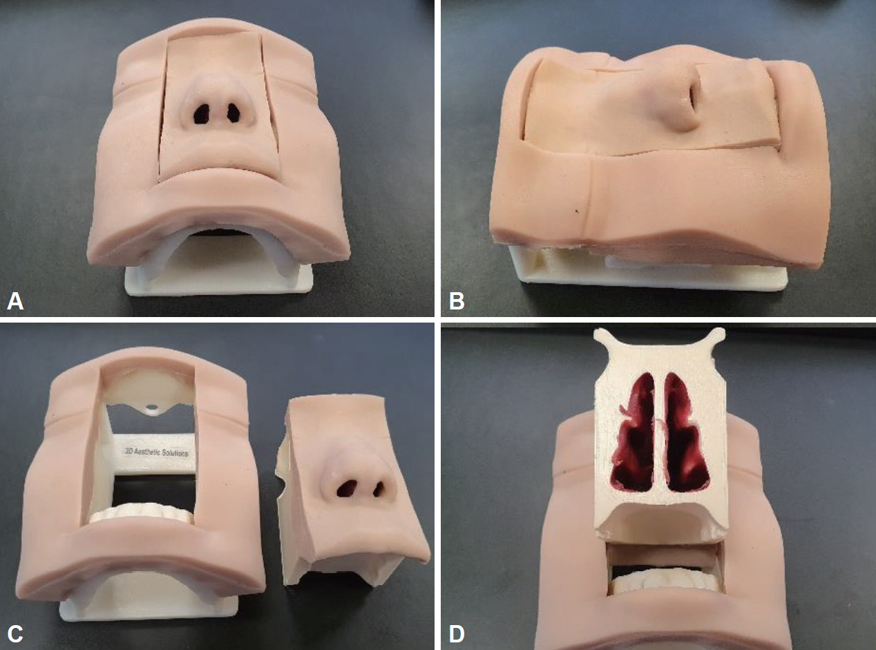
Fig. 1. Demonstrative photos of a silicone nose model (Simulare Medical Corp., Ontario, Canada). A: Frontal view. B: Lateral view. C: Separated view between a facial part for the support and a nasal part for the operation. D: Posterior view.

Fig. 2. Demonstrative photos of incision using a silicone nose model. A: Marking for inverted V incision. B: Mid-columellar incision. C: Marginal incision along the caudal margin of lower lateral cartilage. D: Lateral columellar incision connected with previous marginal incision and mid-columellar incision.
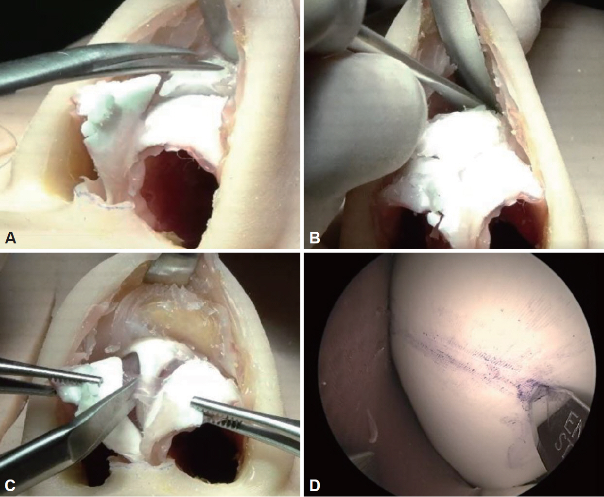
Fig. 3. Demonstrative photos of flap elevation and septal cartilage harvest using a silicone nose model. A: Supraperichondrial dissection. B: Subperiosteal dissection. C: Dissection of the membranous septum. D: Endoscopic view of septal cartilage harvest using a 0-degree rigid telescope.
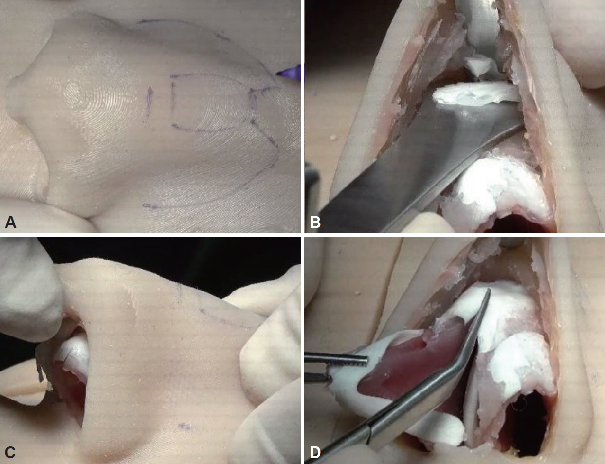
Fig. 4. Demonstrative photos of humpectomy using a silicone nose model. A: Design of osteotomy. B: En-bloc resection of a cartilaginous and bony hump. C: Rasping. D: Division of upper lateral cartilage and nasal septum.
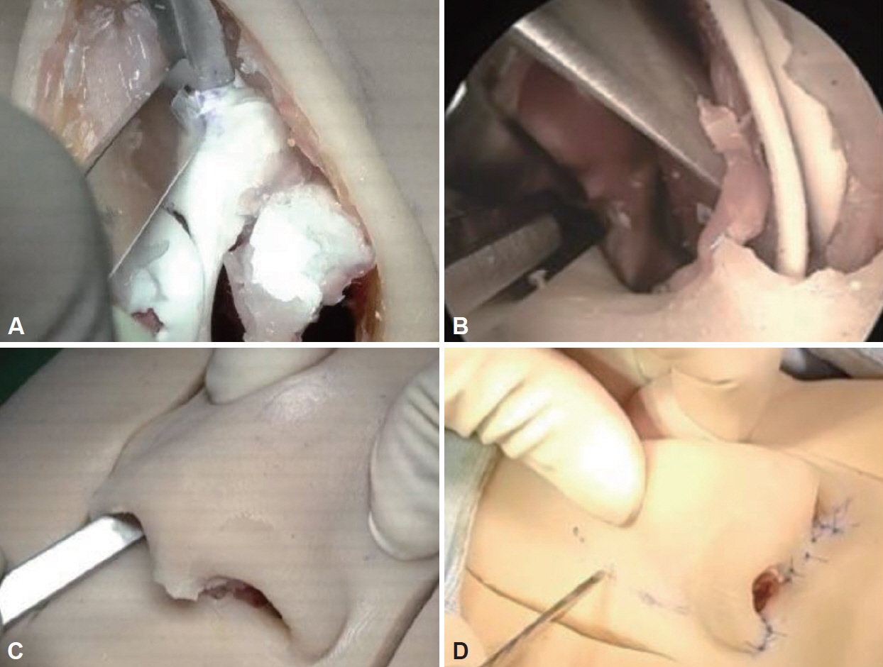
Fig. 5. Demonstrative photos of medial and lateral osteotomy using a silicone nose model. A: Medial osteotomy. B: Intra-nasal view of lateral osteotomy using a 0-degree rigid telescope. C: Outside view of the lateral osteotomy. D: Outside view of percutaneous osteotomy using 2 mm osteotome.

Fig. 6. Demonstrative photos of suture technique using a silicone nose model. A: Transdomal suture. B: Interdomal suture.

Fig. 7. Demonstrative photos of cartilage grafting using a silicone nose model. A: Onlay graft. B: Shield graft. C: Spreader graft. D: Columellar strut. E: Septal extension graft (caudal septal extension type). F: Septal extension graft (extended spreader type).

Fig. 8. Representative photos of dorsal augmentation with a silicone implant. A: Carving of a silicone implant. B: Insertion of a silicone implant into a dorsal pocket.

Fig. 9. Demonstrative photos of dorsal augmentation with diced cartilage. A: Dicing of cartilage. B: Molding with tissue glue using a commercially available mold (Jang Cartilage Mold, Nextcore Co., Ltd., Ulsan, Republic of Korea).
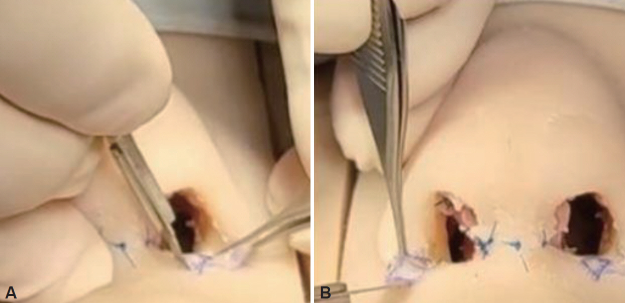
Fig. 10. Demonstrative photos of alar base resection. A: Nostril sill excision. B: Alar wedge resection.
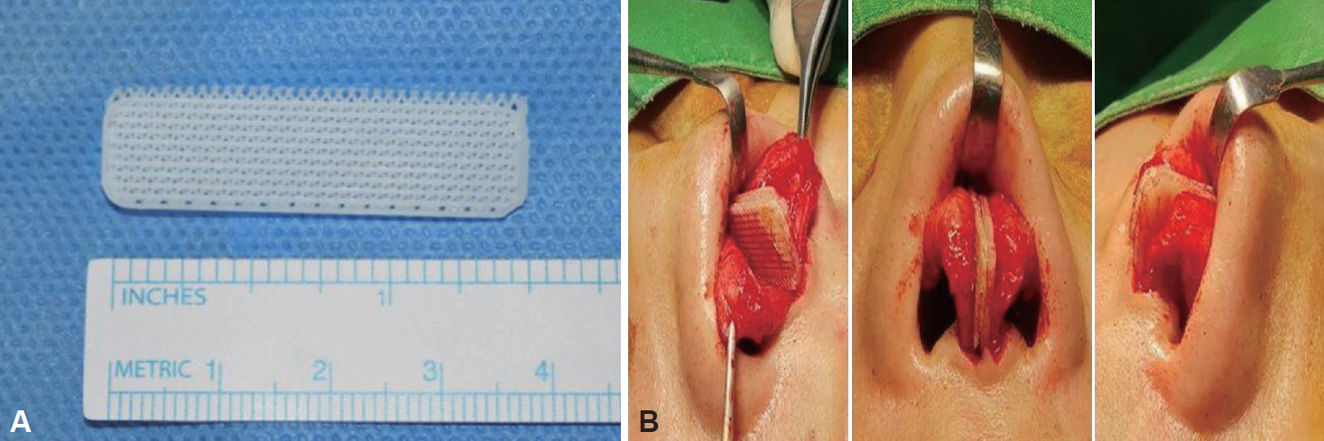
Fig. 11. Representative photos of PCL mesh. A: Demonstrative picture. B: Surgical view of the septal extension graft combined with PCL mesh bilaterally (From Kim, et al. Laryngoscope 2020;130(7):1680-5 [28]., with permission of Wiley). PCL, polycarprolactone.
Cited by 1 articles
-
The Effect of a Dummy Nose Model as an Educational Tool for Rhinoplasty Beginners
Minju Kim, Seung-No Hong, Soo Whan Kim, Tae-Bin Won
Korean J Otorhinolaryngol-Head Neck Surg. 2024;67(12):608-613. doi: 10.3342/kjorl-hns.2023.01088.
Reference
-
1. Jang YJ. Rhinoseptoplasty. 2nd ed. Seoul: Koonja;2013. p. 115.2. Jones N. The nose and paranasal sinuses physiology and anatomy. Adv Drug Deliv Rev. 2001; 51(1-3):5–19.
Article3. Neves JC, Zholtikov V, Cakir B, Coşkun E, Arancibia-Tagle D. Rhinoplasty dissection planes (subcutaneous, sub-SMAS, supra-perichondral, and sub-perichondral) and soft tissues management. Facial Plast Surg. 2021; 37(1):2–11.
Article4. Jin HR. Korean rhinoplasty: surgical techniques and case illustration. 1st ed. Seoul: Ilchogak;2013. p. 72.5. Jeong J, Terence G, Kim J. Understanding the anatomy of the transverse nasalis aponeurotic fibers and its importance in Asian rhinoplasty. Ann Plast Surg. 2018; 81(5):516–22.
Article6. Rohrich RJ, Dauwe PB, Pulikkottil BJ, Pezeshk RA. The importance of the anterior septal angle in the open dorsal approach to rhinoplasty. Plast Reconstr Surg. 2017; 139(3):604–12.
Article7. Kim JS, Khan NA, Song HM, Jang YJ. Intraoperative measurements of harvestable septal cartilage in rhinoplasty. Ann Plast Surg. 2010; 65(6):519–23.
Article8. Locketz GD, Lozada KN, Becker DG. Osteotomies-when, why, and how? Facial Plast Surg. 2020; 36(1):57–65.
Article9. Gerecci D, Perkins SW. The use of spreader grafts or spreader flaps-or not-in hump reduction rhinoplasty. Facial Plast Surg. 2019; 35(5):467–75.
Article10. Pereira Nunes D, Tinoco C, Oliveira E Carmo D, Paço J. Intermediate osteotomies in rhinoplasty: a new perspective. Eur Arch Otorhinolaryngol. 2017; 274(7):2953–8.
Article11. Farrior RT. The osteotomy in rhinoplasty. Laryngoscope. 1978; 88(9 Pt 1):1449–59.
Article12. Jang YJ, Wang JH, Sinha V, Lee BJ. Percutaneous root osteotomy for correction of the deviated nose. Am J Rhinol. 2007; 21(4):515–9.
Article13. VanKoevering KK, Rosko AJ, Moyer JS. Osteotomies demystified. Facial Plast Surg Clin North Am. 2017; 25(2):201–10.
Article14. Jang TY, Choi YS, Jung YG, Kim KT, Kim KS, Choi JS. Effect of nasal tip surgery on asian noses using the transdomal suture technique. Aesthetic Plast Surg. 2007; 31(2):174–8.
Article15. Guyuron B. Dynamics in rhinoplasty. Plast Reconstr Surg. 2000; 105(6):2257–9.
Article16. Pasinato R, Mocelin M, Berger CA. Nose tip refinement using interdomal suture in caucasian nose. Int Arch Otorhinolaryngol. 2012; 16(3):391–5.
Article17. Jin HR, Won TB. Nasal tip augmentation in Asians using autogenous cartilage. Otolaryngol Head Neck Surg. 2009; 140(4):526–30.
Article18. Jang YJ, Kim SH. Tip Grafting for the Asian nose. Facial Plast Surg Clin North Am. 2018; 26(3):343–56.
Article19. Jang YJ, Min JY, Lau BC. A multilayer cartilaginous tip-grafting technique for improved nasal tip refinement in Asian rhinoplasty. Otolaryngol Head Neck Surg. 2011; 145(2):217–22.
Article20. Johnson JB. Spreader-graft fixation. Plast Reconstr Surg. 1989; 84(3):540–1.
Article21. Cingi C, Bayar Muluk N, Winkler A, Thomas JR. Nasal tip grafts. J Craniofac Surg. 2018; 29(7):1914–21.
Article22. Kim JH, Song JW, Park SW, Oh WS, Lee JH. Effective septal extension graft for asian rhinoplasty. Arch Plast Surg. 2014; 41(1):3–11.
Article23. Na HG, Jang YJ. Dorsal augmentation using alloplastic implants. Facial Plast Surg. 2017; 33(2):189–94.
Article24. Kim IS. Augmentation rhinoplasty using silicone implants. Facial Plast Surg Clin North Am. 2018; 26(3):285–93.
Article25. Ledo TO, Ramos HHA, Buba CM, Webster G, de Lima JT Jr, de Paiva DL, et al. Outcome of free diced cartilage grafts in rhinoplasty: a systematic review. Facial Plast Surg. 2021; 37(1):117–21.
Article26. Tasman AJ. Dorsal augmentation-diced cartilage techniques: the diced cartilage glue graft. Facial Plast Surg. 2017; 33(2):179–88.
Article27. Kim DH, Yun WS, Shim JH, Park KH, Choi D, Park MI, et al. Clinical application of 3-dimensional printing technology for patients with nasal septal deformities: a multicenter study. JAMA Otolaryngol Head Neck Surg. 2018; 144(12):1145–52.
Article28. Kim SH, Choi JY. Surgical outcomes and complications of septal extension graft supported by 3D printed polycaprolactone plate. Laryngoscope. 2020; 130(7):1680–5.
Article
- Full Text Links
-
- Actions
-
Cited
- CITED
-
- Close
- Share
-
- Similar articles
-
- Management of Prolapsed Silicone Tube Inserted for Treatment of Nasolacrimal Duct Obstruction
- Correction fo the Short Nose: Relocation of the Alar Cartilge Using Silicone Implants
- Revision rhinoplasty for contracted nose in Asia
- Simple Correction Method of Binder`s Syndrome
- The Effect of a Dummy Nose Model as an Educational Tool for Rhinoplasty Beginners
