Standards and Guidelines for Reporting Diagnostic Test Results in Acute Leukemia Patients: Bone Marrow Examination, Flow Cytometry, and Cytogenetic/Molecular Genetics Tests
- Bang HI1
- Kim IS2
- Park SH3
- Lee ST4
- Hwang SM5
- Huh J6
- Huh HJ7
- Song J4
- Park R1
- Lee YJ8
- Kim Y9
- Kong SY
 10
10 - The Committee for Standardization in Korean Society for Laboratory Hematology
- Affiliations
-
- 1Department of Laboratory Medicine, Soonchunghyang University Seoul Hospital, Seoul, Korea
- 2Department of Laboratory Medicine, Pusan National University Yangsan Hospital, Yangsan, Korea
- 3Department of Laboratory Medicine, University of Ulsan College of Medicine and Ulsan University Hospital, Ulsan, Korea
- 4Department of Laboratory Medicine, Yonsei University College of Medicine, Seoul, Korea
- 5Department of Laboratory Medicine, Seoul National University Bundang Hospital, Seongnam, Korea
- 6Department of Laboratory Medicine, Ewha Womans University, College of Medicine, Mokdong Hospital, Seoul, Korea
- 7Department of Laboratory Medicine, Dongkuk University Ilsan Hospital, Goyang, Korea
- 8Department of Laboratory Medicine, Wonkwang University Hospital, Iksan, Korea
- 9Department of Laboratory Medicine, Catholic University Seoul St. Mary’s Hospital, Seoul, Korea
- 10Department of Laboratory Medicine, National Cancer Center Hospital, Goyang, Korea
- KMID: 2525774
- DOI: http://doi.org/10.47429/lmo.2021.11.1.1
Abstract
- Reports on hematological neoplasms are produced in various formats in different laboratories. For best patient care, standardization of reports adopting a format that provides concise and clear information is necessary. To this end, the Diagnostic Hematology Standardization Committee, organized by the Korean Society for Laboratory Hematology, has proposed a standardized format for reporting diagnostic test results for acute leukemia patients. It is hoped that this standardization will broadly improve communication with clinicians and improve patient care.
Keyword
Figure
-
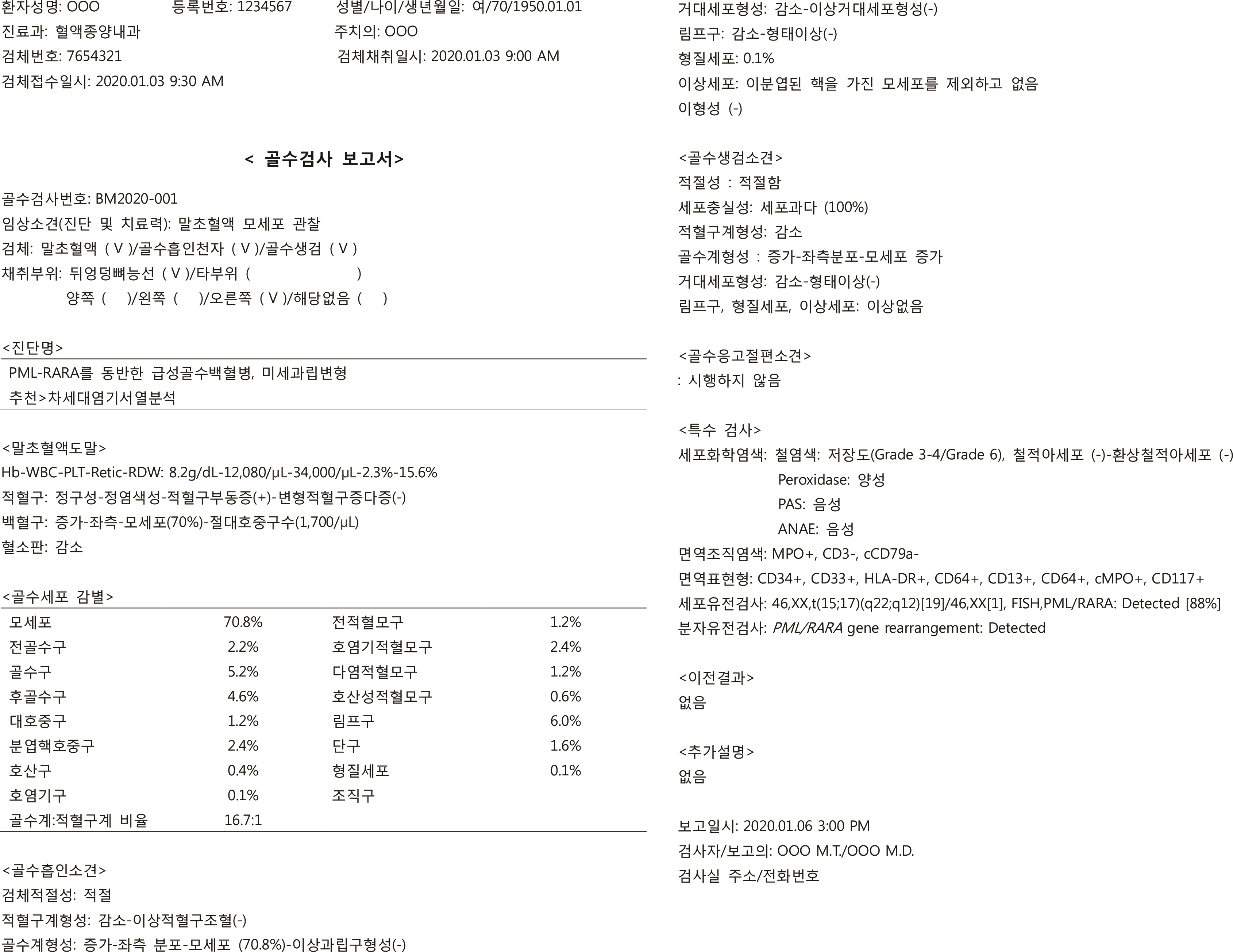
Fig. 1 Sample bone marrow report for a patient diagnosed with acute myeloid leukemia, PML-RARA according to consensus guidelines.
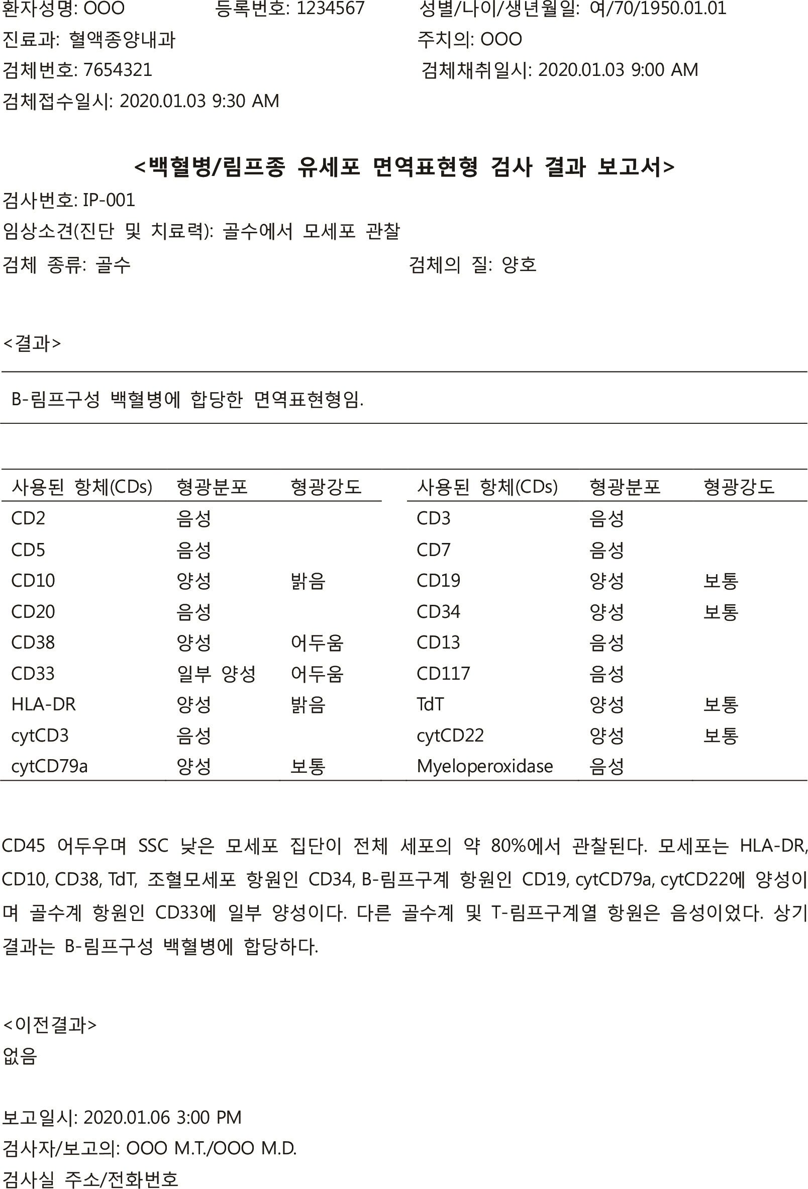
Fig. 2 Sample leukemia/lymphoma flow cytometric immunophenotyping report for a patient diagnosed with B-lymphoblastic leukemia according to consensus guidelines.
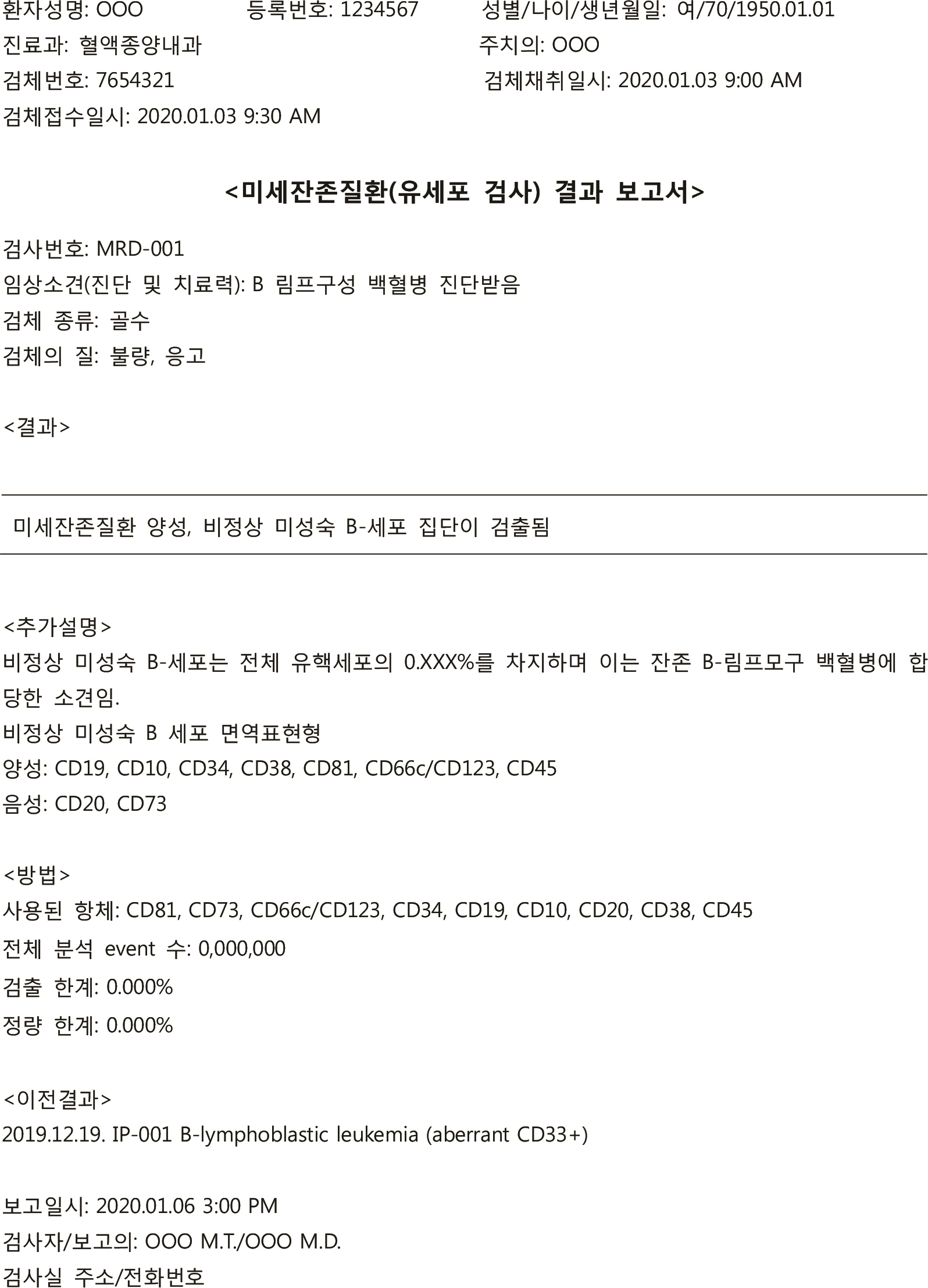
Fig. 3 Sample minimal residual disease flow cytometric immunophenotyping report for a patient receiving follow up care for B-lymphoblastic leukemia, according to consensus guidelines.
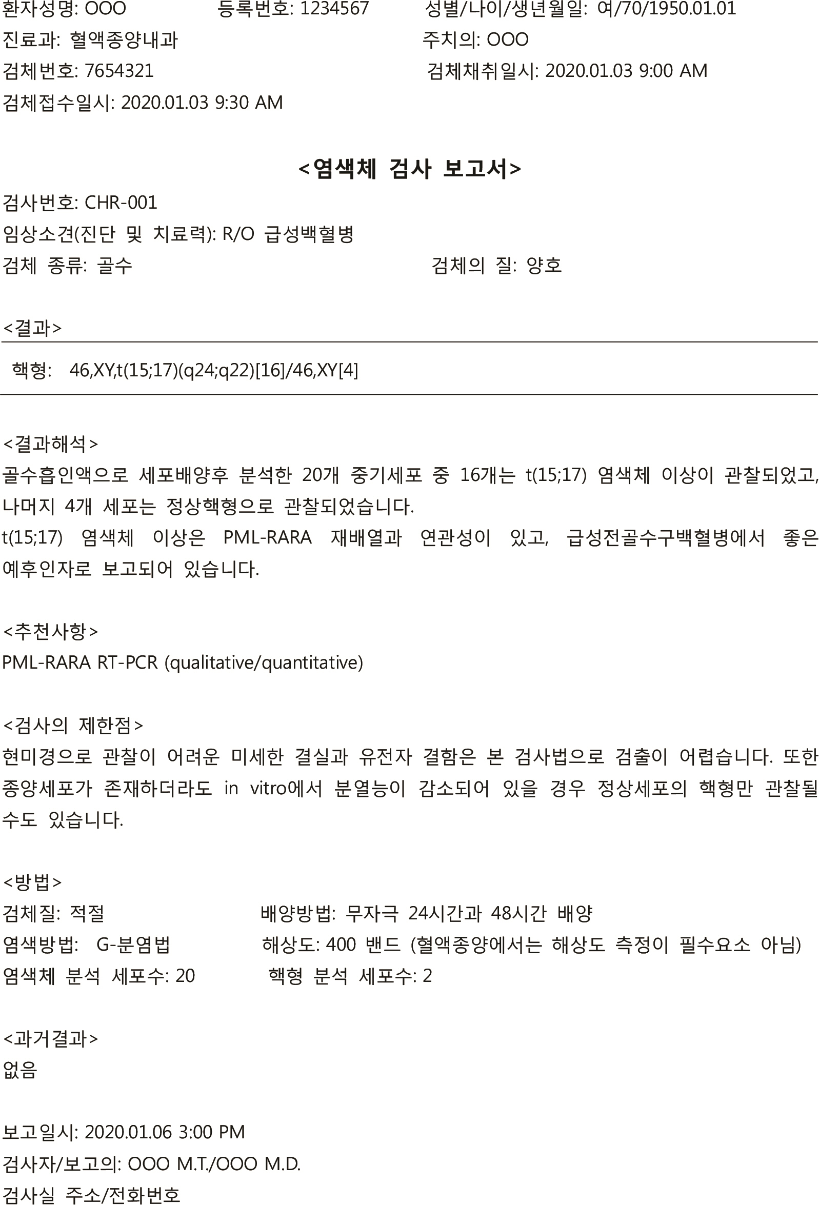
Fig. 4 Sample chromosome analysis report for a patient diagnosed with acute myeloid leukemia, PML-RARA according to consensus guidelines.
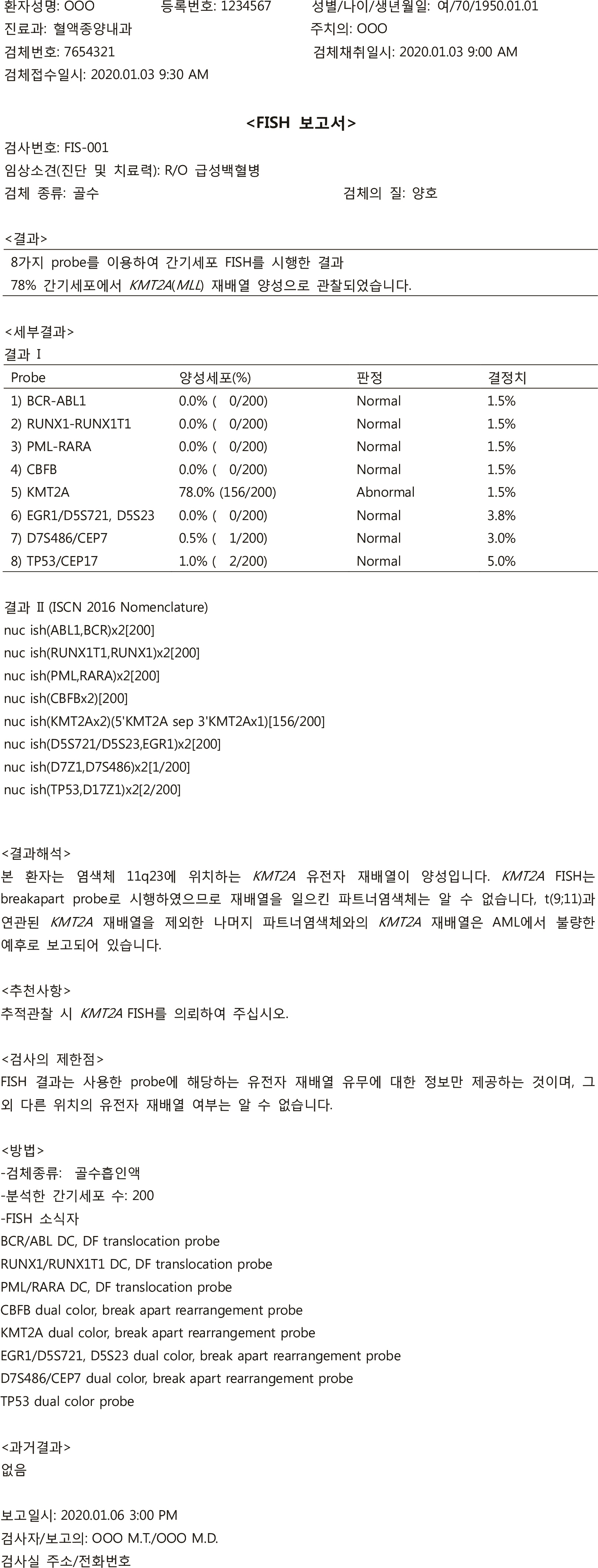
Fig. 5 Sample FISH report for a patient with KMT2A rearrangement according to consensus guidelines.
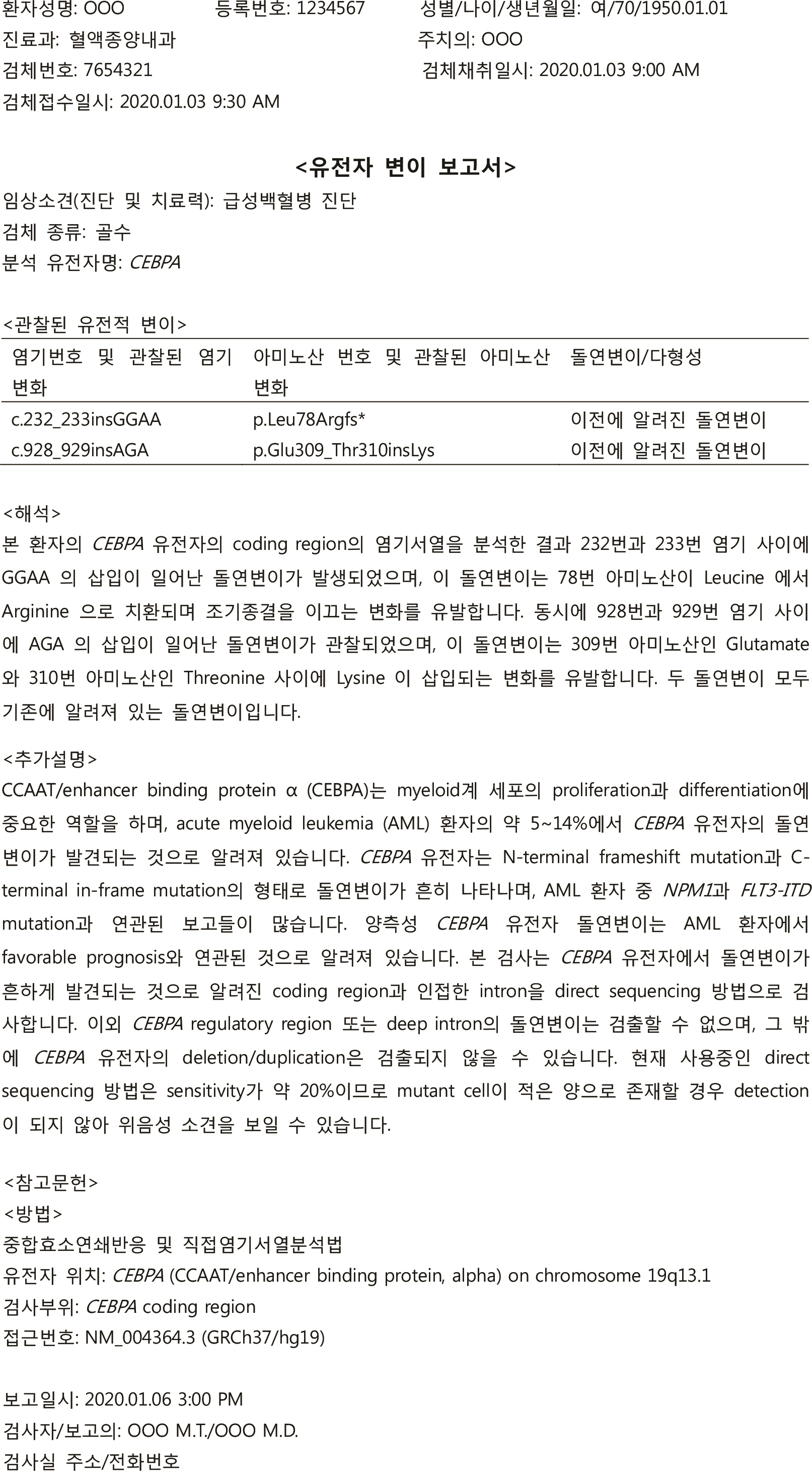
Fig. 6 Sample gene mutation test report for a patient with CEBPA mutation according to consensus guidelines.
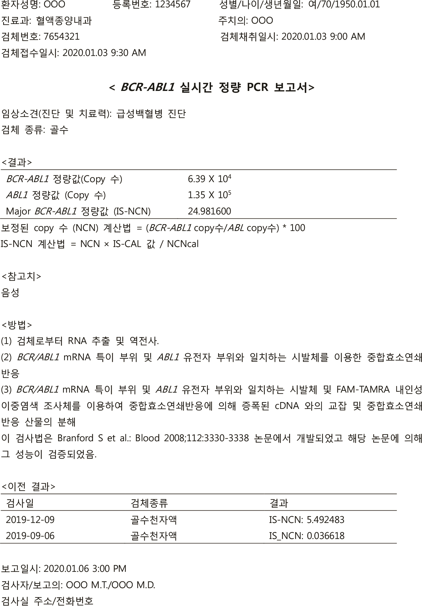
Fig. 7 Sample BCR-ABL1 real-time quantitative PCR test report for a patient according to consensus guidelines.
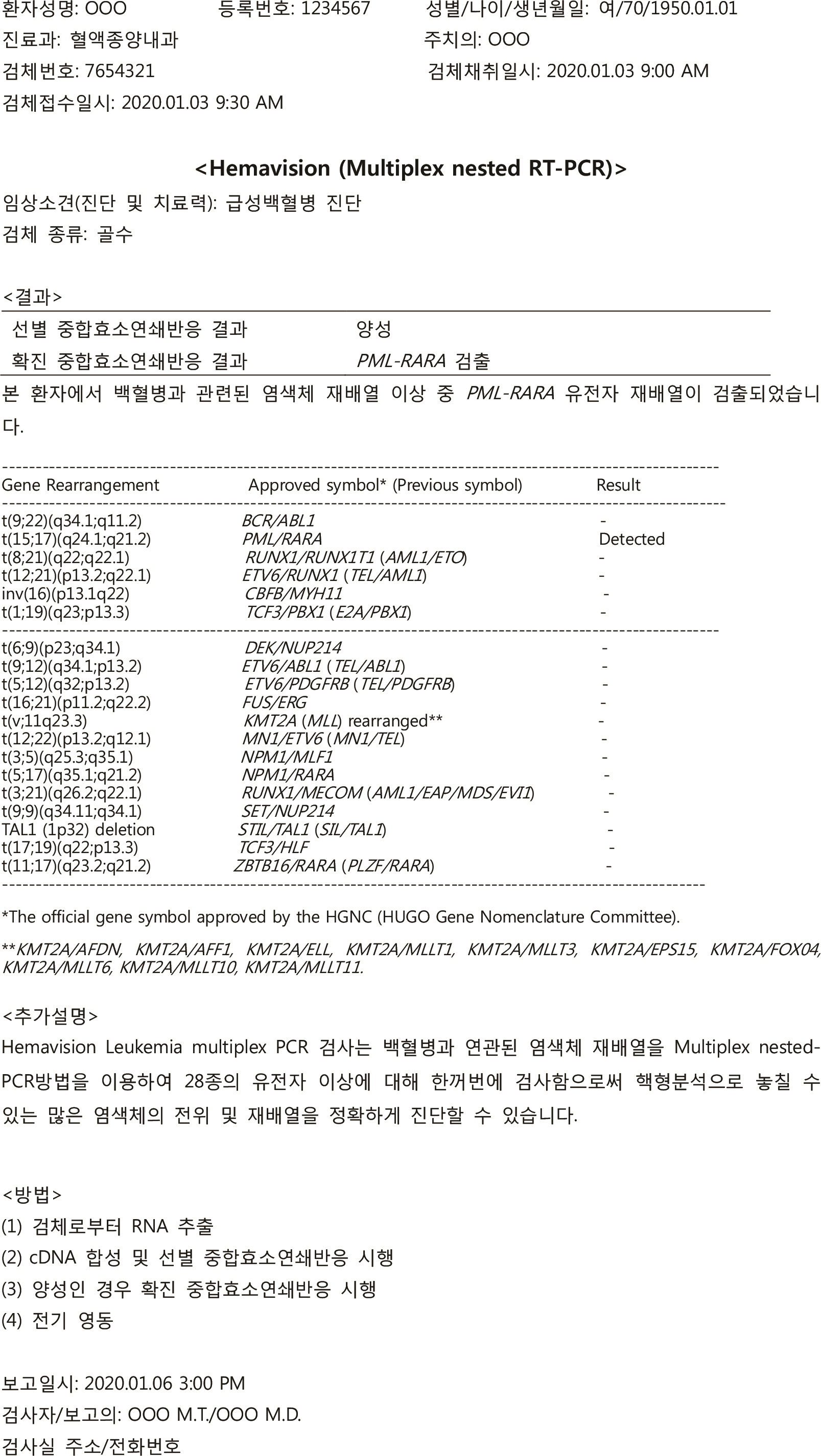
Fig. 8 Sample Hemavision, multiplex nested RT-PCR test report for a patient according to consensus guidelines.
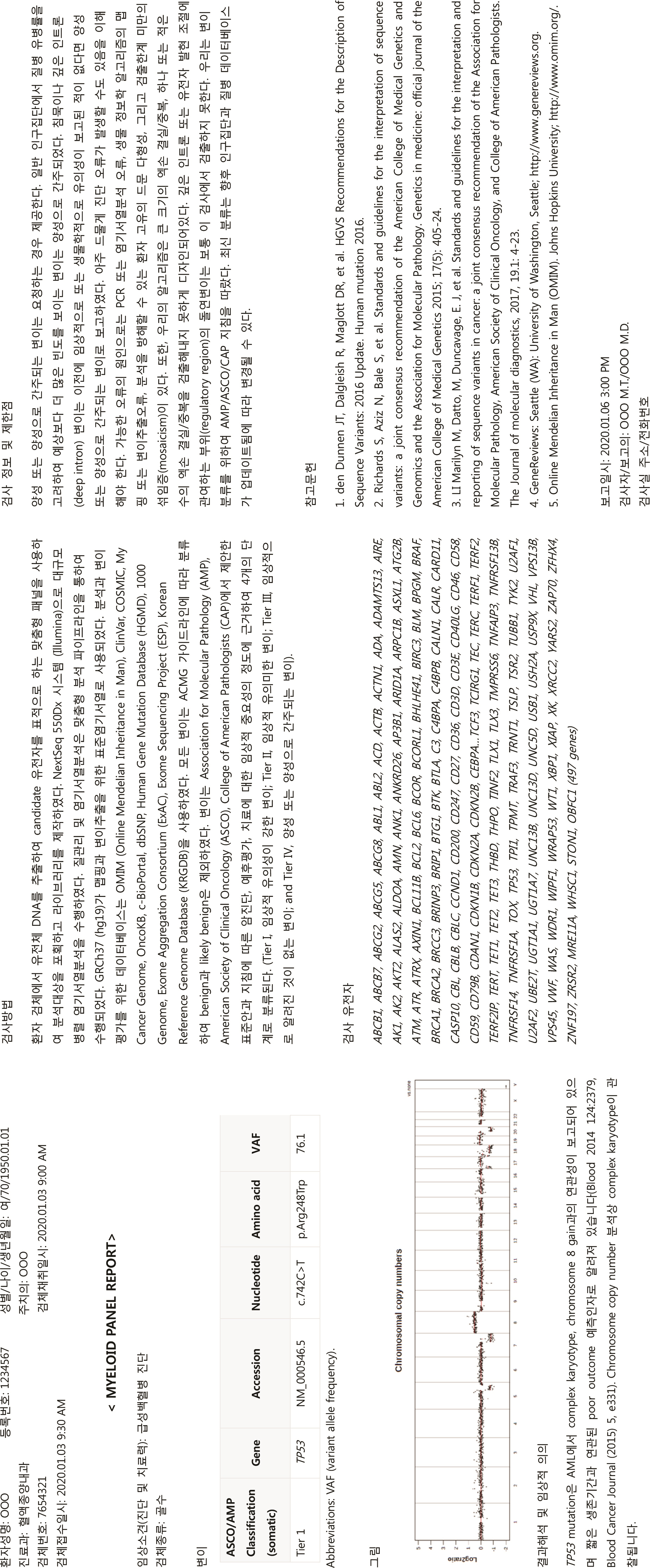
Fig. 9 Sample next-generation sequencing test report for a patient according to consensus guidelines.
Cited by 1 articles
-
Body Fluid Analysis for Cellular Composition Using Manual Methods: Current Status and Clinical Laboratory Guidelines in Korea (2021)
Hae In Bang, Hyun-Young Kim, Saeam Shin, Ja Young Lee, In-Suk Kim, Young-Uk Cho, Ji Myung Kim, Myung-Geun Shin, Jeong Nyeo Lee, Sang Mee Hwang, Sun-Young Kong
Lab Med Online. 2022;12(4):262-268. doi: 10.47429/lmo.2022.12.4.262.
Reference
-
1. Swerdlow SH, Campo E, editors. 2017. WHO classification of tumours of haematopoietic and lymphoid tissues. Revised 4th ed. International Agency for Research on Cancer;Lyon, France:2. Sever C, Abbott CL, de Baca ME, Khoury JD, Perkins SL, Reichard KK, et al. 2016; Bone marrow synoptic reporting for hematologic neoplasms: Guideline from the College of American Pathologists Pathology and Laboratory Quality Center. Arch Pathol Lab Med. 140:932–49. DOI: 10.5858/arpa.2015-0450-SA. PMID: 26905483.3. Lee SH, Erber WN, Porwit A, Tomonaga M, Peterson LC. 2008; ICSH guidelines for the standardization of bone marrow specimens and reports. Int J Lab Hematol. 30:349–64. DOI: 10.1111/j.1751-553X.2008.01100.x. PMID: 18822060.4. Wood BL, Arroz M, Barnett D, DiGiuseppe J, Greig B, Kussick SJ, et al. 2007; 2006 Bethesda International Consensus recommendations on the immunophenotypic analysis of hematolymphoid neoplasia by flow cytometry: optimal reagents and reporting for the flow cytometric diagnosis of hematopoietic neoplasia. Cytometry B Clin Cytom. 72:S14–22. DOI: 10.1002/cyto.b.20363. PMID: 17803189.5. Clinical and Laboratory Standards Institute. 2007. Clinical flow cytometric analysis of neoplastic hematolymphoid cells;Approved guideline-Second edition. CLSI document H43-A2. Clinical and Laboratory Standards Institute;Wayne, PA:6. Claustres M, Kožich V, Dequeker E, Fowler B, Hehir-Kwa JY, Miller K, et al. 2014; Recommendations for reporting results of diagnostic genetic testing (biochemical, cytogenetic and molecular genetic). Eur J Hum Genet. 22:160–70. DOI: 10.1038/ejhg.2013.125. PMID: 23942201. PMCID: PMC3895644.7. Mikhail FM, Heerema NA, Rao KW, Burnside RD, Cherry AM, Cooley LD. 2016; Section E6.1-6.4 of the ACMG technical standards and guidelines: chromosome studies of neoplastic blood and bone marrow-acquired chromosomal abnormalities. Genet Med. 18:635–42. DOI: 10.1038/gim.2016.50. PMID: 27124785.8. Kwon JA, Kim YG, Park G, Kim JM, Cho YU, Huh J, et al. 2019; Recommendation for the peripheral blood cell morphology report. Lab Med Online. 9:115–25. DOI: 10.3343/lmo.2019.9.3.115.9. Arber DA, Orazi A, Hasserjian R, Thiele J, Borowitz MJ, Le Beau MM, et al. 2016; The 2016 revision to the World Health Organization classification of myeloid neoplasms and acute leukemia. Blood. 127:2391–405. DOI: 10.1182/blood-2016-03-643544. PMID: 27069254.10. Béné M, Nebe T, Bettelheim P, Buldini B, Bumbea H, Kern W, et al. 2011; Immunophenotyping of acute leukemia and lymphoproliferative disorders: a consensus proposal of the European LeukemiaNet Work Package 10. Leukemia. 25:567–74. DOI: 10.1038/leu.2010.312. PMID: 21252983.11. Theunissen P, Mejstrikova E, Sedek L, van der Sluijs-Gelling AJ, Gaipa G, Bartels M, et al. 2017; Standardized flow cytometry for highly sensitive MRD measurements in B-cell acute lymphoblastic leukemia. Blood. 129:347–57. DOI: 10.1182/blood-2016-07-726307. PMID: 27903527. PMCID: PMC5291958.12. Wood BL. 2016; Principles of minimal residual disease detection for hematopoietic neoplasms by flow cytometry. Cytometry B Clin Cytom. 90:47–53. DOI: 10.1002/cyto.b.21239. PMID: 25906832.13. Schuurhuis GJ, Heuser M, Freeman S, Béné M-C, Buccisano F, Cloos J, et al. 2018; Minimal/measurable residual disease in AML: a consensus document from the European LeukemiaNet MRD Working Party. Blood. 131:1275–91. DOI: 10.1182/blood-2017-09-801498. PMID: 29330221. PMCID: PMC5865231.14. Rack KA, van den Berg E, Haferlach C, Beverloo HB, Costa D, Espinet B, et al. 2019; European recommendations and quality assurance for cytogenomic analysis of haematological neoplasms. Leukemia. 33:1851–67. DOI: 10.1038/s41375-019-0378-z. PMID: 30696948. PMCID: PMC6756035.15. McGowan-Jordan J, Simons A, Schmid M. 2016; ISCN: an international system for human cytogenomic nomenclature (2016). Cytogenetic and Genome Research. 149:Basel, Switzerland;1–2.16. Cross NCP, White HE, Evans PAS, Hancock J, Copland M, Milojkovic D, et al. 2018; Consensus on BCR-ABL1 reporting in chronic myeloid leukaemia in the UK. Br J Haematol. 182:777–88. DOI: 10.1111/bjh.15542. PMID: 30125955. PMCID: PMC6175193.17. Ohgami RS, Arber DA. 2013; Challenges in consolidated reporting of hematopoietic neoplasms. Surg Pathol Clin. 6:795–806. DOI: 10.1016/j.path.2013.08.001. PMID: 26839198.18. Richards S, Aziz N, Bale S, Bick D, Das S, Gastier-Foster J, et al. 2015; Standards and guidelines for the interpretation of sequence variants: a joint consensus recommendation of the American College of Medical Genetics and Genomics and the Association for Molecular Pathology. Genet Med. 17:405–24. DOI: 10.1038/gim.2015.30. PMID: 25741868. PMCID: PMC4544753.19. Li MM, Datto M, Duncavage EJ, Kulkarni S, Lindeman NI, Roy S, et al. 2017; Standards and guidelines for the interpretation and reporting of sequence variants in cancer: a joint consensus recommendation of the Association for Molecular Pathology, American Society of Clinical Oncology, and College of American Pathologists. J Mol Diagn. 19:4–23. DOI: 10.1016/j.jmoldx.2016.10.002. PMID: 27993330. PMCID: PMC5707196.
- Full Text Links
-
- Actions
-
Cited
- CITED
-
- Close
- Share
-
- Similar articles
-
- Measurements of treatment response in childhood acute leukemia
- Discrepant Immunophenotypic Characteristics between the Lymph Node and Bone Marrow in Two Mixed-Phenotype Acute Leukemia Patients
- Diagnostic Approach of Acute Leukemia in Children with Bone and Joint Pain
- Acute Megakaryoblastic Leukemia with CD41a-/CD61-/CD42a+ Blasts in an Infant with Down Syndrome
- Relation among the Tests and Comparison of Positivity of Tests for Multi-Drug Resistance in Newly Diagnosed Acute Leukemia
