Multilocular cystic hemangioma of the liver mimicking mucinous cystic neoplasm: a case report
- Affiliations
-
- 1Department of Radiology, Biomedical Research Institute and Pusan National University Hospital, Pusan National University School of Medicine, Busan, Korea
- 2Department of Pathology, Biomedical Research Institute and Pusan National University Hospital, Pusan National University School of Medicine, Busan, Korea
- 3Department of Surgery, Biomedical Research Institute and Pusan National University Hospital, Pusan National University School of Medicine, Busan, Korea
- KMID: 2525028
- DOI: http://doi.org/10.12701/yujm.2021.00969
Abstract
- Hepatic hemangiomas infrequently exhibit atypical imaging features, which may cause diagnostic confusion with hepatic malignancies and lead to unnecessary surgery. We report a rare case of multilocular cystic hemangioma of the liver mimicking a mucinous cystic neoplasm of the liver in a 48-year-old female, focusing on computed tomography and magnetic resonance imaging features and their differential diagnosis.
Keyword
Figure
-
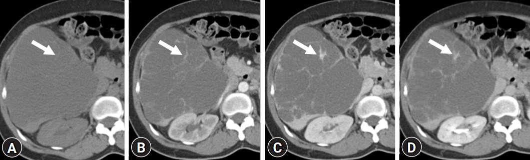
Fig. 1. Computed tomography (CT) findings of a multilocular cystic hemangioma of the liver in a 48-year-old female. Contrast-enhanced dynamic-phase abdominal CT scans show a huge septated cystic mass in the right lobe of the liver. (A) Nonenhanced, (B) arterial phase, (C) portal venous phase, and (D) delayed phase. Enhancement of the septa within the mass (arrows) is noted.
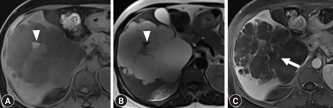
Fig. 2. Magnetic resonance imaging findings of a multilocular cystic hemangioma of the liver in a 48-year-old female. (A) T1-weighted and (B) T2-weighted images show a multiseptated cystic mass containing variable signal intensity. Most locules present as T1-hypointensity and T2-hyperintensity, identical to signal intensity of fluid. Some T1 hyperintense locules present as T2-hypointensity, suggesting subacute hemorrhage (arrowheads). (C) Contrast-enhanced fat-suppressed T1-weighted image shows septal enhancement (arrow).
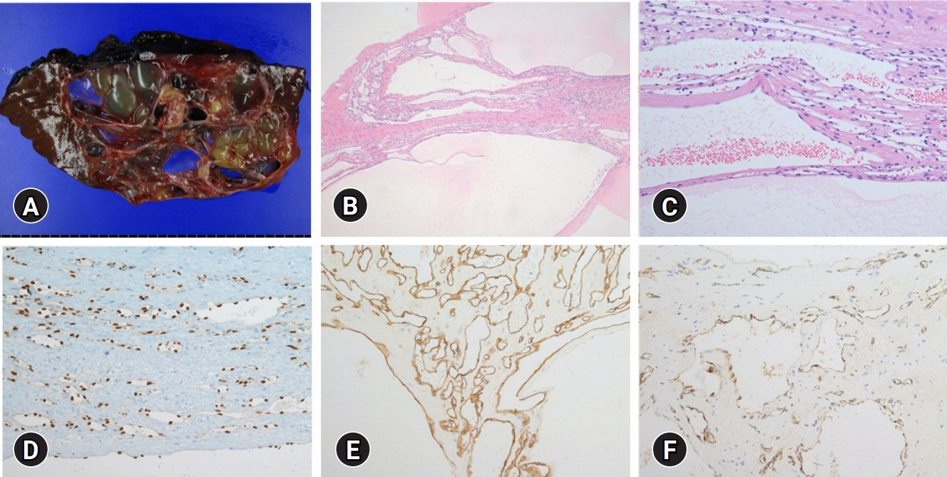
Fig. 3. Histopathologic findings of a multilocular cystic hemangioma of the liver in a 48-year-old female. (A) The cut section of the gross specimen shows multiple cystic loculi separated by multiple septa. (B) Low-power magnification shows a multilocular cystic mass composed of medium to small-sized cystic space (hematoxylin and eosin [H&E] stain, ×40). (C) High-power magnification shows the cystic wall lined by flattened or cuboidal endothelial cells without cytologic atypia and supported by fibrotic stroma (H&E stain, ×200). On immunohistochemical staining, the endothelial cells are positive for vascular endothelial cell markers such as (D) ETS-related gene (ERG), (E) CD34, and (F) factor VIII (immunohistochemical stain, ×200 [D-F]), but negative for biliary epithelial cell markers such as cytokeratin 7 (CK 7) and CK 19 (not shown).
Reference
-
References
1. Caseiro-Alves F, Brito J, Araujo AE, Belo-Soares P, Rodrigues H, Cipriano A, et al. Liver haemangioma: common and uncommon findings and how to improve the differential diagnosis. Eur Radiol. 2007; 17:1544–54.2. Klotz T, Montoriol PF, Da Ines D, Petitcolin V, Joubert-Zakeyh J, Garcier JM. Hepatic haemangioma: common and uncommon imaging features. Diagn Interv Imaging. 2013; 94:849–59.3. Vilgrain V, Boulos L, Vullierme MP, Denys A, Terris B, Menu Y. Imaging of atypical hemangiomas of the liver with pathologic correlation. Radiographics. 2000; 20:379–97.4. Jang HJ, Kim TK, Lim HK, Park SJ, Sim JS, Kim HY, et al. Hepatic hemangioma: atypical appearances on CT, MR imaging, and sonography. AJR Am J Roentgenol. 2003; 180:135–41.5. Hihara T, Araki T, Katou K, Odashima H, Ounishi H, Kachi K, et al. Cystic cavernous hemangioma of the liver. Gastrointest Radiol. 1990; 15:112–4.6. Hussain MZ, Ohtomo K, Hihara T, Uchiyama G, Ainoda T, Yamamoto M, et al. Multilocular cystic hemangioma: CT and MR appearance. Radiat Med. 1992; 10:206–9.7. Nakachi A, Shiraishi M, Shimoji H, Tomori T, Oshiro T, Muto Y. Multicystic cavernous hemangioma of the liver: report of a case including diagnostic imaging and pathologic correlation. Radiat Med. 1998; 16:209–12.8. Cha EY, Kim KW, Choi YJ, Song JS, Cho KJ, Lee MG. Multicystic cavernous haemangioma of the liver: ultrasonography, CT, MR appearances and pathological correlation. Br J Radiol. 2008; 81:e37–9.9. Qian LJ, Zhu J, Zhuang ZG, Xia Q, Liu Q, Xu JR. Spectrum of multilocular cystic hepatic lesions: CT and MR imaging findings with pathologic correlation. Radiographics. 2013; 33:1419–33.10. Borhani AA, Wiant A, Heller MT. Cystic hepatic lesions: a review and an algorithmic approach. AJR Am J Roentgenol. 2014; 203:1192–204.
- Full Text Links
-
- Actions
-
Cited
- CITED
-
- Close
- Share
-
- Similar articles
-
- Large, Multilocular Cystic Mass in the Uterine Cervix Mimicking Adenoma Malignum
- A case of multilocular cystic renal cell carcinoma mistaken for multilocular renal cyst
- Pedunculated mucinous cystic neoplasm of the liver: a case report
- Multilocular Cystic Renal Cell Carcinoma: A case report
- Oncocytic Type Intraductal Papillary Mucinous Neoplasm Mimicking Mucinous Cystic Neoplasm of the Pancreas: A Case Report
