Comparison of soft tissue changes between incisor tipping and translation after premolar extraction
- Affiliations
-
- 1Department of Orthodontics, Yonsei University College of Dentistry, Seoul, Korea
- 2Department of Orthodontics, Institute of Craniofacial Deformity, Yonsei University College of Dentistry, Seoul, Korea
- KMID: 2524901
- DOI: http://doi.org/10.4041/kjod.2022.52.1.42
Abstract
Objective
This study compared soft tissue changes after extraction of the four premolars followed by maximum retraction of the anterior teeth according to the type of anterior teeth movement: tipping and translation.
Methods
Patients who had undergone orthodontic treatment involving the extraction of four premolars were retrospectively selected and divided into either the tipping (n = 27) or translation (n = 26) groups based on the retraction of the incisor root apex and the axis changes of the incisors during the treatment period. Lateral pre- and post-treatment cephalograms were analyzed.
Results
There were no significant differences between the tipping and translation groups before treatment. The retraction amounts of the root apex of the upper and lower incisors in the tipping group were 0.33 and 0.26 mm, respectively, and 5.02 and 5.31 mm, respectively, in the translation group (p < 0.001). The posterior movements of soft tissue points A and B in the tipping group were 0.61 and 1.25 mm, respectively, and 1.10 and 3.25 mm, respectively, in the translation group (p < 0.01). The mentolabial sulcus angle increased by 5.89° in the tipping group, whereas it decreased by 8.13° in the translation group (p < 0.001).
Conclusions
An increased amount of retraction of the incisor root apex led to the increased posterior movement of soft tissue points A and B, and this appeared more distinct in cases involving the lower incisor and lower lip.
Keyword
Figure
-

Figure 1 Cephalometric landmarks and reference planes. S, sella; N, nasion; A, point A; B, point B; Pog, pogonion; Me, menton; U1e, upper central incisor edge; U1c, the center of resistance of the upper central incisor; U1a, root apex of the upper central incisor; L1e, lower central incisor edge; L1c, the center of resistance of the lower central incisor; L1a, root apex of the lower central incisor; Sn, subnasale; A’, soft tissue point A; Ls, labrale superioris; ULa, the most anterior point of upper lip; Stms, stomion superioris; Stmi, stomion inferioris; LLa, the most anterior point of lower lip; Li, labrale inferioris; B’, soft tissue point B; Pog’, soft tissue pogonion; Me’, soft tissue menton; HRP, horizontal reference plane; VRP, vertical reference plane.
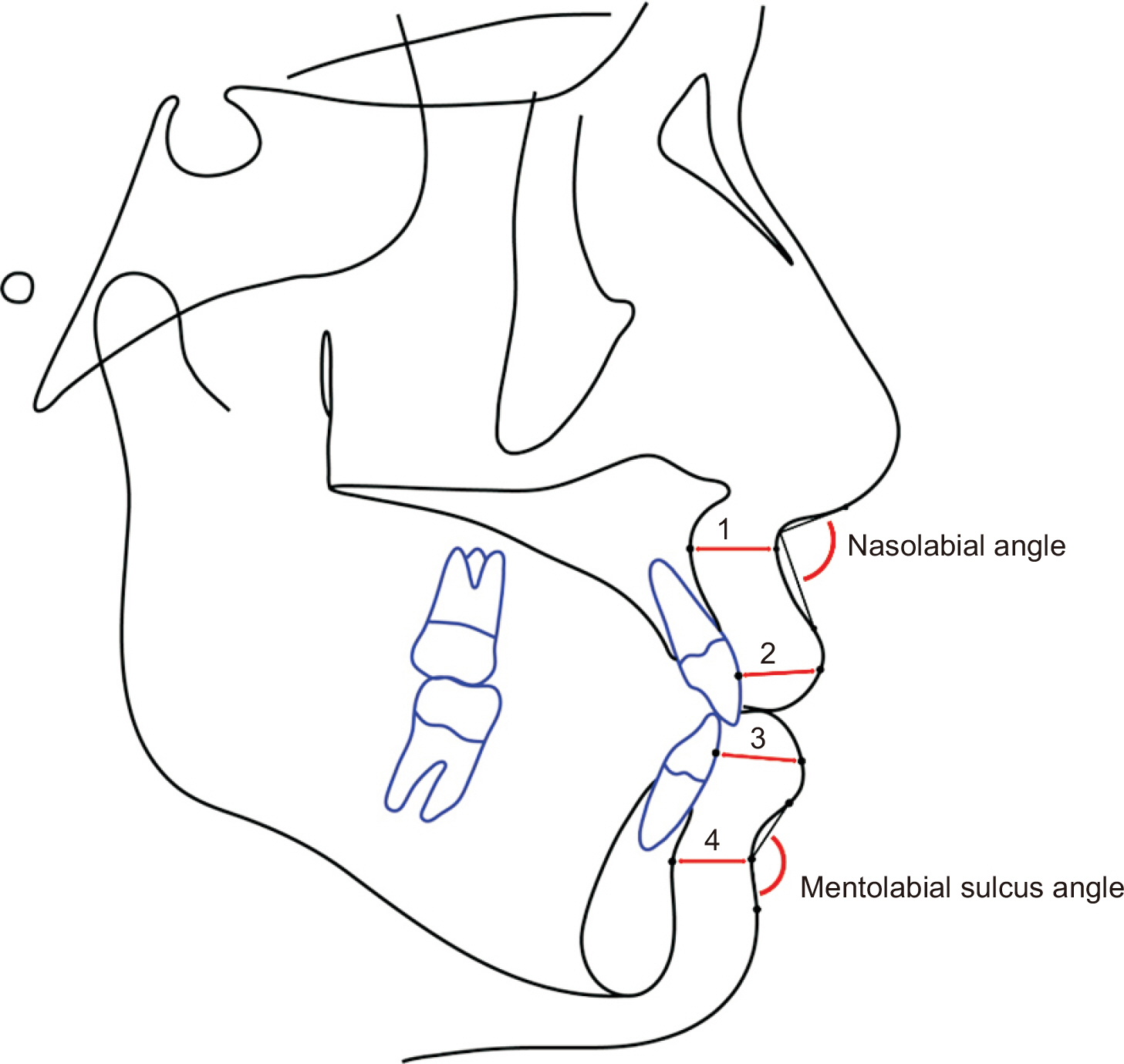
Figure 2 Additional cephalometric measurements. 1, basal upper lip thickness (distance between the A point and soft tissue point A); 2, upper lip thickness (the shortest distance between the labial surface of the upper central incisor and ULa); 3, lower lip thickness (the shortest distance between the labial surface of the lower central incisor and LLa); 4, basal lower lip thickness (the distance between point B and soft tissue point B).
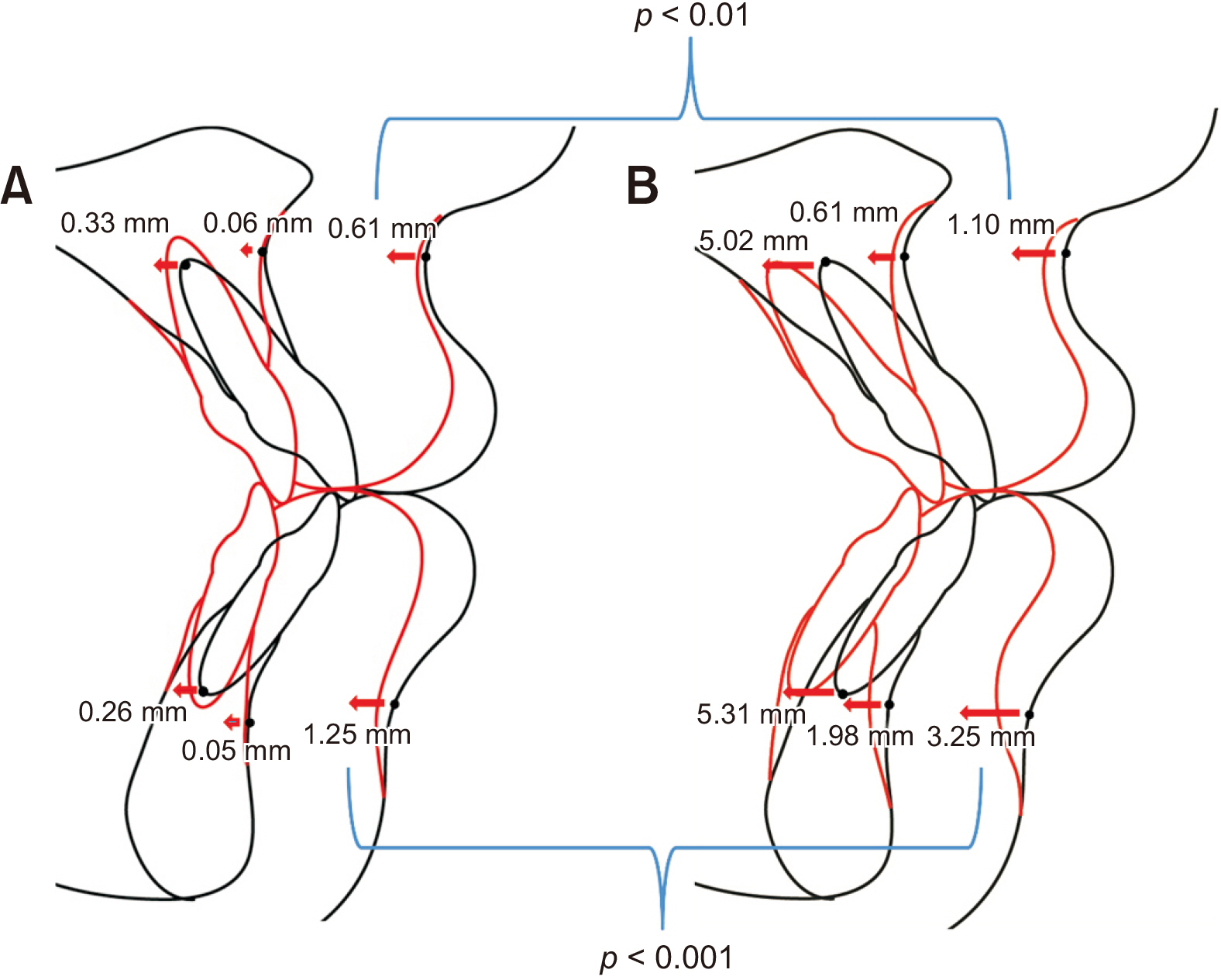
Figure 3 Comparison of treatment changes between the two groups. A, Tipping group. B, Translation group.
Reference
-
1. Hayashida H, Ioi H, Nakata S, Takahashi I, Counts AL. 2011; Effects of retraction of anterior teeth and initial soft tissue variables on lip changes in Japanese adults. Eur J Orthod. 33:419–26. DOI: 10.1093/ejo/cjq095. PMID: 20966067.
Article2. Bravo LA. 1994; Soft tissue facial profile changes after orthodontic treatment with four premolars extracted. Angle Orthod. 64:31–42. DOI: 10.1043/0003-3219(1994)064<0031:STFPCA>2.0.CO;2. PMID: 8172393.3. Kim K, Choi SH, Choi EH, Choi YJ, Hwang CJ, Cha JY. 2017; Unpredictability of soft tissue changes after camouflage treatment of Class II division 1 malocclusion with maximum anterior retraction using miniscrews. Angle Orthod. 87:230–8. DOI: 10.2319/042516-332.1. PMID: 27768390. PMCID: PMC8384362.
Article4. Caplan MJ, Shivapuja PK. 1997; The effect of premolar extractions on the soft-tissue profile in adult African American females. Angle Orthod. 67:129–36. DOI: 10.1043/0003-3219(1997)067<0129:TEOPEO>2.3.CO;2. PMID: 9107377.5. Talass MF, Talass L, Baker RC. 1987; Soft-tissue profile changes resulting from retraction of maxillary incisors. Am J Orthod Dentofacial Orthop. 91:385–94. DOI: 10.1016/0889-5406(87)90391-X. PMID: 3472457.
Article6. Hershey HG. 1972; Incisor tooth retraction and subsequent profile change in postadolescent female patients. Am J Orthod. 61:45–54. DOI: 10.1016/0002-9416(72)90175-3. PMID: 4500186.
Article7. Kuroda S, Yamada K, Deguchi T, Kyung HM, Takano-Yamamoto T. 2009; Class II malocclusion treated with miniscrew anchorage: comparison with traditional orthodontic mechanics outcomes. Am J Orthod Dentofacial Orthop. 135:302–9. DOI: 10.1016/j.ajodo.2007.03.038. PMID: 19268827.
Article8. Antoszewska-Smith J, Sarul M, Łyczek J, Konopka T, Kawala B. 2017; Effectiveness of orthodontic miniscrew implants in anchorage reinforcement during en-masse retraction: a systematic review and meta-analysis. Am J Orthod Dentofacial Orthop. 151:440–55. DOI: 10.1016/j.ajodo.2016.08.029. PMID: 28257728.
Article9. Vibhute PJ. 2011; Optimizing anterior en masse retraction with miniscrew anchorage. Case Rep Dent. 2011:475638. DOI: 10.1155/2011/475638. PMID: 22567438. PMCID: PMC3335551.
Article10. Kim SJ, Kim JW, Choi TH, Lee KJ. 2014; Combined use of miniscrews and continuous arch for intrusive root movement of incisors in Class II division 2 with gummy smile. Angle Orthod. 84:910–8. DOI: 10.2319/080713-587.1. PMID: 24512532. PMCID: PMC8641270.
Article11. Park HS, Kwon TG. 2004; Sliding mechanics with microscrew implant anchorage. Angle Orthod. 74:703–10. DOI: 10.1043/0003-3219(2004)074<0703:SMWMIA>2.0.CO;2. PMID: 15529508.12. Kim JH, Gansukh O, Amarsaikhan B, Lee SJ, Kim TW. 2011; Comparison of cephalometric norms between Mongolian and Korean adults with normal occlusions and well-balanced profiles. Korean J Orthod. 41:42–50. DOI: 10.4041/kjod.2011.41.1.42.
Article13. Diels RM, Kalra V, DeLoach N Jr, Powers M, Nelson SS. 1995; Changes in soft tissue profile of African-Americans following extraction treatment. Angle Orthod. 65:285–92. DOI: 10.1043/0003-3219(1995)065<0285:CISTPO>2.0.CO;2. PMID: 7486243.14. Bergman RT, Waschak J, Borzabadi-Farahani A, Murphy NC. 2014; Longitudinal study of cephalometric soft tissue profile traits between the ages of 6 and 18 years. Angle Orthod. 84:48–55. DOI: 10.2319/041513-291.1. PMID: 23834271. PMCID: PMC8683052.
Article15. Lee KJ, Kim SJ. 2018; Advanced biomechanics for total arch movement and non-surgical treatment for hyperdivergent faces. Semin Orthod. 24:83–94. DOI: 10.1053/j.sodo.2018.01.008.
Article16. Sharma JN. 2010; Skeletal and soft tissue point A and B changes following orthodontic treatment of Nepalese Class I bimaxillary protrusive patients. Angle Orthod. 80:91–6. DOI: 10.2319/010409-6.1. PMID: 19852646.
Article17. Zhang S, Chen W, Ding S, Han H, Yu Z. 2015; Skelate changes induced by orthodontic in class II division 1 by CBCT: a long-term follow-up prospective study. Int J Clin Exp Med. 8:11312–6. PMID: 26379941. PMCID: PMC4565324.18. Huang YP, Li WR. 2015; Correlation between objective and subjective evaluation of profile in bimaxillary protrusion patients after orthodontic treatment. Angle Orthod. 85:690–8. DOI: 10.2319/070714-476.1. PMID: 25347046. PMCID: PMC8611736.
Article19. Ning F, Duan YZ. 2010; Camouflage treatment in adult skeletal Class III cases by extraction of two lower premolars. Korean J Orthod. 40:349–57. DOI: 10.4041/kjod.2010.40.5.349.
Article20. Martinez P, Bellot-Arcís C, Llamas JM, Cibrian R, Gandia JL, Paredes-Gallardo V. 2017; Orthodontic camouflage versus orthognathic surgery for class III deformity: comparative cephalometric analysis. Int J Oral Maxillofac Surg. 46:490–5. DOI: 10.1016/j.ijom.2016.12.001. PMID: 28034574.
Article21. Georgalis K, Woods MG. 2015; A study of Class III treatment: orthodontic camouflage vs orthognathic surgery. Aust Orthod J. 31:138–48. DOI: 10.21307/aoj-2020-148. PMID: 26999886.
Article22. Yogosawa F. 1990; Predicting soft tissue profile changes concurrent with orthodontic treatment. Angle Orthod. 60:199–206. DOI: 10.1043/0003-3219(1990)060<0199:PSTPCC>2.0.CO;2. PMID: 2389852.23. Brock RA 2nd, Taylor RW, Buschang PH, Behrents RG. 2005; Ethnic differences in upper lip response to incisor retraction. Am J Orthod Dentofacial Orthop. 127:683–91. quiz 755DOI: 10.1016/j.ajodo.2004.01.026. PMID: 15953893.
Article24. Dong Y, Huang L, Feng Z, Bai S, Wu G, Zhao Y. 2012; Influence of sex and body mass index on facial soft tissue thickness measurements of the Northern Chinese adult population. Forensic Sci Int. 222:396.e1–7. DOI: 10.1016/j.forsciint.2012.06.004. PMID: 22738738.
Article
- Full Text Links
-
- Actions
-
Cited
- CITED
-
- Close
- Share
-
- Similar articles
-
- A comparative study of pre- and post-treatment cephalometric measurements: Upper premolar extraction only vs. upper/lower premolar extraction groups
- Changes in soft tissue chin resulting from premolar extraction and incisor retraction in adult female patients.
- Three dimensional finite element analysis for reaction to molar uprighting spring
- A comparative study of pre- and post-treatment cephalometric measurements : extraction vs. non-extraction groups of Class I malocclusion
- Spatial changes of the upper dentition following en-masse space closure: A comparison between first and second premolar extraction
