Giant liver teratoma with gliosis peritonei treated by right extended hepatectomy: Overview and case report
- Affiliations
-
- 1Department of Surgical Oncology, Soft Tissue, Bone Tumors and Digestive Tract Tumors, Oncology Hospital, XXI Century National Medical Center, Mexican Institute of Social Security, Mexico City, Mexico
- 2Department of Pathology, Oncology Hospital, XXI Century National Medical Center, Mexican Institute of Social Security, Mexico City, Mexico
- KMID: 2523061
- DOI: http://doi.org/10.14701/ahbps.2021.25.4.544
Abstract
- Germ cell tumors (GCTs) are considered as extragonadal if there is no evidence of a primary tumor in the testes or ovaries. GCTs can be classified as seminomas, non-seminomatous, mature teratomas, and immature teratomas based upon histology. Mature teratomas are generally found in prepuberal children. Less than 1% of them have been reported in the gastrointestinal tract and liver. Liver teratomas are extremely rare. There are only 11 cases reported in adults up to 2018. Isolated liver metastasis of ovarian teratoma is also very rare. We present a case of a late metachronous recurrence of liver cystic teratoma with gliosis peritonei in a female adult treated by a right extended hepatectomy along with a literature review.
Keyword
Figure
-
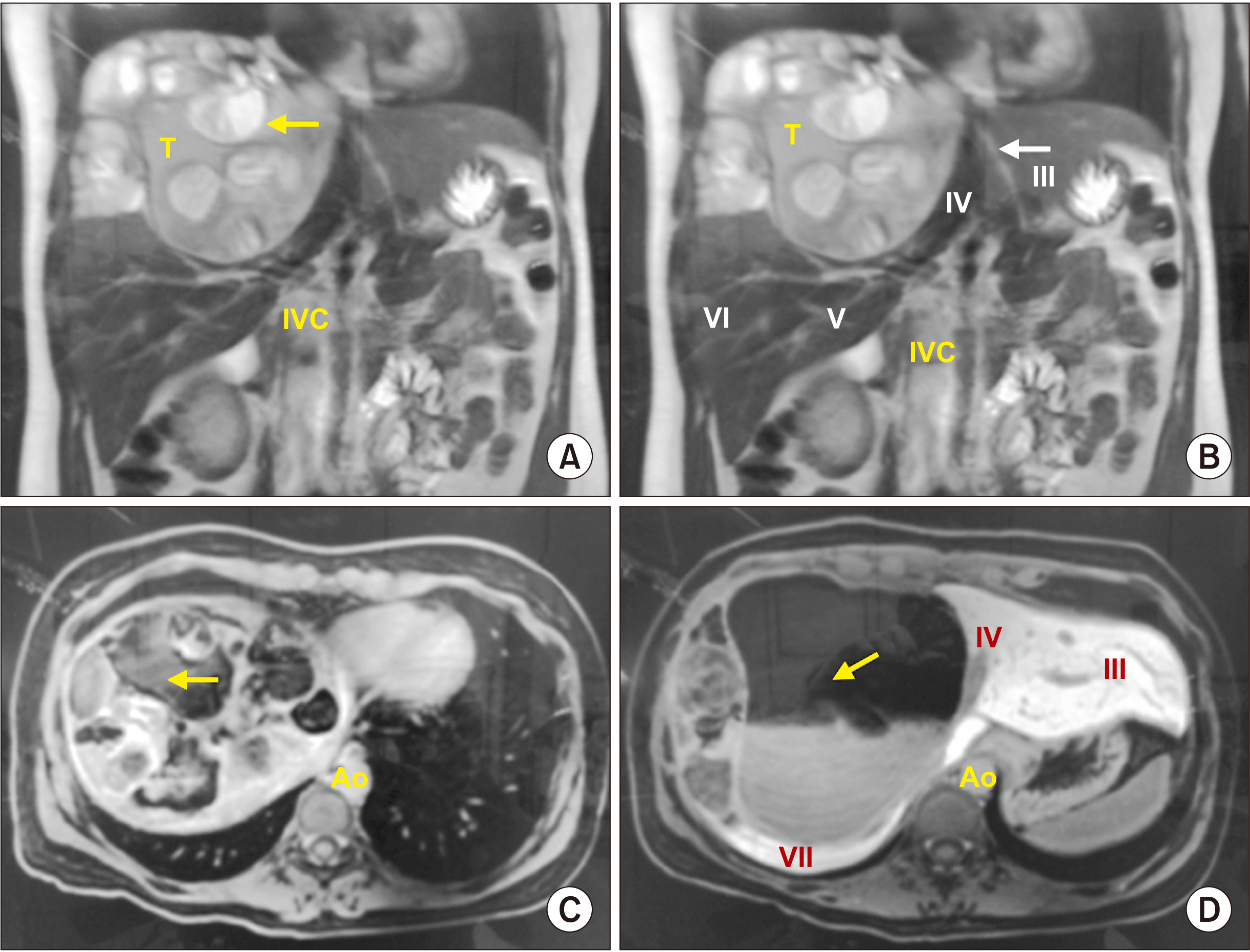
Fig. 1 Magnetic resonance imaging (MRI) demonstrating a liver mass with multiple distinct intensities. Cystic and solid features involving right liver at IV and VIII segments of 160 mm × 180 mm × 90 mm are shown. (A) Coronal MRI in T2 with hypo-intense component suggesting bone calcifications (arrow). (B) Coronal MRI in T2 demonstrating heterogeneous mass in the right liver lobe (IV, V, and VIII), and free liver segments (arrow). (C) Axial MRI in T1 with hypo-intense cystic component (arrow). (D) Axial MRI in T1 with another perspective of cystic component of the tumor (arrow), showing Couinaud liver segments in contact with tumor (roman numeral). IVC, inferior vena cava; Ao, aorta.

Fig. 2 A 29-year-old female computed tomography in arterial phase. (A) Coronal plane that demonstrating liver mass extension (T), involving the IV, V, and VIII liver segments. Low attenuation in the middle of the mass highly suggestive of fat and hypodense image suggestive of hair (arrow). (B) Low attenuation in the middle of the mass highly suggestive of fat and hypodense image suggestive of hair (arrow). (C) Axial plane showing Rokitansky nodule (arrow). (D) Axial plane where sebaceous tissue is identified (arrow), with features suggestive of liver teratoma.

Fig. 3 Trans-operative photo after surgical excision of liver teratoma. (A) Surgical limit of the neoplasia lateral to IV liver segment (arrow) (II, III), residual liver Couinaud segments from left liver (LL), right colon (RC), and stomach (S) are shown. (B) Pearly areas over pelvic recess compatible with gliomatosis peritonei (arrows) and right ovary (Ro).

Fig. 4 Surgical specimen product of right extended hepatectomy. (A) Liver teratoma (T) involving IV, V, and VIII liver segments, compression of falciform ligament (arrow), and inferior vena cava (IVC). (B) Upper view demonstrating the extension to IV and VIII liver segments with falciform ligament (arrows). (C) Inferior view showing the tumor extension (T) to the liver segments (IV, VIII), gallbladder (Gb), cystic duct (green arrow), right branch of right liver artery (red arrow), and right branch of portal vein (blue arrow). RL, right liver lobe; CBD, common bile duct; PV, portal vein; PLA, principal liver artery.
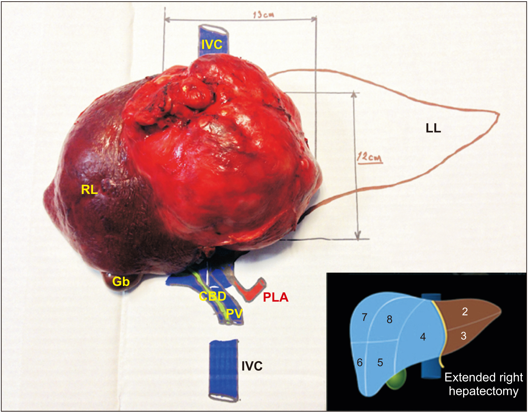
Fig. 5 Specimen mounted in a model after surgical excision to illustrate their anatomical relationships, tumor size, and proper orientation of resected liver segments. IVC, inferior vena cava; RL, right liver lobe; Gb, gallbladder; CBD, common bile duct; PV, portal vein; PLA, principal liver artery; LL, left liver lobe.

Fig. 6 Photomicrograph with H&E stain. (A) High power field (HPF) 10×. Photomicrograph showing liver parenchyma and cystic lesion with keratin. (B) HPF 20×. Photomicrograph showing mature neuroectodermal elements: sebaceous glands, apocrine metaplasia and epithelia lining of cylindrical ciliated epithelium. (C) HPF 20×. Neuroectodermal elements with rosette formation, ined by crowded basophil cells with hyperchromatic nuclei. (D) HPF 20×. Immature mesenchymal elements: adipose tissue and stroma. (E) HPF 20×. Neuroglia and dystrophic calcification of the psammoma type in gliomatosis peritonei. (F) HPF 10×. Squamous epithelium in transition with immature elements of pigmented neuroepithelium.
Reference
-
1. McKenney JK, Heerema-McKenney A, Rouse RV. 2007; Extragonadal germ cell tumors: a review with emphasis on pathologic features, clinical prognostic variables, and differential diagnostic considerations. Adv Anat Pathol. 14:69–92. DOI: 10.1097/PAP.0b013e31803240e6. PMID: 17471115.
Article2. Ronchi A, Cozzolino I, Montella M, Panarese I, Zito Marino F, Rossetti S, et al. 2019; Extragonadal germ cell tumors: not just a matter of location. A review about clinical, molecular and pathological features. Cancer Med. 8:6832–6840. DOI: 10.1002/cam4.2195. PMID: 31568647. PMCID: PMC6853824.
Article3. Ashley DJ. 1973; Origin of teratomas. Cancer. 32:390–394. DOI: 10.1002/1097-0142(197308)32:2<390::AID-CNCR2820320216>3.0.CO;2-W. PMID: 4722920.
Article4. Davidson AJ, Hartman DS, Goldman SM. 1989; Mature teratoma of the retroperitoneum: radiologic, pathologic, and clinical correlation. Radiology. 172:421–425. DOI: 10.1148/radiology.172.2.2664866. PMID: 2664866.
Article5. Winter TC 3rd, Freeny P. 1993; Hepatic teratoma in an adult. Case report with a review of the literature. J Clin Gastroenterol. 17:308–310. DOI: 10.1097/00004836-199312000-00009. PMID: 8308217.6. Glenn OA, Barkovich AJ. 1996; Intracranial germ cell tumors: a comprehensive review of proposed embryologic derivation. Pediatr Neurosurg. 24:242–251. DOI: 10.1159/000121046. PMID: 8933567.
Article7. Chaganti RS, Houldsworth J. 2000; Genetics and biology of adult human male germ cell tumors. Cancer Res. 60:1475–1482. PMID: 10749107.8. Hailemariam S, Engeler DS, Bannwart F, Amin MB. 1997; Primary mediastinal germ cell tumor with intratubular germ cell neoplasia of the testis--further support for germ cell origin of these tumors: a case report. Cancer. 79:1031–1036. DOI: 10.1002/(SICI)1097-0142(19970301)79:5<1031::AID-CNCR21>3.0.CO;2-1. PMID: 9041167.9. Chaganti RS, Rodriguez E, Mathew S. 1994; Origin of adult male mediastinal germ-cell tumours. Lancet. 343:1130–1132. DOI: 10.1016/S0140-6736(94)90235-6. PMID: 7910232.
Article10. Gonzalez-Crussi F. 1982. Extragonadal teratoma. Armed Forces Institute of Pathology;Washington, DC:11. Vicus D, Beiner ME, Clarke B, Klachook S, Le LW, Laframboise S, et al. 2011; Ovarian immature teratoma: treatment and outcome in a single institutional cohort. Gynecol Oncol. 123:50–53. DOI: 10.1016/j.ygyno.2011.06.037. PMID: 21764111.
Article12. Ramkumar J, Best A, Gurung A, Dufresne AM, Melich G, Vikis E, et al. 2018; Resection of ruptured hepatic teratoma in an adult. Int J Surg Case Rep. 53:414–419. DOI: 10.1016/j.ijscr.2018.11.032. PMID: 30567058. PMCID: PMC6259038.
Article13. Caspi B, Appelman Z, Rabinerson D, Zalel Y, Tulandi T, Shoham Z. 1997; The growth pattern of ovarian dermoid cysts: a prospective study in premenopausal and postmenopausal women. Fertil Steril. 68:501–505. DOI: 10.1016/S0015-0282(97)00228-8. PMID: 9314922.
Article14. Sinha A, Ewies AA. 2016; Ovarian mature cystic teratoma: challenges of surgical management. Obstet Gynecol Int. 2016:2390178. DOI: 10.1155/2016/2390178. PMID: 27110246. PMCID: PMC4823513.
Article15. Cöl C. 2003; Immature teratoma in both mediastinum and liver of a 21-year-old female patient. Acta Med Austriaca. 30:26–28. DOI: 10.1046/j.1563-2571.2003.02024.x. PMID: 12558563.
Article16. Rahmat K, Vijayananthan A, Abdullah B, Amin S. 2006; Benign teratoma of the liver: a rare cause of cholangitis. Biomed Imaging Interv J. 2:e20. DOI: 10.2349/biij.2.3.e20. PMID: 21614237. PMCID: PMC3097637.
Article17. Silva DS, Dominguez M, Silvestre F, Calhim I, Daniel J, Teixeira M, et al. 2007; Liver teratoma in an adult. Eur Surg. 39:372–375. DOI: 10.1007/s10353-007-0370-0.
Article18. Certo M, Franca M, Gomes M, Machado R. 2008; Liver teratoma. Acta Gastroenterol Belg. 71:275–279. PMID: 18720943.19. Karlo C, Leschka S, Dettmer M, Breitenstein S, Stolzmann P. 2009; Hepatic teratoma and peritoneal gliomatosis: a case report. Cases J. 2:9302. DOI: 10.1186/1757-1626-2-9302. PMID: 20062626. PMCID: PMC2803966.
Article20. Madan M, Arora R, Singh J, Kaur A. 2010; Mature cystic teratoma of the liver in an adult female. Indian J Pathol Microbiol. 53:872–873. DOI: 10.4103/0377-4929.72022. PMID: 21045458.
Article21. Harris B, De Simone N. 2011; Primary mature cystic teratoma of the liver: report of a rare case. Am J Clin Med. 8:57–59.22. Gupta R, Bansal K, Manchanda V, Gupta R. 2013; Mature cystic teratoma of liver. APSP J Case Rep. 4:13. PMID: 24040591. PMCID: PMC3754398.23. Martin LC, Papadatos D, Michaud C, Thomas J. 2004; Best cases from the AFIP: liver teratoma. Radiographics. 24:1467–1471. DOI: 10.1148/rg.245035209. PMID: 15371619.24. Robboy SJ, Scully RE. 1970; Ovarian teratoma with glial implants on the peritoneum. An analysis of 12 cases. Hum Pathol. 1:643–653. DOI: 10.1016/S0046-8177(70)80062-4. PMID: 5521737.25. Hill DA, Dehner LP, White FV, Langer JC. 2000; Gliomatosis peritonei as a complication of a ventriculoperitoneal shunt: case report and review of the literature. J Pediatr Surg. 35:497–499. DOI: 10.1016/S0022-3468(00)90221-5. PMID: 10726696.
Article26. Dadmanesh F, Miller DM, Swenerton KD, Clement PB. 1997; Gliomatosis peritonei with malignant transformation. Mod Pathol. 10:597–601. PMID: 9195578.27. Liang L, Zhang Y, Malpica A, Ramalingam P, Euscher ED, Fuller GN, et al. 2015; Gliomatosis peritonei: a clinicopathologic and immunohistochemical study of 21 cases. Mod Pathol. 28:1613–1620. DOI: 10.1038/modpathol.2015.116. PMID: 26564007. PMCID: PMC4682736.
Article28. Fortt RW, Mathie IK. 1969; Gliomatosis peritonei caused by ovarian teratoma. J Clin Pathol. 22:348–353. DOI: 10.1136/jcp.22.3.348. PMID: 5784983. PMCID: PMC474087.
Article29. Best DH, Butz GM, Moller K, Coleman WB, Thomas DB. 2004; Molecular analysis of an immature ovarian teratoma with gliomatosis peritonei and recurrence suggests genetic independence of multiple tumors. Int J Oncol. 25:17–25. DOI: 10.3892/ijo.25.1.17. PMID: 15201985.
Article30. Harada M, Osuga Y, Fujimoto A, Fujimoto A, Fujii T, Yano T, et al. 2013; Predictive factors for recurrence of ovarian mature cystic teratomas after surgical excision. Eur J Obstet Gynecol Reprod Biol. 171:325–328. DOI: 10.1016/j.ejogrb.2013.09.004. PMID: 24070501.
Article31. Kaiser HE. Gorelik EL, editor. 1989. Metastasis of teratomas. Metastasis /dissemination. Springer;Dordrecht: p. 275–281. DOI: 10.1007/978-94-009-2534-2_22.
Article32. Kommoss F, Emond J, Hast J, Talerman A. 1990; Ruptured mature cystic teratoma of the ovary with recurrence in the liver and colon 17 years later. A case report. J Reprod Med. 35:827–831. PMID: 2213748.33. Chen L, Nelson SD, Berek JS. 1999; Recurrent mature cystic teratoma presenting as a perihepatic mass. Obstet Gynecol. 94(5 Pt 2):856. DOI: 10.1016/S0029-7844(99)00490-1. PMID: 10546767.
Article34. Okino H, Koga Y, Tsuneyoshi M, Takeda S. 2006; Metachronous mature cystic teratomas in the left ovary and bilateral diaphragm: report of a case. Surg Today. 36:1012–1014. DOI: 10.1007/s00595-006-3299-1. PMID: 17072726.
Article35. Li X, Zhu D, Lv LI, Yu J. 2016; An uncommon recurrence of an immature teratoma: a case report. Oncol Lett. 11:2453–2456. DOI: 10.3892/ol.2016.4254. PMID: 27073497. PMCID: PMC4812457.
Article
- Full Text Links
-
- Actions
-
Cited
- CITED
-
- Close
- Share
-
- Similar articles
-
- A Case of Gliomatosis Peritonei
- A case of Gliomatosis Peritonei after 10 years following Operation of Immature Teratoma of the Ovary
- Low-grade Immature Teratoma of the Ovary with Gliomatosis Peritonei: A case report
- Case report of solitary giant hepatic lymphangioma
- One Case Report of Giant Sacrococcygeal Teratoma in Infancy
