Diagnosis and surgical treatment of a rare hepatic angiomyolipoma with internal hemorrhage
- Affiliations
-
- 1Department of Surgery, Seoul National University Bundang Hospital, Seoul National University College of Medicine, Seongnam, Korea
- 2Department of Pathology, Seoul National University Bundang Hospital, Seoul National University College of Medicine, Seongnam, Korea
- KMID: 2523059
- DOI: http://doi.org/10.14701/ahbps.2021.25.4.532
Abstract
- Although angiomyolipoma (AML) is commonly found in the kidney, its appearance in the liver is rare. The first hepatic AML was reported by Ishak in 1976. Since then, there have been various reports of AML. An AML is a tumour affecting adipocytes, muscle tissues, and blood vessels. Hepatic AML has been associated with tuberous sclerosis. Its spontaneous occurrence has also been noticed. It can have a varied presentation with some being asymptomatic, others presenting with a dull aching pain, while some with disastrous consequences due to rupture and torrential bleeding intra-abdominally. Herein, we present a case of a 47-year-old female with a large hepatic AML having an internal hemorrhage that caused changes in hepatic arteries. In our case, magnetic resonance imaging was unable to establish a diagnosis. Intraoperatively, AML caused dilatation and engorgement of vessels around the porta. Immediately post resection, vessel dilatation and engorgement were reduced on table. Another notable feature was that these changes caused no intra-operative or post-operative hemodynamic changes. We report a case of a huge hepatic AML with internal hemorrhage associated with perihepatic vascular changes having a successful surgical treatment.
Keyword
Figure
-
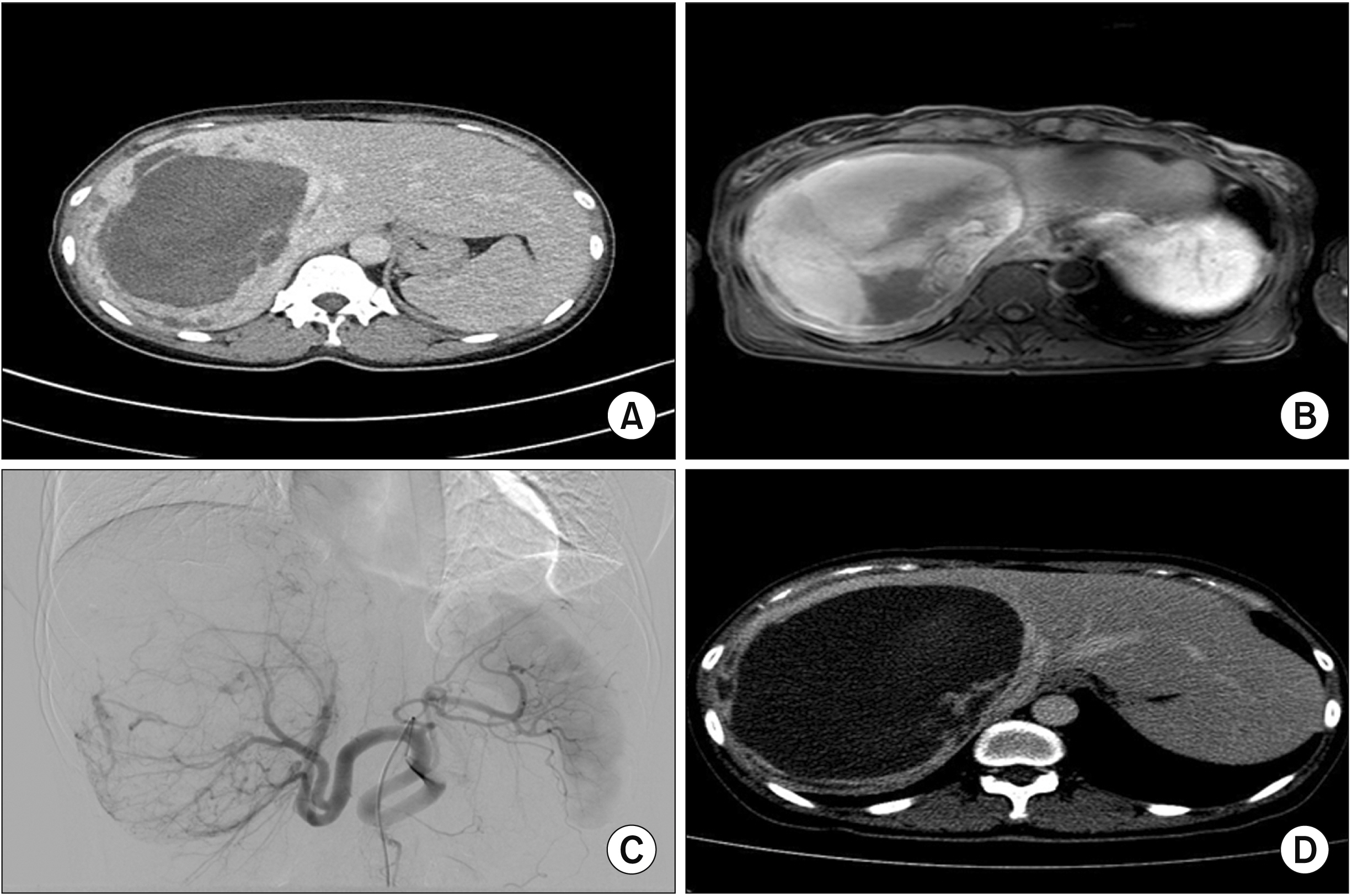
Fig. 1 Preoperative images. Huge cystic tumor or hemorrhage in the right liver on computed tomography (CT) (A), suspected mural nodules inside the cystic tumor on magnetic resonance imaging (B), engorged hepatic artery and huge tumor on angiogram (C), and persistent cystic and solid huge tumor on immediate preoperative CT scan (D).
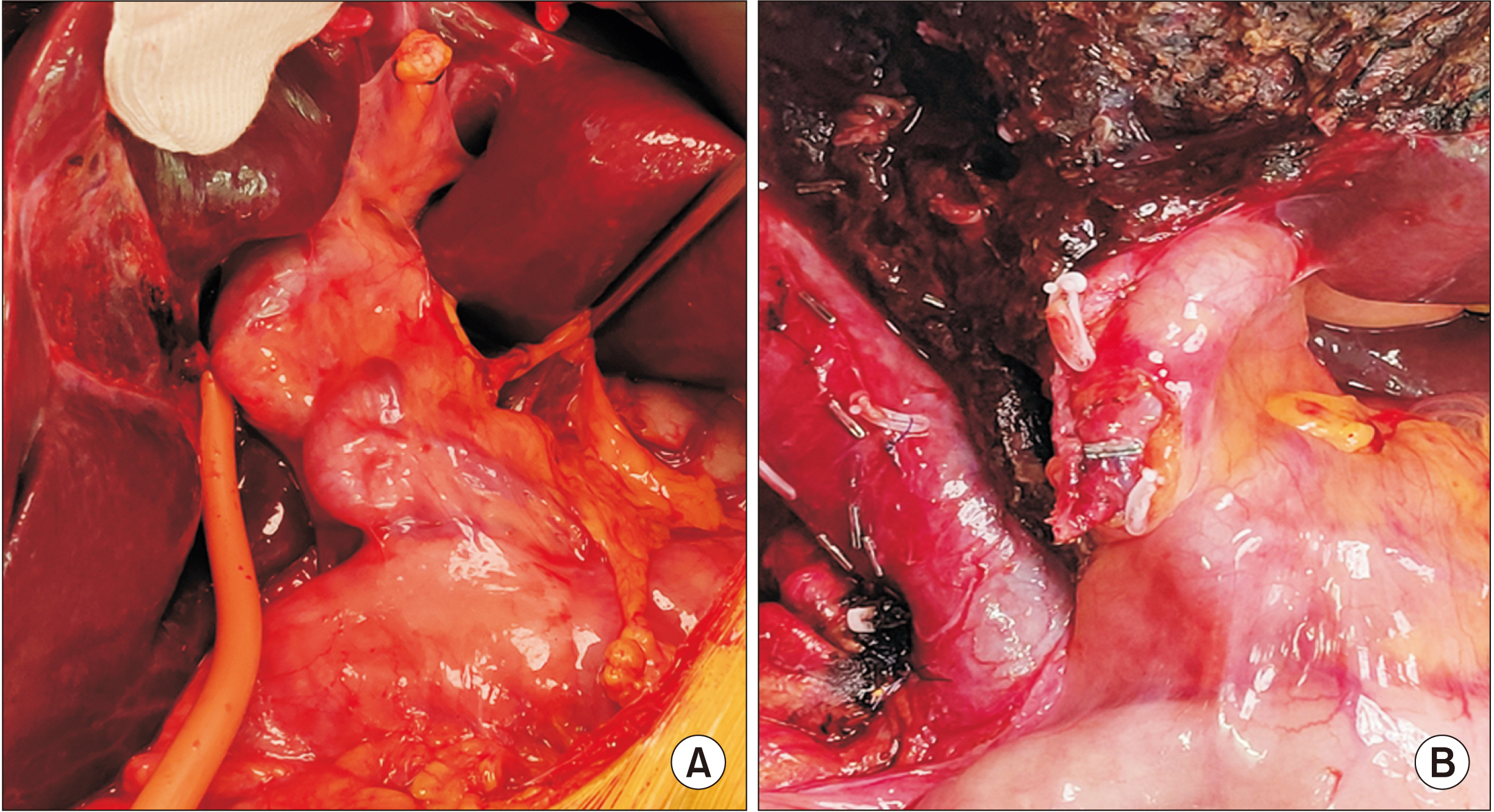
Fig. 2 Intraoperative images showing engorgement of vessels at the porta hepatis (A) and slight reduction of engorgement after right hepatectomy (B).

Fig. 3 Histopathological slides of hepatic angiomyolipoma. Low magnification view shows well delineated mass containing adipose tissue and irregular large blood vessels (A). High power view shows three components of angiomyolipoma: adipose tissue, blood vessels, and epithelioid to spindle cells (B). Immunohistochemistry shows HMB45 expression (C) and smooth muscle actin expression (D) of tumor cells.
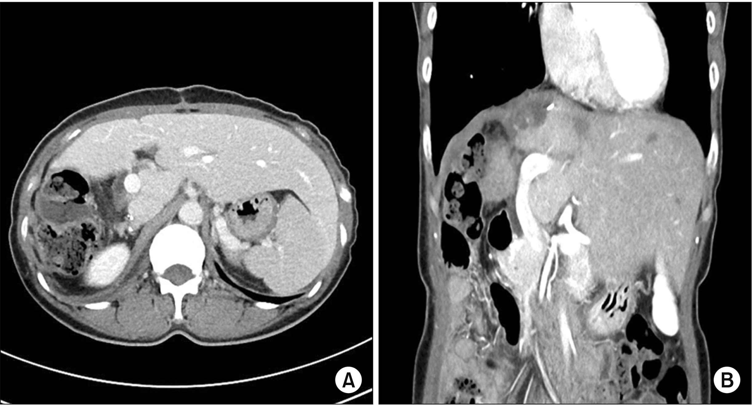
Fig. 4 Follow-up computed tomography scans at six months after surgery showing well regenerated remnant left liver (A) and a further reduction in the size of the left hepatic artery (B).
Reference
-
1. Roncalli M, Sciarra A, Tommaso LD. 2016; Benign hepatocellular nodules of healthy liver: focal nodular hyperplasia and hepatocellular adenoma. Clin Mol Hepatol. 22:199–211. DOI: 10.3350/cmh.2016.0101. PMID: 27189732. PMCID: PMC4946404.
Article2. Bruix J, Han KH, Gores G, Llovet JM, Mazzaferro V. 2015; Liver cancer: approaching a personalized care. J Hepatol. 62(1 Suppl):S144–S156. DOI: 10.1016/j.jhep.2015.02.007. PMID: 25920083. PMCID: PMC4520430.
Article3. Guidi G, Catalano O, Rotondo A. 1997; Spontaneous rupture of a hepatic angiomyolipoma: CT findings and literature review. Eur Radiol. 7:335–337. DOI: 10.1007/s003300050162. PMID: 9087353.
Article4. Blokhin I, Chernina V, Menglibaev M, Kalinin D, Schima W, Karmazanovsky G. 2018; Giant hepatic angiomyolipoma: a case report. BJR Case Rep. 5:20180072. DOI: 10.1259/bjrcr.20180072. PMID: 31131134. PMCID: PMC6519506.
Article5. Huber C, Treutner KH, Steinau G, Schumpelick V. 1996; Ruptured hepatic angiolipoma in tuberous sclerosis complex. Langenbecks Arch Chir. 381:7–9. DOI: 10.1007/BF00184248. PMID: 8717168.
Article6. Kim SH, Kang TW, Lim K, Joh HS, Kang J, Sinn DH. 2017; A case of ruptured hepatic angiomyolipoma in a young male. Clin Mol Hepatol. 23:179–183. DOI: 10.3350/cmh.2016.0027. PMID: 28449573. PMCID: PMC5497672.
Article7. Galant J, Martí-Bonmatí L, Ripollés T, Martinez-Rodrigo J, Ferrer MD. 1995; Hepatic manifestations of tuberous sclerosis studied by US and CT. Eur Radiol. 5:486–491. DOI: 10.1007/BF00208339.
Article8. Lee SJ, Kim SY, Kim KW, Kim JH, Kim HJ, Lee MG, et al. 2016; Hepatic angiomyolipoma versus hepatocellular carcinoma in the noncirrhotic liver on gadoxetic acid-enhanced MRI: a diagnostic challenge. AJR Am J Roentgenol. 207:562–570. DOI: 10.2214/AJR.15.15602. PMID: 27248975.
Article9. Yang L, Xu Z, Dong R, Fan J, Du Y, Zhang Y, et al. 2013; Is surgery necessary for patients with hepatic angiomyolipoma? Retrospective analysis from eight Chinese cases. J Gastroenterol Hepatol. 28:1648–1653. DOI: 10.1111/jgh.12289. PMID: 23731017.10. Du S, Li Y, Mao Y, Sang X, Lu X, Wang W, et al. 2012; Diagnosis and treatment of hepatic angiomyolipoma. Hepatobiliary Surg Nutr. 1:19–24. DOI: 10.3978/j.issn.2304-3881.2012.07.02. PMID: 24570900. PMCID: PMC3924623.11. Goodman ZD, Ishak KG. 1984; Angiomyolipomas of the liver. Am J Surg Pathol. 8:745–750. DOI: 10.1097/00000478-198410000-00003. PMID: 6496843.
Article12. Ding GH, Liu Y, Wu MC, Yang GS, Yang JM, Cong WM. 2011; Diagnosis and treatment of hepatic angiomyolipoma. J Surg Oncol. 103:807–812. DOI: 10.1002/jso.21814. PMID: 21283992.
Article
- Full Text Links
-
- Actions
-
Cited
- CITED
-
- Close
- Share
-
- Similar articles
-
- Multiple Hepatic Angiomyolipoma Detected by Abdominal Ultrasonography
- A Case of Hepatic Angiomyolipoma Showing Different Uptake on F-18 FDG and C-11 Acetate PET
- A case of ruptured hepatic angiomyolipoma in a young male
- Sponataneous Hemorrhage of Renal Angiomyolopoma during Pregnancy
- A case of spontaneous rupture of hepatic angiomyolipoma
