Posterior Fossa Teratomas in Adults : A Systematic Review
- Affiliations
-
- 1Department of Neurosurgery, Asan Medical Center, College of Medicine, University of Ulsan, Seoul, Korea
- 2Department of Pathology, Asan Medical Center, College of Medicine, University of Ulsan, Seoul, Korea
- KMID: 2521988
- DOI: http://doi.org/10.3340/jkns.2020.0343
Abstract
Objective
: The occurrence of posterior fossa teratomas in adulthood is extremely rare. In this study, we aimed to report our experience with two cases of posterior fossa mature teratoma in adults who underwent surgical resection. We also performed a systematic review of published papers available to date.
Methods
: We retrospectively reviewed the electronic medical records of patients who had onset of posterior fossa teratomas in adulthood at our institute between 1995 and 2020. We evaluated the clinical, radiographic, and pathological features of mature teratomas at the posterior fossa in adulthood. Furthermore, we searched the PubMed, EMBASE, and Web of Science database and reviewed published articles.
Results
: We found 507 articles on database review; of them, 102 were duplicates and 389 were excluded based on the inclusion criteria. Finally, 16 cases of posterior fossa from the web search and related articles. Subsequently, we added two cases that underwent surgery at our institute. We analyzed a total of 18 cases of mature teratomas. Headache was the most common (55.6%) symptom. The teratomas showed heterogeneous signals on magnetic resonance imaging. Thirteen patients (72.2%) had lesion at midline, five patients (27.8%) had calcification. Surgical resection was performed in all patients. No studies reported recurrence after resection.
Conclusion
: The occurrence of posterior fossa teratomas in adulthood is difficult to diagnose at the initial stage. Radiographic diagnosis alone can lead to misdiagnosis. Pathological confirmation is essential. Surgical resection is a curative option for posterior fossa teratomas in adulthood.
Keyword
Figure
-

Fig. 1. Flow chart of the literature review process.

Fig. 2. A : Initial radiographic image showing an oval mass with a heterogeneous signal on T1- and T2-weighted imaging at quadrigeminal cistern. Computed tomography showing focal calcification and lipid component. B : After a 16-year follow-up period, the mass had increased in size and nodular strong enhancement appeared. Gd : gadolinium, CT : computed tomography.
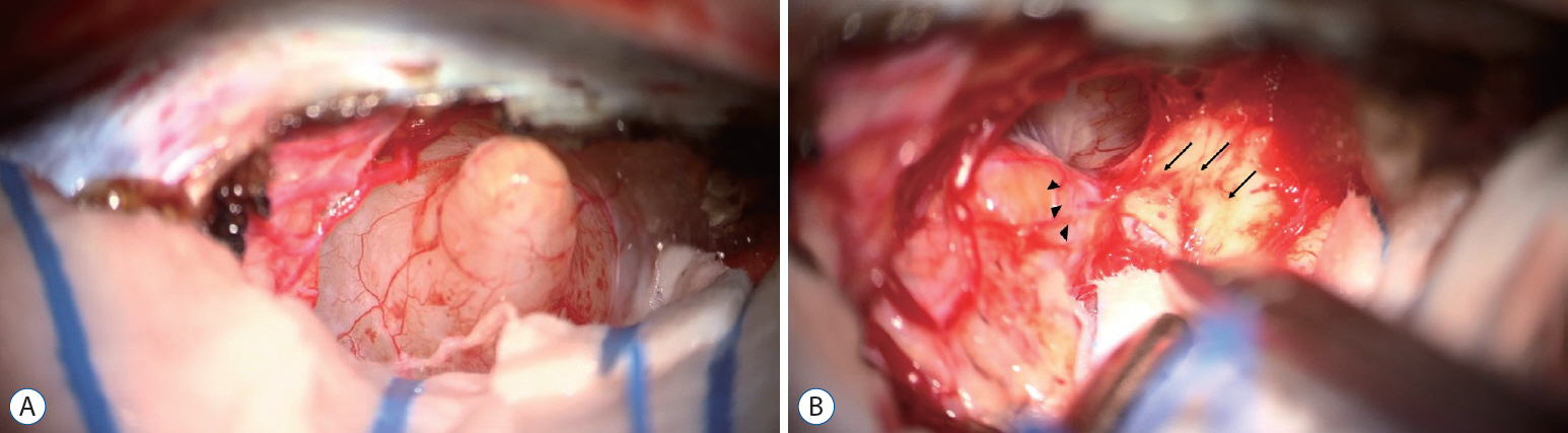
Fig. 3. A : Microscopic view through the occipital transtentorial approach. The tentorium was coagulated and cut. The mass was soft, fragile, and capsulated. B : Severe adhesion was noted between the quadrigeminal plate (black arrows) and the mass (arrowheads). Numerous small vessels were surrounding the mass.

Fig. 4. Image of the mass located at the posterior cerebellum (midline). The image shows heterogeneous signal intensity without peritumoral edema.
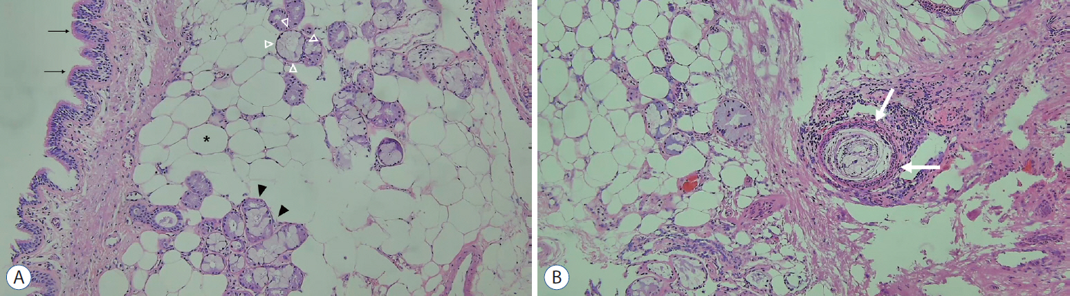
Fig. 5. A : The ciliated columnar epithelium (black arrows), adipose tissue (asterisk), and salivary gland tissue (white and black arrowheads) are observed. The salivary gland tissue shows serous acini (black arrowheads) and mucinous acini (white arrowheads) (H&E stain, ×100). B : A tumor with fully differentiated three germ-layer derivates—the hair follicle (white arrows), adipose tissue, and skin appendages (H&E stain, ×100).
Reference
-
References
1. Arseni C, Dănăilă L, Nicola N, Georgian M, Istrati C. Intracranial teratomas. Acta Neurochir (Wien). 20:37–51. 1969.
Article2. Barkley AS, Kuo CH, Leary SES, Ojemann JG, Susarla SM. Unusual radiographic presentation of intracranial mature teratoma and resection via supraorbital approach. World Neurosurg. 122:81–84. 2019.
Article3. Beschorner R, Schittenhelm J, Bueltmann E, Ritz R, Meyermann R, Mittelbronn M. Mature cerebellar teratoma in adulthood. Neuropathology. 29:176–180. 2009.
Article4. Bohara M, Yonezawa H, Karki P, Bakhtiar Y, Hirano H, Kitazono I, et al. Mature posterior fossa teratoma mimicking dermoid cyst. Brain Tumor Pathol. 30:262–265. 2013.
Article5. Buetow PC, Smirniotopoulos JG, Done S. Congenital brain tumors: a review of 45 cases. AJNR Am J Neuroradiol. 11:793–799. 1990.
Article6. Clack TD, McGillicuddy JE, Wolf GT. Labyrinthine functional tests and a case of midline cerebellar teratoma. Am J Otol. 9:481–488. 1988.7. Coulibaly O, El Kacemi I, Fatemi N, Gana R, Saïdi A, Maaqili R, et al. Mature posterior fossa teratoma mimicking infratentorial meningioma: a case report. Neurochirurgie. 58:40–43. 2012.
Article8. Drapkin AJ, Rose WS, Pellmar MB. Mature teratoma in the fourth ventricle of an adult: case report and review of the literature. Neurosurgery. 21:404–410. 1987.
Article9. Goyal N, Kakkar A, Singh PK, Sharma MC, Chandra PS, Mahapatra AK, et al. Intracranial teratomas in children: a clinicopathological study. Childs Nerv Syst. 29:2035–2042. 2013.
Article10. Harada K, Okamoto H, Fujioka Y, Kiya K, Mukada K, Uozumi T, et al. Teratoma in the fourth ventricle of an elderly adult--case report. Neurol Med Chir (Tokyo). 24:499–503. 1984.11. Jennings MT, Gelman R, Hochberg F. Intracranial germ-cell tumors: natural history and pathogenesis. J Neurosurg. 63:155–167. 1985.
Article12. Kyritsis AP. Management of primary intracranial germ cell tumors. J Neurooncol. 96:143–149. 2010.
Article13. Labauge R, Pagès M, Pagès A, Ségnarbieux F. Cerebellar teratoma in adults. Contribution of x-ray computed tomography and magnetic resonance imaging. Rev Neurol (Paris). 146:310–312. 1990.14. Lee YH, Park EK, Park YS, Shim KW, Choi JU, Kim DS. Treatment and outcomes of primary intracranial teratoma. Childs Nerv Syst. 25:1581–1587. 2009.
Article15. Liu Z, Lv X, Wang W, An J, Duan F, Feng X, et al. Imaging characteristics of primary intracranial teratoma. Acta Radiol. 55:874–881. 2014.
Article16. Matsutani M, Sano K, Takakura K, Fujimaki T, Nakamura O, Funata N, et al. Primary intracranial germ cell tumors: a clinical analysis of 153 histologically verified cases. J Neurosurg. 86:446–455. 1997.
Article17. Noudel R, Vinchon M, Dhellemmes P, Litré CF, Rousseaux P. Intracranial teratomas in children: the role and timing of surgical removal. J Neurosurg Pediatr. 2:331–338. 2008.
Article18. Park KB, Park HS, Lee JI, Suh YL. Mature teratoma in the cerebellar hemisphere of an adult. J Korean Neurosurg Soc. 41:180–181. 2007.
Article19. Pöschl J, Berger F, Kretzschmar H, Schüller U. A 59-year-old man with two cerebellar lesions and disturbed cerebellar morphology. Brain Pathol. 25:790–791. 2015.
Article20. Sanyal P, Barui S, Mathur S, Basak U. A case of mature cystic teratoma arising from the fourth ventricle. Case Rep Pathol. 2013:702424. 2013.
Article21. Saura H, Beppu T, Matsuura H, Asahi S, Uesugi N, Sasaki M, et al. Intractable yawning associated with mature teratoma of the supramedial cerebellum. J Neurosurg. 121:387–389. 2014.
Article22. Strang RR. Teratomas of the posterior cranial fossa. Zentralbl Neurochir. 20:359–372. 1960.23. Zavanone M, Alimehmeti R, Campanella R, Ram-Pini P, Locatelli M, Egidi M, et al. Cerebellar mature teratoma in adulthood. J Neurosurg Sci. 46:35–38. discussion 38. 2002.24. Zhang S, Wang X, Liu X, Hui X. Mature teratoma in cerebellopontine angle in a 70-year-old female: a rare tumor with exceptional location, age, and presentation. Neurol India. 60:660–661. 2012.
Article
- Full Text Links
-
- Actions
-
Cited
- CITED
-
- Close
- Share
-
- Similar articles
-
- Teratoma in the Posterior Cranial Fossa
- Hyperdense Cystic Teratoma in Posterior Cranial Fossa
- Posterior mediastinal teratoma - Diagnosis by computerized tomography and ultrasonography
- Two Cases of Presacral Teratomas in Adult
- Hemangiopericytoma of the Posterior Fossa: A Case Report and Review of the Literature
