Hemangiopericytoma of the Posterior Fossa: A Case Report and Review of the Literature
- Affiliations
-
- 1Department of Neurosurgery, Maryknoll Medical Center, Busan, Korea. yunsuk.kim@gmail.com
- KMID: 2048482
- DOI: http://doi.org/10.14791/btrt.2013.1.2.95
Abstract
- Intracranial hemangiopericytoma is unusual, and those occurring in the posterior fossa is extremely rare; we report such a rare case of hemangiopericytoma of the posterior fossa. The radiologic findings and gross characteristics of hemangiopericytomas are sometimes quite similar to those of meningiomas. Although extremely rare, the operator should be aware of the existence of this disorder to dexterously manage the aggressive nature and high vascular tendency of hemangiopericytomas. The radiological features and histological findings in this case are discussed in this study.
Keyword
MeSH Terms
Figure
-
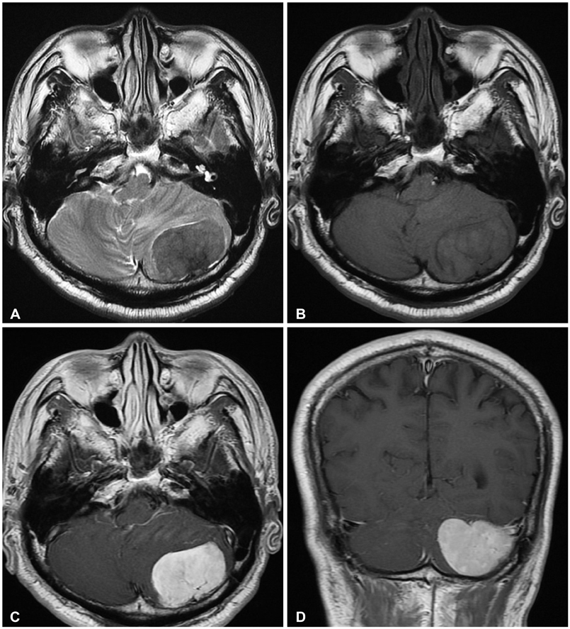
Fig. 1 Pre-operative axial T1 (A) and T2 (B) magnetic resonance images show a tumor mass in the left cerebellum compressing the 4th ventricle with surrounding edema. Gd-enhanced T1 axial (C) and coronal (D) images reveal a well-enhancing lesion with dural attachment adjacent to the junction of the transverse and sigmoid sinuses.

Fig. 2 A photomicrograph of the tumor specimen shows vascular space surrounded by proliferating oval to spindle-shaped pericytes (A) (hematoxylin and eosin stain; ×400), prominent vasculature (B) (hematoxylin and eosin stain; ×100) represented by staghorn pattern (arrow) and tumor cells strongly expressing CD-34 (C) (immunohistochemistry; ×200).

Fig. 3 Post-operative axial T1 (A) and coronal T1 (B) magnetic resonance images taken within two weeks of the surgery show the resection cavity with residual tumor tissue adherent to the transverse sinus.
Reference
-
1. Kim JH, Kwon TH, Kim JH, Park YK, Chung YG, Chung HS. Meningeal hemangiopericytoma: study of 6 cases and review of the literatures. J Korean Neurosurg Soc. 2006; 39:32–35.2. Fountas KN, Kapsalaki E, Kassam M, et al. Management of intracranial meningeal hemangiopericytomas: outcome and experience. Neurosurg Rev. 2006; 29:145–153.
Article3. Tashjian VS, Khanlou N, Vinters HV, Canalis RF, Becker DP. Hemangiopericytoma of the cerebellopontine angle: a case report and review of the literature. Surg Neurol. 2009; 72:290–295.
Article4. Louis DN, Ohgaki H, Wiestler OD, et al. The 2007 WHO classification of tumours of the central nervous system. Acta Neuropathol. 2007; 114:97–109.
Article5. Chiechi MV, Smirniotopoulos JG, Mena H. Intracranial hemangiopericytomas: MR and CT features. AJNR Am J Neuroradiol. 1996; 17:1365–1371.6. Liu G, Chen ZY, Ma L, Lou X, Li SJ, Wang YL. Intracranial hemangiopericytoma: MR imaging findings and diagnostic usefulness of minimum ADC values. J Magn Reson Imaging. 2013; [Epub ahead of print].
Article7. Spatola C, Privitera G. Recurrent intracranial hemangiopericytoma with extracranial and unusual multiple metastases: case report and review of the literature. Tumori. 2004; 90:265–268.
Article8. Guthrie BL, Ebersold MJ, Scheithauer BW, Shaw EG. Meningeal hemangiopericytoma: histopathological features, treatment, and long-term follow-up of 44 cases. Neurosurgery. 1989; 25:514–522.
Article9. Alén JF, Lobato RD, Gómez PA, et al. Intracranial hemangiopericytoma: study of 12 cases. Acta Neurochir (Wien). 2001; 143:575–586.
Article
