Ann Clin Neurophysiol.
2021 Oct;23(2):69-81. 10.14253/acn.2021.23.2.69.
New approach of using cortico-cortical evoked potential for functional brain evaluation
- Affiliations
-
- 1Department of Neurology, Samsung Medical Center, Sungkyunkwan University School of Medicine, Seoul, Korea
- KMID: 2521884
- DOI: http://doi.org/10.14253/acn.2021.23.2.69
Abstract
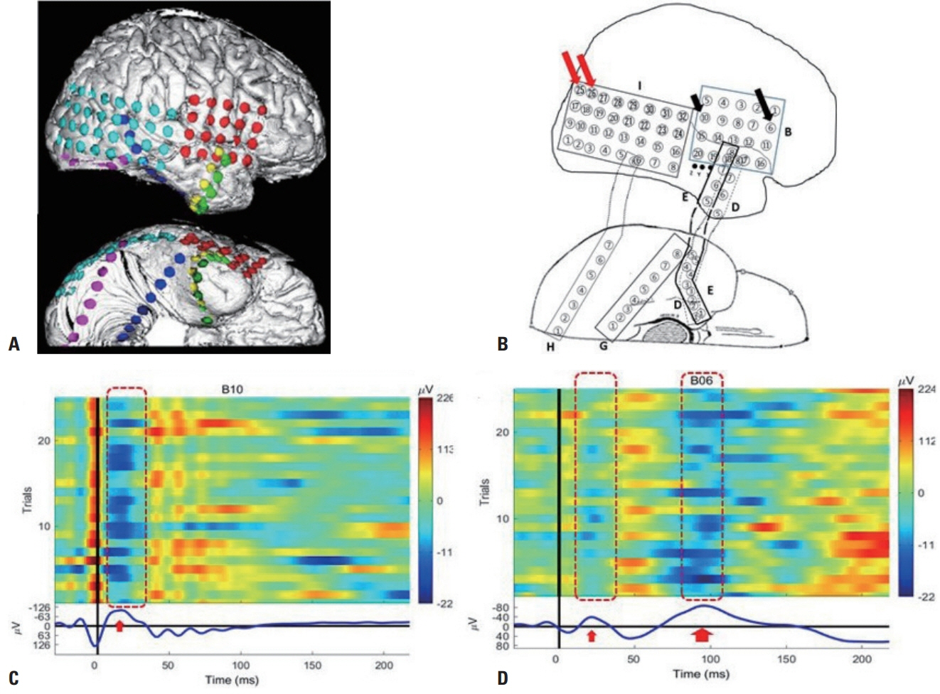
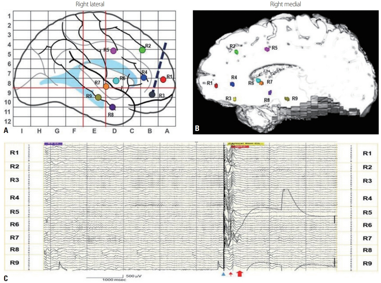
- Cortico-cortical evoked potential (CCEP) mapping is a rapidly developing method for visualizing the brain network and estimating cortical excitability. The CCEP comprises the early N1 component the occurs at 10-30 ms poststimulation, indicating anatomic connectivity, and the late N2 component that appears at < 200 ms poststimulation, suggesting long-lasting effective connectivity. A later component at 200-1,000 ms poststimulation can also appear as a delayed response in some studied areas. Such delayed responses occur in areas with changed excitability, such as an epileptogenic zone. CCEP mapping has been used to examine the brain connections causally in functional systems such as the language, auditory, and visual systems as well as in anatomic regions including the frontoparietal neocortices and hippocampal limbic areas. Task-based CCEPs can be used to measure behavior. In addition to evaluations of the brain connectome, single-pulse electrical stimulation (SPES) can reflect cortical excitability, and so it could be used to predict a seizure onset zone. CCEP brain mapping and SPES investigations could be applied both extraoperatively and intraoperatively. These underused electrophysiologic tools in basic and clinical neuroscience might be powerful methods for providing insight into measures of brain connectivity and dynamics. Analyses of CCEPs might enable us to identify causal relationships between brain areas during cortical processing, and to develop a new paradigm of effective therapeutic neuromodulation in the future.
Figure
Reference
-
1. van den Heuvel MP, Sporns O. Network hubs in the human brain. Trends Cogn Sci. 2013; 17:683–696.
Article2. van Diessen E, Diederen SJ, Braun KP, Jansen FE, Stam CJ. Functional and structural brain networks in epilepsy: what have we learned? Epilepsia. 2013; 54:1855–1865.
Article3. Raichle ME. A paradigm shift in functional brain imaging. J Neurosci. 2009; 29:12729–12734.
Article4. Sporns O. Structure and function of complex brain networks. Dialogues Clin Neurosci. 2013; 15:247–262.
Article5. Lacruz ME, García Seoane JJ, Valentin A, Selway R, Alarcón G. Frontal and temporal functional connections of the living human brain. Eur J Neurosci. 2007; 26:1357–1370.
Article6. Conner CR, Ellmore TM, DiSano MA, Pieters TA, Potter AW, Tandon N. Anatomic and electro-physiologic connectivity of the language system: a combined DTI-CCEP study. Comput Biol Med. 2011; 41:1100–1109.
Article7. Koubeissi MZ, Lesser RP, Sinai A, Gaillard WD, Franaszczuk PJ, Crone NE. Connectivity between perisylvian and bilateral basal temporal cortices. Cereb Cortex. 2012; 22:918–925.
Article8. Swann NC, Cai W, Conner CR, Pieters TA, Claffey MP, George JS, et al. Roles for the pre-supplementary motor area and the right inferior frontal gyrus in stopping action: electrophysiological responses and functional and structural connectivity. Neuroimage. 2012; 59:2860–2870.
Article9. Kubota Y, Enatsu R, Gonzalez-Martinez J, Bulacio J, Mosher J, Burgess RC, et al. In vivo human hippocampal cingulate connectivity: a corticocortical evoked potentials (CCEPs) study. Clin Neurophysiol. 2013; 124:1547–1556.
Article10. Matsuzaki N, Juhász C, Asano E. Cortico-cortical evoked potentials and stimulation-elicited gamma activity preferentially propagate from lower- to higher-order visual areas. Clin Neurophysiol. 2013; 124:1290–1296.
Article11. Enatsu R, Gonzalez-Martinez J, Bulacio J, Kubota Y, Mosher J, Burgess RC, et al. Connections of the limbic network: a corticocortical evoked potentials study. Cortex. 2015; 62:20–33.
Article12. Usami K, Milsap GW, Korzeniewska A, Collard MJ, Wang Y, Lesser RP, et al. Cortical responses to input from distant areas are modulated by local spontaneous alpha/beta oscillations. Cereb Cortex. 2019; 29:777–787.
Article13. Kobayashi K, Matsumoto R, Usami K, Matsuhashi M, Shimotake A, Kikuchi T, et al. Cortico-cortical evoked potential by single-pulse electrical stimulation is a generally safe procedure. Clin Neurophysiol. 2021; 132:1033–1040.
Article14. Takeyama H, Matsumoto R, Usami K, Nakae T, Kobayashi K, Shimotake A, et al. Human entorhinal cortex electrical stimulation evoked short-latency potentials in the broad neocortical regions: evidence from cortico-cortical evoked potential recordings. Brain Behav. 2019; 9:e01366.
Article15. Keller CJ, Honey CJ, Mégevand P, Entz L, Ulbert I, Mehta AD. Mapping human brain networks with cortico-cortical evoked potentials. Philos Trans R Soc Lond B Biol Sci. 2014; 369:20130528.
Article16. Keller CJ, Bickel S, Entz L, Ulbert I, Milham MP, Kelly C, et al. Intrinsic functional architecture predicts electrically evoked responses in the human brain. Proc Natl Acad Sci U S A. 2011; 108:10308–10313.
Article17. Fox MD, Raichle ME. Spontaneous fluctuations in brain activity observed with functional magnetic resonance imaging. Nat Rev Neurosci. 2007; 8:700–711.
Article18. Kobayashi M, Pascual-Leone A. Transcranial magnetic stimulation in neurology. Lancet Neurol. 2003; 2:145–156.
Article19. Hallett M. Transcranial magnetic stimulation: a primer. Neuron. 2007; 55:187–199.
Article20. Purpura DP, Pool JL, Ransohoff J, Frumin MJ, Housepian EM. Observations on evoked dendritic potentials of human cortex. Electroencephalogr Clin Neurophysiol. 1957; 9:453–459.
Article21. Fritsch B, Reis J, Martinowich K, Schambra HM, Ji Y, Cohen LG, et al. Direct current stimulation promotes BDNF-dependent synaptic plasticity: potential implications for motor learning. Neuron. 2010; 66:198–204.
Article22. Medeiros LF, de Souza IC, Vidor LP, de Souza A, Deitos A, Volz MS, et al. Neurobiological effects of transcranial direct current stimulation: a review. Front Psychiatry. 2012; 3:110.
Article23. Paulus W. Transcranial electrical stimulation (tES-tDCS; tRNS, tACS) methods. Neuropsychol Rehabil. 2011; 21:602–617.24. Matsumoto R, Nair DR, LaPresto E, Najm I, Bingaman W, Shibasaki H, et al. Functional connectivity in the human language system: a cortico-cortical evoked potential study. Brain. 2004; 127(Pt 10):2316–2330.
Article25. Prime D, Rowlands D, O'Keefe S, Dionisio S. Considerations in performing and analyzing the responses of cortico-cortical evoked potentials in stereo-EEG. Epilepsia. 2018; 59:16–26.
Article26. Merrill DR, Bikson M, Jefferys JG. Electrical stimulation of excitable tissue: design of efficacious and safe protocols. J Neurosci Methods. 2005; 141:171–198.
Article27. Goldring S, Harding GW, Gregorie EM. Distinctive electrophysiological characteristics of functionally discrete brain areas: a tenable approach to functional localization. J Neurosurg. 1994; 80:701–709.
Article28. Vincent M, Rossel O, Hayashibe M, Herbet G, Duffau H, Guiraud D, et al. The difference between electrical microstimulation and direct electrical stimulation-towards new opportunities for innovative functional brain mapping? Rev Neurosci. 2016; 27:231–258.29. Mandonnet E, Dadoun Y, Poisson I, Madadaki C, Froelich S, Lozeron P. Axono-cortical evoked potentials: a proof-of-concept study. Neurochirurgie. 2016; 62:67–71.
Article30. Yamao Y, Matsumoto R, Kunieda T, Arakawa Y, Kobayashi K, Usami K, et al. Intraoperative dorsal language network mapping by using single-pulse electrical stimulation. Hum Brain Mapp. 2014; 35:4345–4361.
Article31. Ezure K, Oshima T. Lateral spread of neuronal activity within the motor cortex investigated with intracellular responses to distant epicortical stimulation. Jpn J Physiol. 1985; 35:223–249.
Article32. Steriade M, Amzica F. Intracortical and corticothalamic coherency of fast spontaneous oscillations. Proc Natl Acad Sci U S A. 1996; 93:2533–2538.
Article33. Creutzfeldt OD, Watanabe S, Lux HD. Relations between EEG phenomena and potentials of single cortical cells. I. Evoked responses after thalamic and erpicortical stimulation. Electroencephalogr Clin Neurophysiol. 1966; 20:1–18.34. Mehta AD, Ulbert I, Schroeder CE. Intermodal selective attention in monkeys. II: physiological mechanisms of modulation. Cereb Cortex. 2000; 10:359–370.
Article35. Keller CJ, Honey CJ, Entz L, Bickel S, Groppe DM, Toth E, et al. Corticocortical evoked potentials reveal projectors and integrators in human brain networks. J Neurosci. 2014; 34:9152–9163.
Article36. Tamura Y, Ogawa H, Kapeller C, Prueckl R, Takeuchi F, Anei R, et al. Passive language mapping combining real-time oscillation analysis with cortico-cortical evoked potentials for awake craniotomy. J Neurosurg. 2016; 125:1580–1588.
Article37. Araki K, Terada K, Usui K, Usui N, Araki Y, Baba K, et al. Bidirectional neural connectivity between basal temporal and posterior language areas in humans. Clin Neurophysiol. 2015; 126:682–688.
Article38. Panesar SS, Yeh FC, Jacquesson T, Hula W, Fernandez-Miranda JC. A quantitative tractography study into the connectivity, segmentation and laterality of the human inferior longitudinal fasciculus. Front Neuroanat. 2018; 12:47.
Article39. Shimotake A, Matsumoto R, Ueno T, Kunieda T, Saito S, Hoffman P, et al. Direct exploration of the role of the ventral anterior temporal lobe in semantic memory: cortical stimulation and local field potential evidence from subdural grid electrodes. Cereb Cortex. 2015; 25:3802–3817.
Article40. Mikuni N, Miyamoto S, Ikeda A, Satow T, Taki J, Takahashi J, et al. Subtemporal hippocampectomy preserving the basal temporal language area for intractable mesial temporal lobe epilepsy: preliminary results. Epilepsia. 2006; 47:1347–1353.
Article41. Nakae T, Matsumoto R, Kunieda T, Arakawa Y, Kobayashi K, Shimotake A, et al. Connectivity gradient in the human left inferior frontal gyrus: intraoperative cortico-cortical evoked potential study. Cereb Cortex. 2020; 30:4633–4650.
Article42. Enatsu R, Gonzalez-Martinez J, Bulacio J, Mosher JC, Burgess RC, Najm I, et al. Connectivity of the frontal and anterior insular network: a cortico-cortical evoked potential study. J Neurosurg. 2016; 125:90–101.
Article43. Dionisio S, Mayoglou L, Cho SM, Prime D, Flanigan PM, Lega B, et al. Connectivity of the human insula: a cortico-cortical evoked potential (CCEP) study. Cortex. 2019; 120:419–442.
Article44. Bou Assi E, Rihana S, Nguyen DK, Sawan M. Effective connectivity analysis of iEEG and accurate localization of the epileptogenic focus at the onset of operculo-insular seizures. Epilepsy Res. 2019; 152:42–51.
Article45. Schmahmann JD, Pandya DN, Wang R, Dai G, D'Arceuil HE, de Crespigny AJ, et al. Association fibre pathways of the brain: parallel observations from diffusion spectrum imaging and autoradiography. Brain. 2007; 130(Pt 3):630–653.
Article46. Yakovlev PI. Motility, behavior and the brain; stereodynamic organization and neural coordinates of behavior. J Nerv Ment Dis. 1948; 107:313–335.47. Papez JW. A proposed mechanism of emotion. 1937. J Neuropsychiatry Clin Neurosci. 1995; 7:103–112.48. Yamao Y, Suzuki K, Kunieda T, Matsumoto R, Arakawa Y, Nakae T, et al. Clinical impact of intraoperative CCEP monitoring in evaluating the dorsal language white matter pathway. Hum Brain Mapp. 2017; 38:1977–1991.
Article49. Valentín A, Alarcón G, Honavar M, García Seoane JJ, Selway RP, Polkey CE, et al. Single pulse electrical stimulation for identification of structural abnormalities and prediction of seizure outcome after epilepsy surgery: a prospective study. Lancet Neurol. 2005; 4:718–726.
Article50. Valentín A, Anderson M, Alarcón G, Seoane JJ, Selway R, Binnie CD, et al. Responses to single pulse electrical stimulation identify epileptogenesis in the human brain in vivo. Brain. 2002; 125(Pt 8):1709–1718.51. Kamada K, Kapeller C, Takeuchi F, Gruenwald J, Guger C. Tailor-Made surgery based on functional networks for intractable epilepsy. Front Neurol. 2020; 11:73.
Article52. Flanagan D, Valentín A, García Seoane JJ, Alarcón G, Boyd SG. Single-pulse electrical stimulation helps to identify epileptogenic cortex in children. Epilepsia. 2009; 50:1793–1803.
Article53. Lega B, Dionisio S, Flanigan P, Bingaman W, Najm I, Nair D, et al. Cortico-cortical evoked potentials for sites of early versus late seizure spread in stereoelectroencephalography. Epilepsy Res. 2015; 115:17–29.
Article54. Enatsu R, Piao Z, O'Connor T, Horning K, Mosher J, Burgess R, et al. Cortical excitability varies upon ictal onset patterns in neocortical epilepsy: a cortico-cortical evoked potential study. Clin Neurophysiol. 2012; 123:252–260.
Article55. Usami K, Matsumoto R, Kobayashi K, Hitomi T, Shimotake A, Kikuchi T, et al. Sleep modulates cortical connectivity and excitability in humans: direct evidence from neural activity induced by single-pulse electrical stimulation. Hum Brain Mapp. 2015; 36:4714–4729.
Article56. Spencer SS. Neural networks in human epilepsy: evidence of and implications for treatment. Epilepsia. 2002; 43:219–227.
Article57. Kundu B, Davis TS, Philip B, Smith EH, Arain A, Peters A, et al. A systematic exploration of parameters affecting evoked intracranial potentials in patients with epilepsy. Brain Stimul. 2020; 13:1232–1244.
Article58. Parker CS, Clayden JD, Cardoso MJ, Rodionov R, Duncan JS, Scott C, et al. Structural and effective connectivity in focal epilepsy. Neuroimage Clin. 2017; 17:943–952.
Article59. Klamer S, Rona S, Elshahabi A, Lerche H, Braun C, Honegger J, et al. Multimodal effective connectivity analysis reveals seizure focus and propagation in musicogenic epilepsy. Neuroimage. 2015; 113:70–77.
Article60. Martinez-Vargas JD, Strobbe G, Vonck K, van Mierlo P, Castellanos-Dominguez G. Improved localization of seizure onset zones using spatiotemporal constraints and time-varying source connectivity. Front Neurosci. 2017; 11:156.
Article61. van Mierlo P, Carrette E, Hallez H, Raedt R, Meurs A, Vandenberghe S, et al. Ictal-onset localization through connectivity analysis of intracranial EEG signals in patients with refractory epilepsy. Epilepsia. 2013; 54:1409–1418.
Article62. Chaitanya G, Toth E, Pizarro D, Iasemidis L, Murray TA, Riley K, et al. Acute modulation of the limbic network with low and high-frequency stimulation of the human fornix. Epilepsy Behav Rep. 2020; 14:100363.
Article63. Suthana N, Fried I. Percepts to recollections: insights from single neuron recordings in the human brain. Trends Cogn Sci. 2012; 16:427–436.
Article64. Beauchamp MS, Sun P, Baum SH, Tolias AS, Yoshor D. Electrocorticography links human temporoparietal junction to visual perception. Nat Neurosci. 2012; 15:957–959.
Article
- Full Text Links
- Actions
-
Cited
- CITED
-
- Close
- Share
- Similar articles
-
- Intraoperative Language Area Mapping: Cortico-Cortical Evoked Potential
- Intraoperative monitoring of cortico-cortical evoked potentials of the frontal aslant tract in a patient with oligodendroglioma
- Cortico-Cortical Modulation Induced by 1-Hz Repetitive Transcranial Magnetic Stimulation of the Temporal Cortex
- Functional Magnetic Resonance Imaging and Diffusion Tensor Imaging for Language Mapping in Brain Tumor Surgery: Validation With Direct Cortical Stimulation and Cortico–Cortical Evoked Potential
- Evoked Potential Findings in Chronic Renal Failure Patients with Retarded Cognitive Function


