Radiologic Findings of Complicated Alloplastic Implants in the Nasal Dorsum
- Affiliations
-
- 1Department of Otorhinolaryngology-Head and Neck Surgery, National Medical Center, Seoul, Korea
- 2Department of Otolaryngology, Asan Medical Center, University of Ulsan College of Medicine, Seoul, Korea
- KMID: 2519187
- DOI: http://doi.org/10.21053/ceo.2020.01725
Abstract
Objectives
. When performing cosmetic rhinoplasty with alloplastic materials, complications such as implant visualization, inflammation, dislocation, and extrusion should be thoroughly evaluated. Although computed tomography (CT) can provide useful information about the implant status and its interaction with the skin soft tissue envelope (SSTE), the radiologic findings of these interactions have rarely been reported.
Methods
. We retrospectively reviewed the data of 80 patients who underwent facial bone CT or ostiomeatal unit CT at Asan Medical Center between July 2008 and January 2020 for the evaluation of dorsal implants with complications. We reviewed the implantation period, implant dislocation, implant curling or deformation, radiodensity (in Hounsfield units), and nasal bone changes including bone erosion or hyperostosis.
Results
. Of the 80 patients, 67 (83.8%) had silicone implants and 13 (16.2%) had Gore-Tex implants. The radiologic findings of the silicone implants were as follows: maintenance of the implant shape (80.6%), radiolucency (similar density to that of fat tissue) halo (83.6%), and homogeneous attenuation (82.1%). Peri-implant calcification was often found in silicone implants with >20-year implantation periods. The findings of Gore-Tex were as follows: curling or deformation (84.6%), heterogeneous attenuation (84.6%), and consistent peri-implant calcification over time.
Conclusions
. Silicone and Gore-Tex implants have distinctive radiologic features. These findings of alloplastic materials help us to understand how implants behave in the nasal dorsum and how they affect the SSTE.
Figure
-
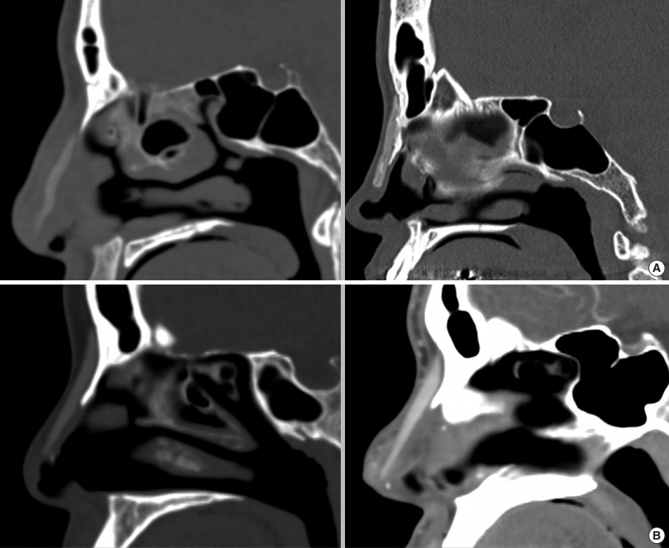
Fig. 1. Shapes of the dorsal alloplastic implants. Gore-Tex implants showed implant curling and deformation in the sagittal plane on computed tomography (A), while silicone implants usually maintained their shape inside the skin soft tissue envelope (B).
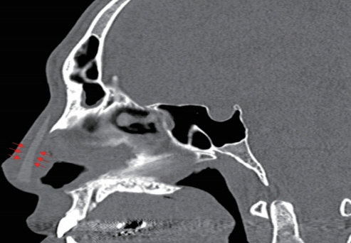
Fig. 2. The peri-implant radiolucent halo found with silicone implants (red arrows).
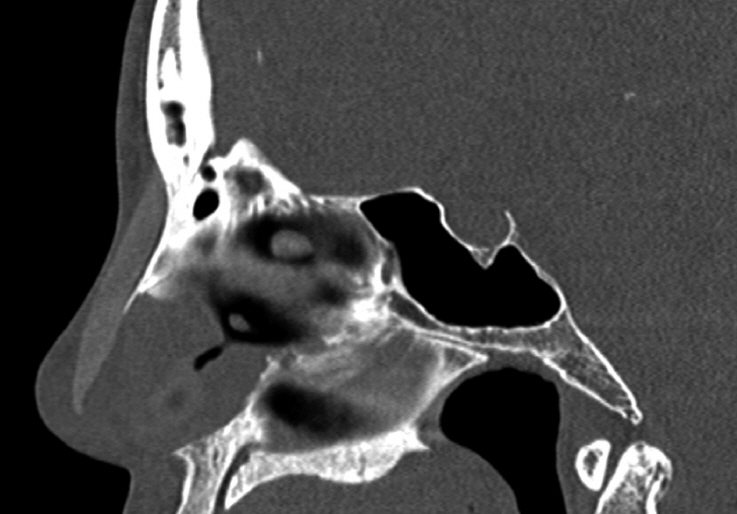
Fig. 3. The intra-implant low attenuation found in Gore-Tex implants.
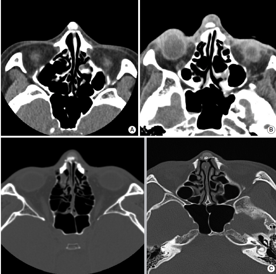
Fig. 4. Erosion of the nasal bone was observed due to (A) a silicone implant (the period of insertion was 25 years), (B) a Gore-Tex implant (the period of insertion was 14 years). (C) Hyperostosis of the nasal bone was observed due to a Gore-Tex implant; the periods of insertion were 4 years (left), and 14 years (right).
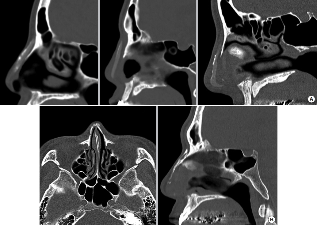
Fig. 5. Peri-implant calcifications of dorsal alloplastic implants. (A) Silicone implants, with periods of insertion of 25 years (left), 40 years (middle), and 18 years (right), (B) Gore-Tex implants (the period of insertion was 15 years).
Reference
-
1. Na HG, Jang YJ. Dorsal augmentation using alloplastic implants. Facial Plast Surg. 2017; Apr. 33(2):189–94.
Article2. Kim IS. Augmentation rhinoplasty using silicone implants. Facial Plast Surg Clin North Am. 2018; Aug. 26(3):285–93.
Article3. Byun JW, Han JY, Ki SH, Lee SH, Shin J, Choi GS. Foreign body type nasal pseudocyst after augmentation rhinoplasty: histopathologically mucin-containing pseudocyst. Am J Dermatopathol. 2017; Jul. 39(7):554–6.
Article4. Sunwoo W, Jung H, Kim DW, Jin HR. Immunohistochemical analysis of capsular contracture in silicone implant rhinoplasty. JAMA Facial Plast Surg. 2017; Sep. 19(5):436–7.
Article5. Jung YG, Kim HY, Dhong HJ, Park KN, Lee HJ, Lim YJ, et al. Ultrasonographic monitoring of implant thickness after augmentation rhinoplasty with expanded polytetrafluoroethylene. Am J Rhinol Allergy. 2009; Jan-Feb. 23(1):105–10.
Article6. Jang TY, Choi JY, Jung DH, Park HJ, Lim SC. Histologic study of Gore-Tex removed after rhinoplasty. Laryngoscope. 2009; Apr. 119(4):620–7.
Article7. Wang JH, Lee BJ, Jang YJ. Use of silicone sheets for dorsal augmentation in rhinoplasty for Asian noses. Acta Otolaryngol Suppl. 2007; Oct. (558):115–20.8. Godin MS, Waldman SR, Johnson CM Jr. Nasal augmentation using Gore-Tex: a 10-year experience. Arch Facial Plast Surg. 1999; AprJun. 1(2):118–21.9. Kim HS, Park SS, Kim MH, Kim MS, Kim SK, Lee KC. Problems associated with alloplastic materials in rhinoplasty. Yonsei Med J. 2014; Nov. 55(6):1617–23.
Article10. Jung DH, Kim BR, Choi JY, Rho YS, Park HJ, Han WW. Gross and pathologic analysis of long-term silicone implants inserted into the human body for augmentation rhinoplasty: 221 revision cases. Plast Reconstr Surg. 2007; Dec. 120(7):1997–2003.
Article11. Moon KC, Lee KI, Lee JS, Kim AR, Dhong ES, Kim DW, et al. Lateonset inflammation in Asian rhinoplasty using alloplastic implants. Aesthetic Plast Surg. 2021; Apr. 45:670–8.
Article12. Jang YJ, Moon BJ. State of the art in augmentation rhinoplasty: implant or graft. Curr Opin Otolaryngol Head Neck Surg. 2012; Aug. 20(4):280–6.13. Schatz CJ, Ginat DT. Imaging features of rhinoplasty. AJNR Am J Neuroradiol. 2014; Feb. 35(2):216–22.
Article14. Schatz CJ, Ginat DT. Imaging of cosmetic facial implants and grafts. AJNR Am J Neuroradiol. 2013; Sep. 34(9):1674–81.
Article
- Full Text Links
-
- Actions
-
Cited
- CITED
-
- Close
- Share
-
- Similar articles
-
- Radiologic Measurement of the Nasal Length and Keystone Area in Relation to Nasal Bony and Septal Cartilaginous Dimensions in Korean Adults
- A Case of Septal Abscess Extending to the Nasal Dorsum
- Problems Associated with Alloplastic Materials in Rhinoplasty
- Augmentation Rhinoplasty Using Calvarial Bone Graft
- Prevalence of complications associated with polymer-based alloplastic materials in nasal dorsal augmentation: a systematic review and meta-analysis
