Endovascular Treatment of Cavernous Sinus Dural Arteriovenous Fistula via Radial Artery and Median Cubital Vein
- Affiliations
-
- 1Department of Radiology, Hospital Sungai Buloh, Selangor, Malaysia
- 2Department of Radiology, Sarawak General Hospital, Kuching, Malaysia
- KMID: 2517748
- DOI: http://doi.org/10.5469/neuroint.2021.00157
Abstract
- Cavernous sinus dural arteriovenous fistula (CS-DAVF) is an arteriovenous shunt where there is fistulous blood flow from the dural arteries from the internal or external carotid artery into the cavernous sinus. The current mainstay of therapy is endovascular treatment. We present a case of restrictive type of CS-DAVF in a 75-year-old male who presented with right eye symptoms. He was treated with embolisation using trans-radial artery access for angiographic runs and a median cubital vein access navigating into the cavernous sinus for coil deployment. This technique completely avoids the conventional technique of a femoral approach and confines all access to the arm. Therefore, there are less risks and complications associated with an arm access, improves patients’ comfort and mobility post procedure. Transradial artery and cubital vein access allows for a safe and convenient alternative technique using the arm as compared with conventional transfemoral approach for treatment of CS-DAVF.
Keyword
Figure
-
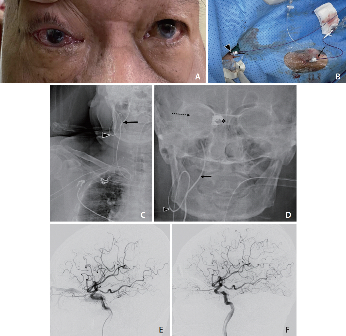
Fig. 1. (A) Pre-operative clinical examination shows presence of right eye proptosis, chemosis, and subconjunctival hemorrhage. The (B) shows upper limb arterial and venous access for transvenous embolization. Trans-radial artery access by puncture of the right radial artery (arrow) and transvenous access by puncture of the right median cubital vein (arrowhead). (C, D) 5 Fr Sims 2 catheter (Merit Medical, South Jordan, UT, USA) within the right internal carotid artery for angiographic runs (arrows). Tip of vertebral catheter at the right facial vein (arrowhead) and microcatheter further navigate into right superior ophthalmic vein and cavernous sinus (dashed arrow) with coils (asterisk) within. (E, F) Digital subtraction angiography of right internal carotid artery (ICA) in lateral view. (E) Pre embolization image shows early filling of contrast media into the cavernous sinus and superior ophthalmic vein from meningohypophyseal branches of right ICA. (F) Post embolization, coils seen within the cavernous sinus and no abnormal flow into the cavernous sinus or superior ophthalmic vein.
Cited by 1 articles
-
Transradial Approach for Neurovascular Interventions : A Literature Review
Hoon Kim, Young Woo Kim, Hyeong Jin Lee, Seon Woong Choi, Sunghan Kim, Jae Sang Oh, Sang-Hyuk Im, Jai Ho Choi, Seong-Rim Kim
J Korean Neurosurg Soc. 2025;68(2):113-126. doi: 10.3340/jkns.2024.0152.
Reference
-
1. Hiramatsu M, Sugiu K, Hishikawa T, Nishihiro S, Kidani N, Takahashi Y, et al. Results of 1940 embolizations for dural arteriovenous fistulas: Japanese Registry of Neuroendovascular Therapy (JR-NET3). J Neurosurg. 2020; 133:166–173.
Article2. Gupta AK, Purkayastha S, Krishnamoorthy T, Bodhey NK, Kapilamoorthy TR, Kesavadas C, et al. Endovascular treatment of direct carotid cavernous fistulae: a pictorial review. Neuroradiology. 2006; 48:831–839.
Article3. Korkmazer B, Kocak B, Tureci E, Islak C, Kocer N, Kizilkilic O. Endovascular treatment of carotid cavernous sinus fistula: a systematic review. World J Radiol. 2013; 5:143–155.
Article4. Suh DC, Lee JH, Kim SJ, Chung SJ, Choi CG, Kim HJ, et al. New concept in cavernous sinus dural arteriovenous fistula: correlation with presenting symptom and venous drainage patterns. Stroke. 2005; 36:1134–1139.5. Ellis JA, Goldstein H, Connolly ES Jr, Meyers PM. Carotid-cavernous fistulas. Neurosurg Focus. 2012; 32:E9.
Article6. Leone G, Renieri L, Enriquez-Marulanda A, Dmytriw AA, Nappini S, Laiso A, et al. Carotid cavernous fistulas and dural arteriovenous fistulas of the cavernous sinus: validation of a new classification according to venous drainage. World Neurosurg. 2019; 128:e621–e631.
Article7. Joshi KC, Beer-Furlan A, Crowley RW, Chen M, Munich SA. Transradial approach for neurointerventions: a systematic review of the literature. J Neurointerv Surg. 2020; 12:886–892.
Article8. Snelling BM, Sur S, Shah SS, Khandelwal P, Caplan J, Haniff R, et al. Transradial cerebral angiography: techniques and outcomes. J Neurointerv Surg. 2018; 10:874–881.
Article9. Zussman BM, Tonetti DA, Stone J, Brown M, Desai SM, Gross BA, et al. Maturing institutional experience with the transradial approach for diagnostic cerebral arteriography: overcoming the learning curve. J Neurointerv Surg. 2019; 11:1235–1238.
Article10. Khanna O, Sweid A, Mouchtouris N, Shivashankar K, Xu V, Velagapudi L, et al. Radial artery catheterization for neuroendovascular procedures. Stroke. 2019; 50:2587–2590.
Article11. Galvan Fernandez J, Martínez-Galdámez M, Schüller Arteaga M, Ortega-Quintanilla J, Hermosín A, Crespo-Vallejo E, et al. Arm-only access for combined transarterial and transvenous neurointerventional procedures. J Neurointerv Surg. 2021; 13:39–41.
Article12. Weinberg JH, Sweid A, Hassan A, Tekle W, Sajja K, Thaete L, et al. Early experience with a novel 088 long sheath in transradial neurointerventions. Clin Neurol Neurosurg. 2021; 202:106510.
Article13. Starke RM, Snelling B, Al-Mufti F, Gandhi CD, Lee SK, Dabus G, Society of NeuroInterventional Surgery, et al. Transarterial and transvenous access for neurointerventional surgery: report of the SNIS Standards and Guidelines Committee. J Neurointerv Surg. 2020; 12:733–741.
Article14. Ramos AD, Sundararajan S, Santillan A, Schwarz JT, Patsalides A. Single arm access venous sinus stenting (SAVeS) technique: technical note. Interv Neuroradiol. 2020; 26:501–505.
Article15. Abecassis IJ, Saini V, Phillips TJ, Osbun JW, Martínez-Galdámez M, Nada A, et al. Upper extremity transvenous access for neuroendovascular procedures: an international multicenter case series. J Neurointerv Surg. 2021; 13:357–362.
Article
- Full Text Links
-
- Actions
-
Cited
- CITED
-
- Close
- Share
-
- Similar articles
-
- Transvenous Embolization of Cavernous Sinus Dural Arteriovenous Fistula Using the Direct Superior Ophthalmic Vein Approach: A Case Report
- Endovascular management of cavernous sinus dural arteriovenous fistulas: Overall review and considerations
- Middle temporal vein access for transvenous embolization of Cavernous sinus dural arteriovenous fistula: A case report and review of literature
- Facilitated Retrograde Access via the Facial Vein for Transvenous Embolization of the Cavernous Sinus Dural Arteriovenous Fistula with Isolated Ophthalmic Venous Drainage
- Superior ophthalmic approach in carotid-cavernous fistula: current concepts in indications, surgical techniques, and case reviews
