Effect of the Number of Projected Images on the Noise Characteristics in Tomosynthesis Imaging
- Affiliations
-
- 1Department of Radiological Technology, Graduate School of Health Sciences, Okayama University, Okayama, Japan
- KMID: 2516964
- DOI: http://doi.org/10.14316/pmp.2021.32.2.50
Abstract
- Purpose
In this study, we investigated the relationship between the noise characteristics and the number of projected images in tomosynthesis using a digital phantom.
Methods
The digital phantom consisted of a columnar phantom in the center of the image and a spherical phantom with a diameter of 80 pixels. A virtual scan was performed, and 128 projected images (Tomo_w/o) of the phantoms were obtained. The image noise according to the Poisson distribution was added to the projected images (Tomo_×1). Furthermore, another projected image with additional noise was prepared (Tomo_×1/2). For each dataset, we created datasets with 64 (half) and 32 (quarter) projections by removing the even-numbered images twice from the 128 (fully) projected images. Tomosynthesis images were reconstructed by filtered back projection (FBP). The modulation transfer function (MTF) was estimated using the sphere method, and the noise power spectrum (NPS) was estimated using the two-dimensional Fourier transform method.
Results
The MTFs did not change between datasets, and the NPSs improved as the number of projected images increased. The noise characteristics of the Tomo_×1_half images were the same as those of the Tomo_×1/2_full.
Conclusions
To achieve a reduction in the patient dose in tomosynthesis acquisition, we recommend reducing the number of projected images rather than reducing the dose per projection.
Keyword
Figure
-
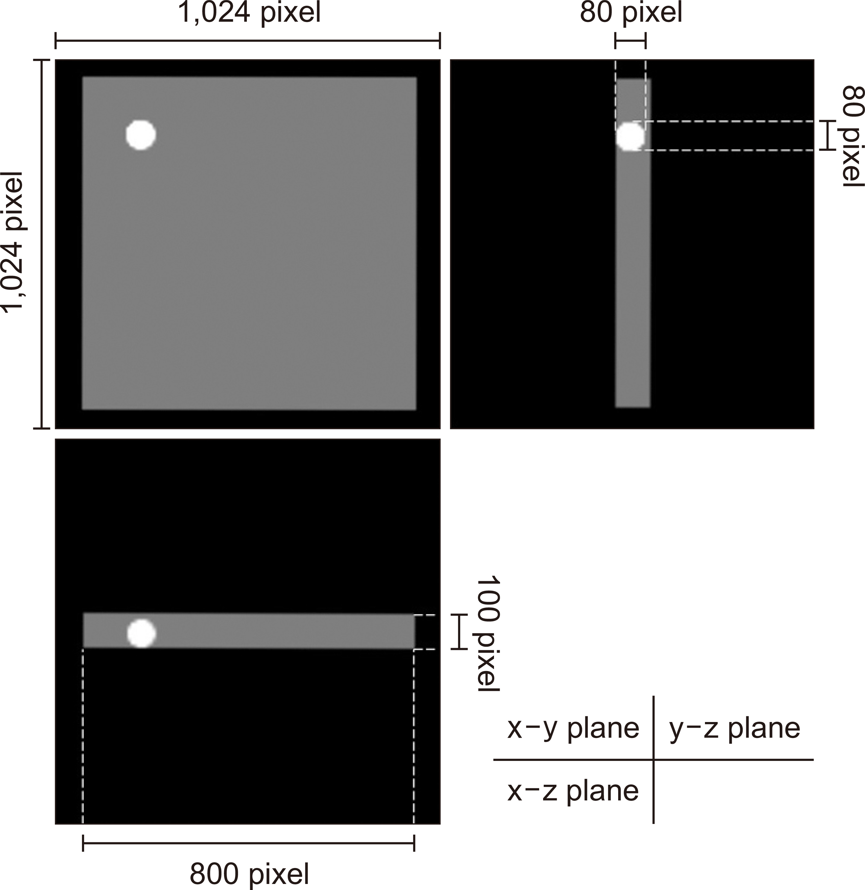
Fig. 1 Schematic of the digital phantom. The pixel values of the columnar and spherical phantoms are 6,000 and 30,000, respectively.
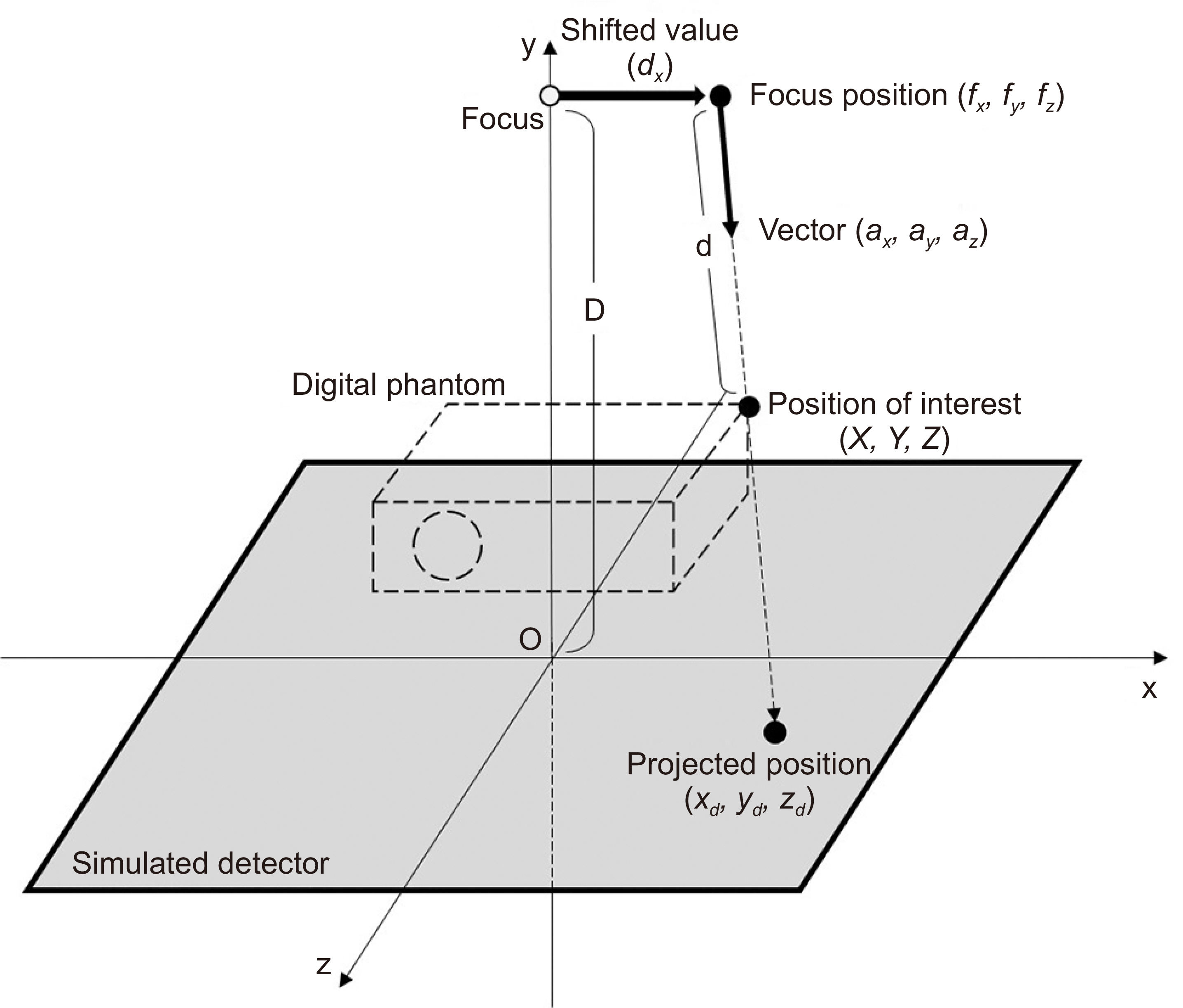
Fig. 2 Schematic of the alignment used for acquiring the projected and tomosynthesis images.
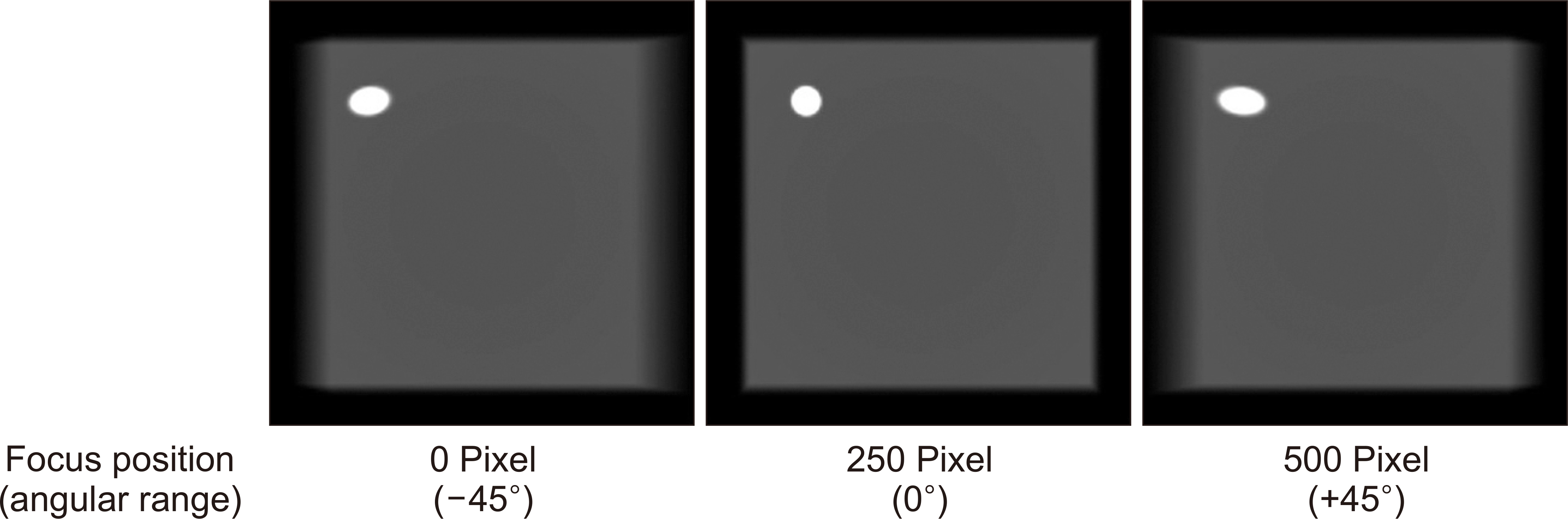
Fig. 3 Angular projected images acquired when the simulated X-ray tube was positioned at ±45° and the center.
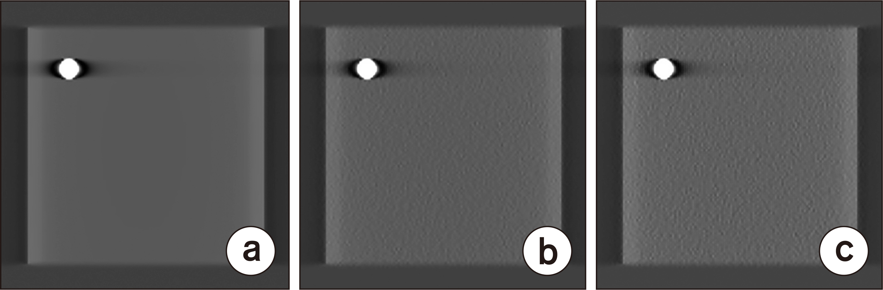
Fig. 4 Tomosynthesis images (a) without image noise (Tomo_w/o), (b) with one-time image noise (Tomo_×1), and (c) with two-time image noise (Tomo_×1/2).
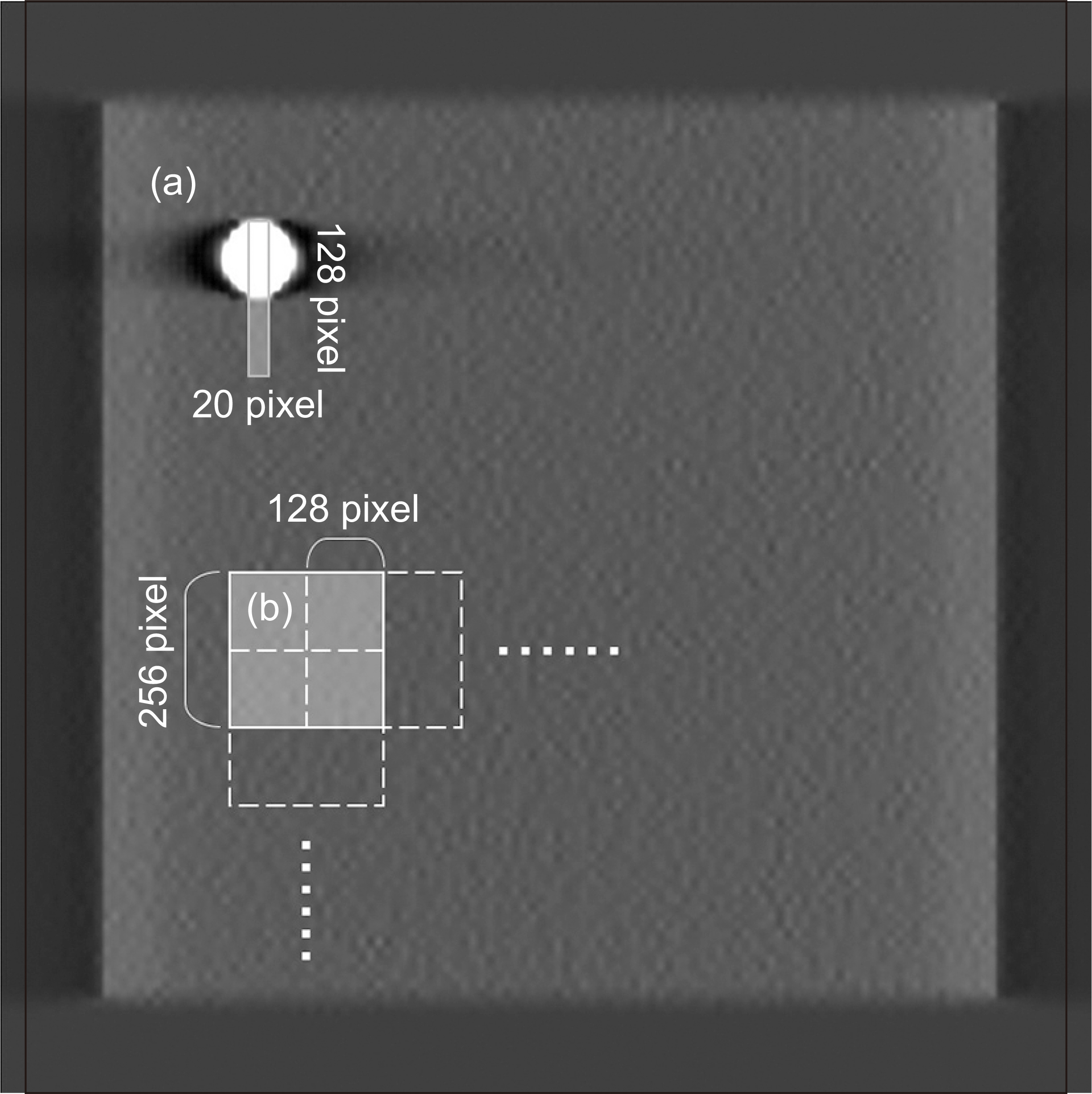
Fig. 5 Region of interest (ROI) positions for (a) modulation transfer function (MTF) and (b) noise power spectrum (NPS).
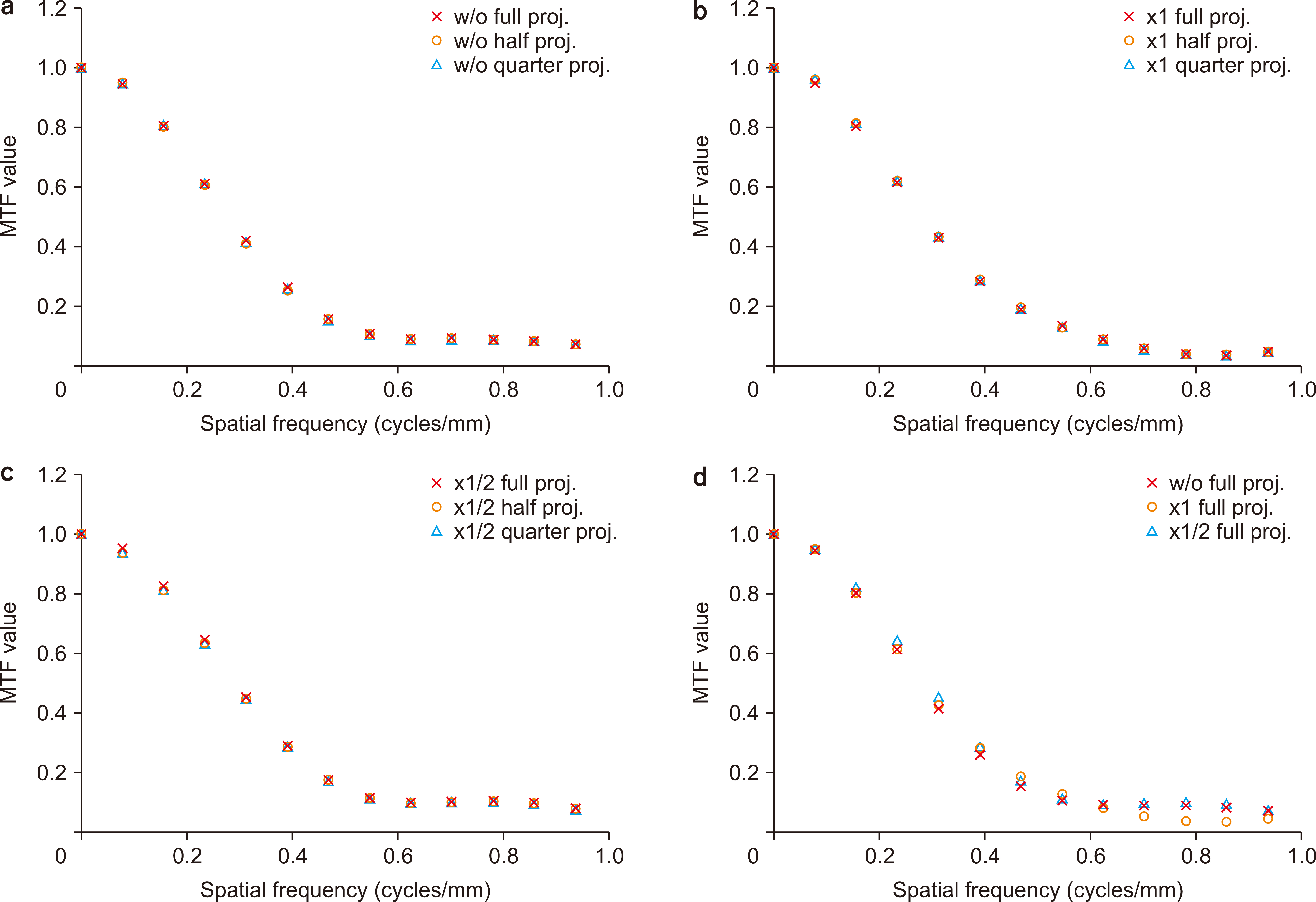
Fig. 6 Results of the modulation transfer functions (MTFs) for (a) Tomo_w/o, (b) Tomo_×1, (c) Tomo_×1/2, and (d) different amounts of image noise. proj., projection.
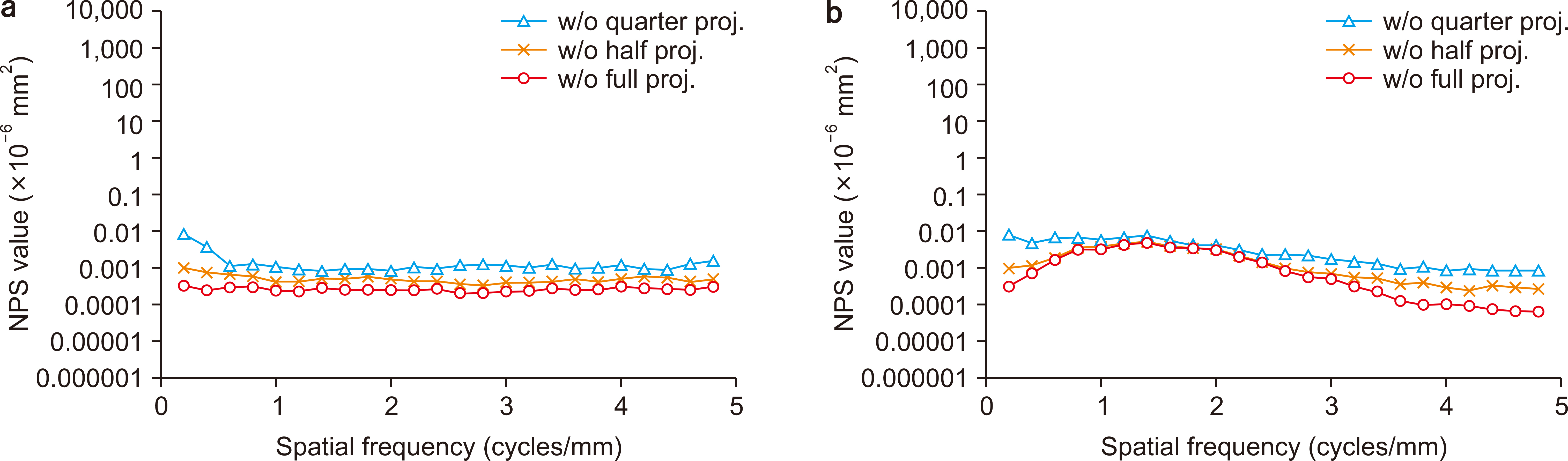
Fig. 7 Noise power spectrum (NPS) of the Tomo_w/o datasets in the (a) vertical and (b) horizontal directions. proj., projection.

Fig. 8 Noise power spectrum (NPS) of the Tomo_×1 and _×1/2 datasets in the (a) vertical and (b) horizontal directions. proj., projection.

Fig. 9 Noise power spectrum (NPS) of the full and half projection of the Tomo_×1 and _×1/2 datasets in the (a) vertical and (b) horizontal directions. proj., projection.
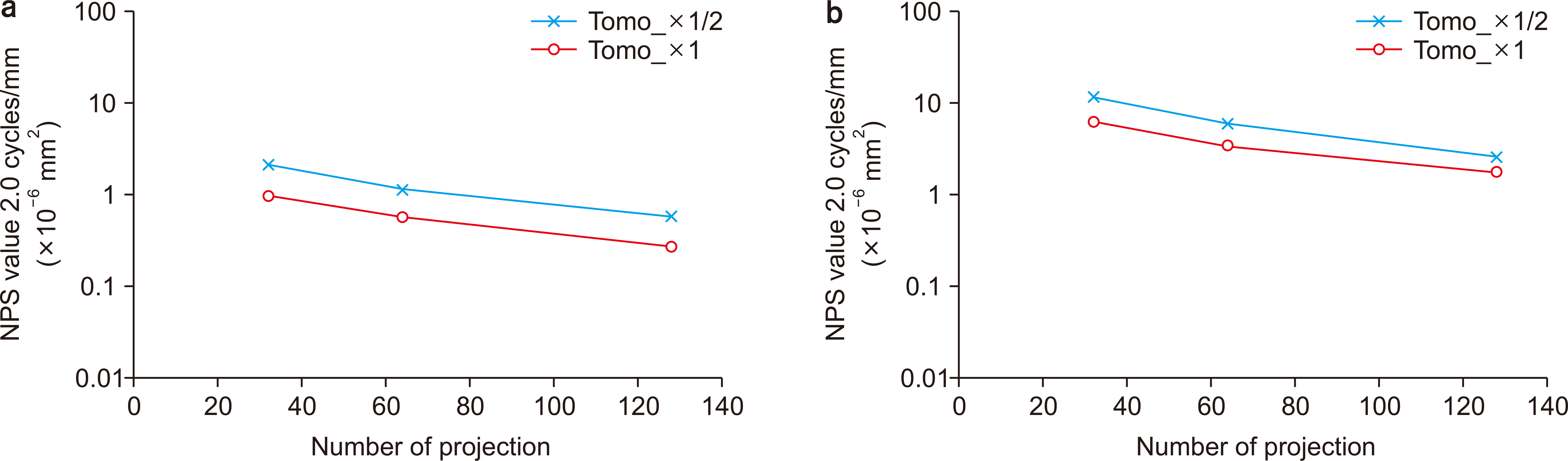
Fig. 10 Relationship between the number of projected images and the noise power spectrum (NPS) value at 2.0 cycles/mm.
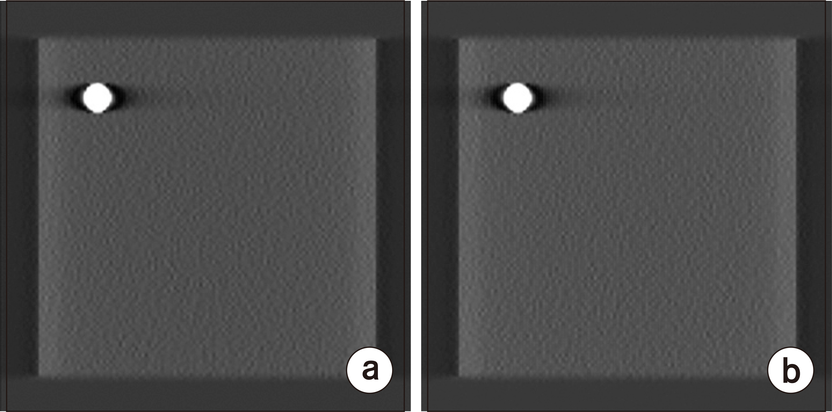
Fig. 11 Samples of (a) the Tomo_×1 half projection and (b) the Tomo_×1/2 full projection.
Cited by 1 articles
-
Estimation of Noise Level and Edge Preservation for Computed Tomography Images: Comparisons in Iterative Reconstruction
Sihwan Kim, Chulkyun Ahn, Woo Kyoung Jeong, Jong Hyo Kim, Minsoo Chun
Prog Med Phys. 2021;32(4):92-98. doi: 10.14316/pmp.2021.32.4.92.
Reference
-
References
1. Grant DG. 1972; Tomosynthesis: a three-dimensional radiographic imaging technique. IEEE Trans Biomed Eng. 19:20–28. DOI: 10.1109/tbme.1972.324154. PMID: 5008409.
Article2. Kim JH, Lee KH, Kim KT, Kim HJ, Ahn HS, Kim YJ, et al. 2016; Comparison of digital tomosynthesis and chest radiography for the detection of pulmonary nodules: systematic review and meta-analysis. Br J Radiol. 89:20160421. DOI: 10.1259/bjr.20160421. PMID: 27759428. PMCID: PMC5604914.
Article3. Ottenin MA, Jacquot A, Grospretre O, Noël A, Lecocq S, Louis M, et al. 2012; Evaluation of the diagnostic performance of tomosynthesis in fractures of the wrist. AJR Am J Roentgenol. 198:180–186. DOI: 10.2214/AJR.11.6374. PMID: 22194495.
Article4. Chong A, Weinstein SP, McDonald ES, Conant EF. 2019; Digital breast tomosynthesis: concepts and clinical practice. Radiology. 292:1–14. DOI: 10.1148/radiol.2019180760. PMID: 31084476. PMCID: PMC6604796.
Article5. Kuwabara N, Takuwa H, Takeuchi M, Kawashima H. 2020; Can digital breast tomosynthesis improve identification of malignant calcifications? Radiol Phys Technol. 13:249–255. DOI: 10.1007/s12194-020-00576-1. PMID: 32681400.
Article6. Marshall NW, Bosmans H. 2012; Measurements of system sharpness for two digital breast tomosynthesis systems. Phys Med Biol. 57:7629–7650. DOI: 10.1088/0031-9155/57/22/7629. PMID: 23123601.
Article7. Olgar T, Kahn T, Gosch D. 2013; Quantitative image quality measurements of a digital breast tomosynthesis system. Rofo. 185:1188–1194. DOI: 10.1055/s-0033-1350106. PMID: 23888475.8. Fukui R, Ishii R, Kishimoto J, Yamato S, Takahata A, Kohama C. 2014; Evaluation of the effect of geometry for measuring section thickness in tomosynthesis. Radiol Phys Technol. 7:141–147. DOI: 10.1007/s12194-013-0243-0. PMID: 24254729.
Article9. Zheng J, Fessler JA, Chan HP. 2019; Effect of source blur on digital breast tomosynthesis reconstruction. Med Phys. 46:5572–5592. DOI: 10.1002/mp.13801. PMID: 31494953. PMCID: PMC6899200.
Article10. van Engen RE, Bosmans H, Bouwman RW, Dance DR, Heid P, Lazzari B, et al. 2018; Protocol for the quality control of the physical and technical aspects of digital breast tomosynthesis systems (version 1.03). EUREF. 30–44.11. Harrison RM, Kotre CJ. 1986; Noise and threshold contrast characteristics of a digital fluorographic system. Phys Med Biol. 31:515–526. DOI: 10.1088/0031-9155/31/5/004. PMID: 3737686.
Article12. Cesarelli M, Bifulco P, Cerciello T, Romano M, Paura L. 2013; X-ray fluoroscopy noise modeling for filter design. Int J Comput Assist Radiol Surg. 8:269–278. DOI: 10.1007/s11548-012-0772-8. PMID: 22718402.
Article13. Samei E, Murphy S, Richard S. 2013; Assessment of multi-directional MTF for breast tomosynthesis. Phys Med Biol. 58:1649–1661. DOI: 10.1088/0031-9155/58/5/1649. PMID: 23422248.
Article14. Dobbins JT 3rd, Samei E, Ranger NT, Chen Y. 2006; Intercomparison of methods for image quality characterization. II. Noise power spectrum. Med Phys. 33:1466–1475. DOI: 10.1118/1.2188819. PMID: 16752581.15. Sechopoulos I, Bliznakova K, Fei B. 2013; Power spectrum analysis of the x-ray scatter signal in mammography and breast tomosynthesis projections. Med Phys. 40:101905. DOI: 10.1118/1.4820442. PMID: 24089907. PMCID: PMC3785536.
Article16. Zhao B, Zhao W. 2008; Three-dimensional linear system analysis for breast tomosynthesis. Med Phys. 35:5219–5232. DOI: 10.1118/1.2996014. PMID: 19175081. PMCID: PMC2673606.
Article17. Fukui R, Shiraishi J. 2019; Application of a pixel-shifted linear interpolation technique for reducing the projection number in tomosynthesis imaging. Radiol Phys Technol. 12:30–39. DOI: 10.1007/s12194-018-0488-8. PMID: 30456708.
Article18. Chawla AS, Lo JY, Baker JA, Samei E. 2009; Optimized image acquisition for breast tomosynthesis in projection and reconstruction space. Med Phys. 36:4859–4869. DOI: 10.1118/1.3231814. PMID: 19994493. PMCID: PMC2773452.
Article
- Full Text Links
-
- Actions
-
Cited
- CITED
-
- Close
- Share
-
- Similar articles
-
- Effect of Bead Device Diameter on Z-Resolution Measurement in Tomosynthesis Images: A Simulation Study
- A Study of Various Filter Setups with FBP Reconstruction for Digital Breast Tomosynthesis
- A Preliminary Results of Acoustic Noise Effect due to Gradient Pulsing in Functional MRI
- Digital Tomosynthesis for Patient Alignment System Using Half-fan Mode CBCT Projection Images
- Removal of Edge Artifact due to Partial Volume Effect in the Adaptive Template Filtering
