Comparative study of the digastric and the stylohyoid muscles between wild boars (Sus scrofa scrofa) and domestic swine (Sus scrofa domesticus): revisiting the gross anatomy
- Magalhães HIR
 1
1 - Barcelos JB2
- Romão FB3
- Borges TRJ2
- Carvalho-Barros RAd4
- Miglino MA1
- e Silva FOC2
- Ribeiro LdA2
- Affiliations
-
- 1Department of Surgery, School of Veterinary Medicine and Animal Sciences, University of São Paulo, São Paulo, Brazil
- 2Animal Anatomy Laboratory, School of Veterinary Medicine and Animal Sciences, Federal University of Uberlândia, Uberlândia, Brazil
- 3Animal Anatomy Laboratory, School of Veterinary Medicine, University Center of Patos de Minas, Patos de Minas, Brazil
- 4Anatomy Laboratory, School of Biological Sciences, Federal University of Catalão, Catalão, Brazil
- KMID: 2516904
- DOI: http://doi.org/10.5115/acb.20.301
Abstract
- Considering Suidae Familie as a perfect and viable experimental biomedical model for research applied to human medicine, it has been sought to describe the comparative anatomy of the digastric and the stylohyoid muscles between boars and domestic swine. Heads of Sus scrofa scrofa and Sus scrofa domesticus were dissected. The digastric muscle presented only one muscle belly as anatomical component of a tendinous origin in the jugular process of the occipital bone, and muscle insertion in the midventral edge of the caudal two thirds of the body of the mandible. Thus, its function is fundamentally associated with the lowering and the retracting of the mandible which, by the way, can deliver greater muscle power at lesser energy expense. For the stylohyoid muscle, the tendinous origin was in the laterocaudal edge of the dorsal third of the stylohyoid bone. The muscle insertion - primarily, was in the lateral and caudal edges from the mid third portion up to the ventral extremity of the thyrohyoid bone, and secondarily as a laterolateral aponeurotic blade which would unite, in a bilateral manner, an insertion that was common to the sternohyoid, the geniohyoid, and the mylohyoid muscles in a median ventral region. This morphology were similar to the two specimens studied expanding the information available, which were completely unknown for the suidae until the moment.
Keyword
Figure
-
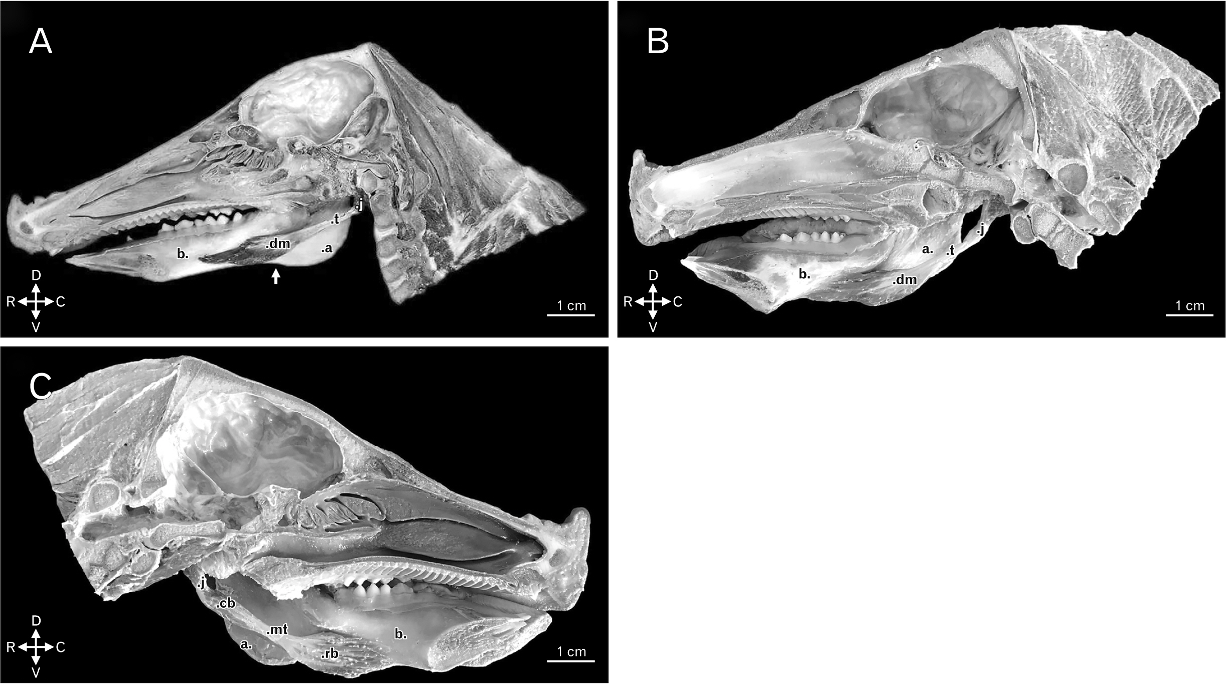
Fig. 1 (A) Medial view of right hemihead of boar, dm and the t of the digastric muscle. Other structures: j, a, b, and angular incisure of the mandible (white arrow). Animal direction: D, V, R, and C. (B) Medial view of right hemihead of domestic swine dm and the t of the digastric muscle. Other structures: j, a, and b. Animal direction: D, V, R, and C. (C) Medial view of left hemihead of domestic swine highlighting the rb and cb bellies, and the mt of the digastric muscle. Other structures: j, a, and b. Animal direction: D, V, R, and C. a, angle of the mandible; b, body of the mandible; C, caudal; cb, caudal belly; D, dorsal; dm, highlighting the single muscle belly; j, jugular process of the occipital bone; mt, middle tendon; R, rostral; rb, rostral belly; t, tendon of origin; V, ventral.
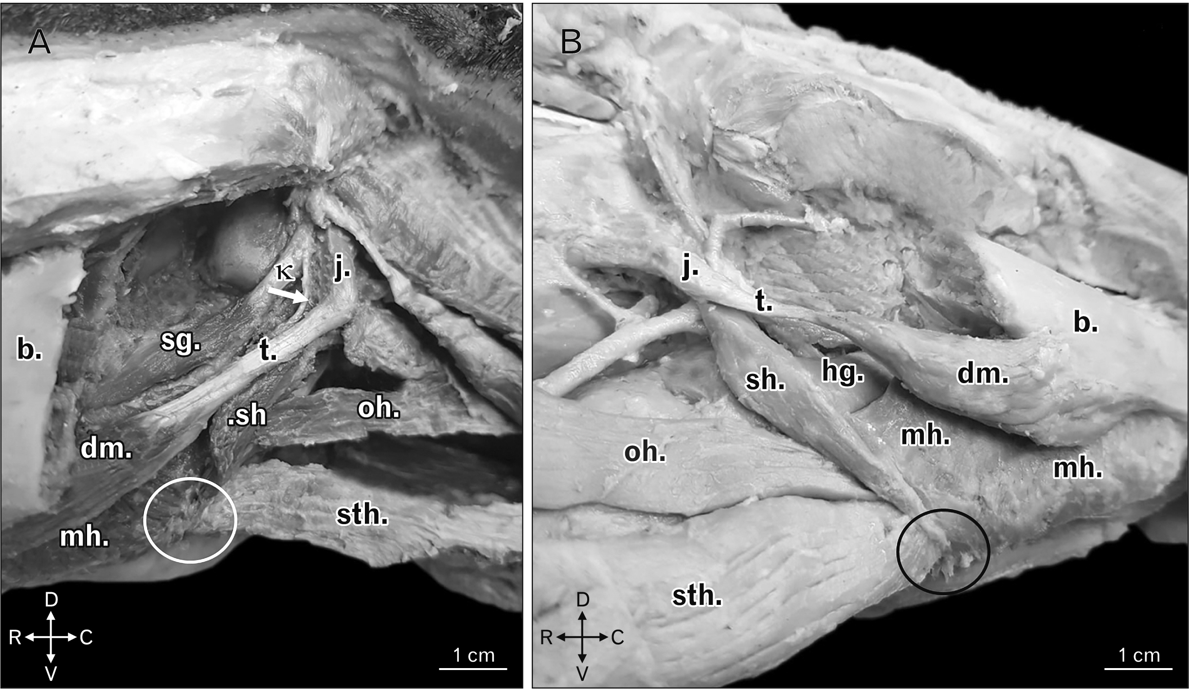
Fig. 2 (A) Left ventrolateral view of boar head highlighting the common insertion, and in the form of an aponeurotic blade (white circle), of sh, sth, and mh. There is also demonstration of the tendon of origin of the stylohyoid muscle (white arrow) as from the κ. Other structures: b, t, dm, sg, oh, and j. Animal direction: D, V, R, and C. (B) Right ventrolateral view of a domestic swine head highlighting the common insertion, and in the form of an aponeurotic blade (black circle), of sh, sth, and mh. Other structures: b, t, dm, hg, oh, and j. Animal direction: D, V, R, and C. b, body of the mandible; C, caudal; D, dorsal; dm, single belly of the digastric muscle; hg, hyoglossus muscle; j, jugular process of the occipital bone; mh, mylohyoid muscles; oh, omohyoid muscle; R, rostral; sg, styloglossus muscle; sh, stylohyoid; sth, sternohyoid; t, tendon of origin of the digastric muscle; V, ventral; κ, laterocaudal edge of the dorsal third of the stylohyoid bone.
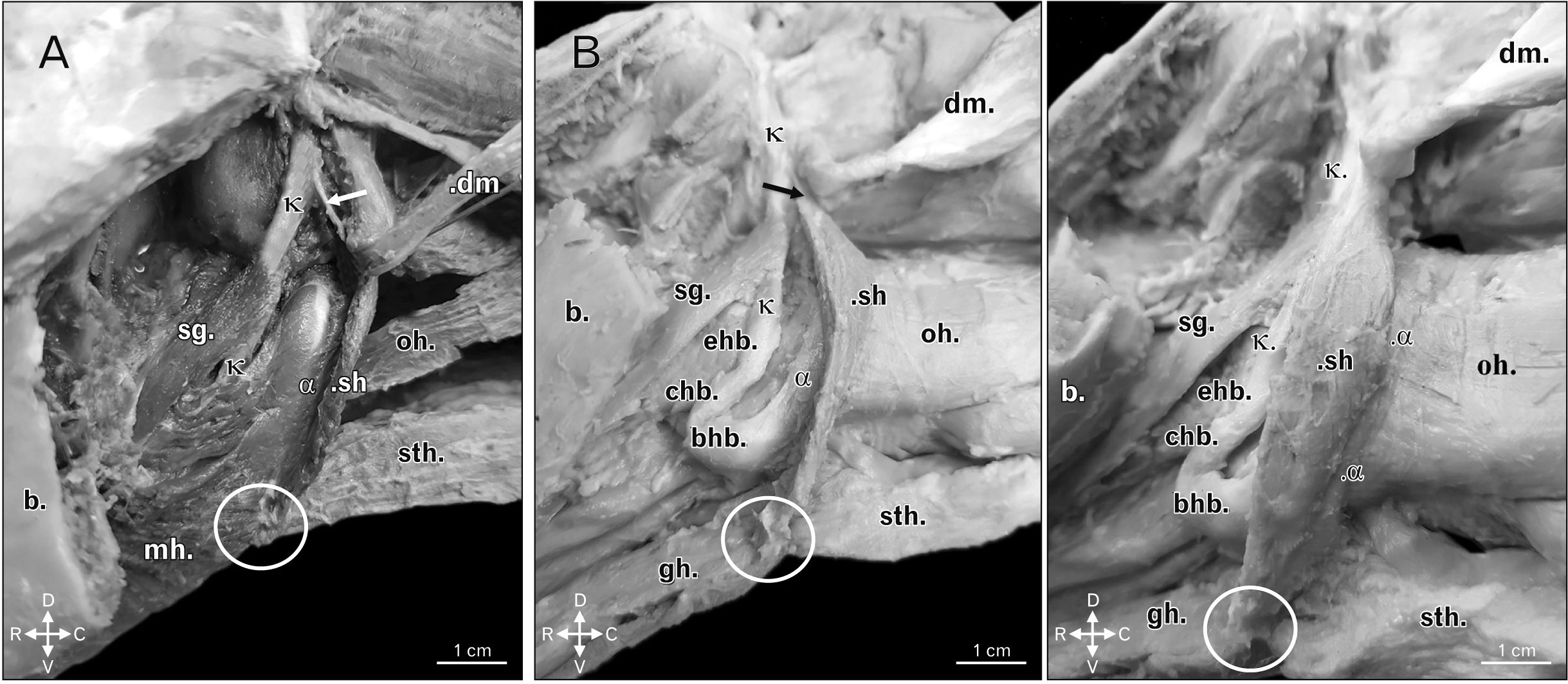
Fig. 3 (A) Left ventrolateral view of boar head highlighting the insertion of the sh in the lateral and caudal edges from the mid third as far as the ventral extremity of the α. There is also demonstration of the tendon of origin of the stylohyoid muscle (white arrow) as from the κ. Other structures: common insertion, and in the form of an aponeurotic blade (white circle), of sh, sth, and the mh, b, dm, oh, and sg. Animal direction: D, V, R, and C. (B) Left lateroventral view of a domestic swine head highlighting the insertion of the sh in the lateral and caudal edges from the mid third as far as the ventral extremity of the α. There is also demonstration of the tendon of origin of the stylohyoid muscle (black arrow) as from the κ. Other structures: common insertion, and in the form of an aponeurotic blade (white circle), of sh, sth, and gh, b, dm, oh, sg, bhb, chb, and ehb. Animal direction: D, V, R, and C. b, body of the mandible; bhb, basihyoid bone; C, caudal; chb, ceratohyoid bone; D, dorsal; dm, digastric muscle open with caudodorsal orientation; ehb, epihyoid bone; hg, hyoglossus muscle; j, jugular process of the occipital bone; mh, mylohyoid muscles; oh, omohyoid muscle; R, rostral; sg, styloglossus muscle; sh, stylohyoid muscle; sth, sternohyoid; t, tendon of origin of the digastric muscle; V, ventral; α, thyrohyoid bone; κ, laterocaudal edge of the dorsal third of the stylohyoid bone.

Fig. 4 (A) Ventral view of a domestic swine head highlighting the common insertion with short and aponeurotic morphology, stretching with laterolateral orientation to be a point of common insertion to muscles positioned in a counterlateral manner, bilaterally uniting the same in a medioventral fashion (white arrow). Other structures: sh’, sh’’, mh, and gh. Animal direction: LL, RL, R, and C. (B) Ventral view of the hyoid bone of a domestic swine highlighting the common insertion with short and aponeurotic morphology of the stylohyoid, the sternohyoid, the geniohyoid, and the mylohyoid muscles (white arrow). Other structures: bhb, chb, and α. Animal direction: LL, RL, R, and C. (C) Ventral view of the hyoid bone of a domestic swine upon removal of the aponeurotic blade of insertion of the stylohyoid, the sternohyoid, the geniohyoid, and the mylohyoid muscles, highlighting the point of attachment of the same (*). Other structures: bhb, chb, and α. Animal direction: LL, RL, R, and C. bhb, basihyoid bone; C, caudal; chb, ceratohyoid bone; gh, geniohyoid muscle; LL, left lateral; mh, mylohyoid muscle open with laterodorsal orientation; R, rostral; RL, right lateral; sh’, left stylohyoid muscle; sh’’, ight stylohyoid muscle; α, thyrohyoid bone.
Reference
-
References
1. Getty R. 1975. Sisson/Grossman: the anatomy of the domestic animals. Saunders;Philadelphia:2. Oliveira C, Teixeira RAP, Conchalo WL. 2004; A contextualized approach to human and comparative anatomy. Núcleos de Ensino. 291–310. Portuguese.3. Matsunaga K, Usui A, Yamaguchi K, Akita K. 2009; An anatomical study of the muscles that attach to the articular disc of the temporomandibular joint. Clin Anat. 22:932–40. DOI: 10.1002/ca.20865. PMID: 19806671.
Article4. Silva ACJ, Ferreira LR, Silva MHM, Cavalcante MG, Monteiro MM, Nascimento HB, Pereira APC, Maschka FG. 2019. Músculos da mastigação: aspectos anatômicos e importância médicocirúrgica (em vídeo) [Internet]. Universidade Federal Rural de Pernambuco;Recife: Available from: http://www.eventosufrpe.com.br/jepex2009/cd/resumos/r1125-1.pdf. cited 2019 Sep 12.5. Marchesan IQ. 1993. Motricidade oral: visão clínica do trabalho fonoaudiológico integrado com outras especialidades. Pancast;São Paulo:6. Sicher H, Du Brul EL. 1977. Anatomia bucal. 6th ed. Guanabara Koogan;Rio de Janeiro:7. Kyllar M, Stembírek J, Putnová I, Stehlík L, Odehnalová S, Buchtová M. 2014; Radiography, computed tomography and magnetic resonance imaging of craniofacial structures in pig. Anat Histol Embryol. 43:435–52. DOI: 10.1111/ahe.12095. PMID: 24261592.
Article8. Papadaki ME, Troulis MJ, Glowacki J, Kaban LB. 2010; A minipig model of maxillary distraction osteogenesis. J Oral Maxillofac Surg. 68:2783–91. DOI: 10.1016/j.joms.2010.06.179. PMID: 20971370.
Article9. Štembírek J, Kyllar M, Putnová I, Stehlík L, Buchtová M. 2012; The pig as an experimental model for clinical craniofacial research. Lab Anim. 46:269–79. DOI: 10.1258/la.2012.012062. PMID: 22969144.
Article10. International Committee on Veterinary Gross Anatomical Nomenclature. 2017. Nomina anatomica veterinaria. 6th ed. World Association of Veterinary Anatomists;Hanover:11. Godinho HP, Cardoso FM, Nascimento JF. 1981. Anatomia dos ruminantes domésticos. UFMG;Belo Horizonte:12. Madeira MC. 2012. Anatomia da face: bases anatomofuncionais para a prática odontológica. 8th ed. Sarvier;Sao Paulo:13. Khan YS, Bordoni B. 2020. Anatomy, head and neck, suprahyoid muscle [Internet]. StatPearls Publishing;Treasure Island, FL: Available from: https://www.ncbi.nlm.nih.gov/books/NBK546710/. cited 2019 Dec 16.14. Budras KD, McCarthy PH, Fricke W, Richter R, Horowitz A, Berg R. 2007. Anatomy of the dog: an illustrated text. 5th ed. Schlütersche;Hannover:15. König HE, Liebich HG. 2020. Veterinary anatomy of domestic animals: textbook and colour atlas. 7th ed. Thieme Medical Publishers;New York: DOI: 10.1055/b-007-167437.16. Pereira KF, Souza DR, Ferreira LS, Ribeiro AR. 2012; Aspectos morfológicos dos músculos da cabeça e pescoço do mão pelada (Procyon cancrivorus). SaBios:. Rev Saúde Biol. 7:1–8.17. Turnbull WD. 1970. Mammalian masticatory apparatus. Field Museum of Natural History;Chicago:18. Scapino RP. 1976; Function of the digastric muscle in carnivores. J Morphol. 150:843–59. DOI: 10.1002/jmor.1051500405. PMID: 30261705.
Article19. Endo H, Sakata S, Arai T, Miyazaki N. 2002; The muscles of mastication in the caspian seal (Phoca caspica). Anat Histol Embryol. 31:262–5. DOI: 10.1046/j.1439-0264.2002.00372.x. PMID: 12484416.
Article20. Tomo S, Tomo I, Townsend GC. 1998; Digastric muscle of the kangaroo: a comparative anatomical study. Anat Rec. 251:346–50. DOI: 10.1002/(SICI)1097-0185(199807)251:3<346::AID-AR10>3.0.CO;2-O. PMID: 9669762.
Article21. Spataru C, Spataru M, Vulpe V, Lazar M. 2013; The peculiarities of the masticator muscles in rodents. Arq Bras Med Vet Zootec. 65:749–56. DOI: 10.1590/S0102-09352013000300021.
Article22. Ferreira JR, Pinto Júnior N, Kajita D, Cirqueira DS, Nogueira DJ. 2005; Study of muscle digastric descriptive and topographical anatomy in primates (Cebus apella, Linnaeus, 1766). Braz J Vet Res Anim Sci. 42:113–21. Portuguese. DOI: 10.11606/issn.1678-4456.bjvras.2005.26441.23. Muhl ZF, Newton JH. 1982; Change of digastric muscle length in feeding rabbits. J Morphol. 171:151–7. DOI: 10.1002/jmor.1051710204. PMID: 7062342.
Article24. Standring S. 2015. Gray's anatomy: the anatomical basis of clinical practice. 41st ed. Churchill Livingstone;Edinburgh:25. Budras KD, Sack WO, Rock S. 2009. Anatomy of the horse. Schlütersche;Hannover:26. Singh B. 2018. Dyce, sack, and Wensing's textbook of veterinary anatomy. 5th ed. Elsevier;St. Louis:27. Evans HE, DeLahunta A, Miller ME. 2017. Guide to the dissection of the dog. 8th ed. Elsevier;St. Louis:28. Moore KL, Dalley AF, Agur AMR. 2018. Clinically oriented anatomy. 8th ed. Lippincott Williams and Wilkins;Philadelphia:29. O'Daniel TG. 2018; Understanding deep neck anatomy and its clinical relevance. Clin Plast Surg. 45:447–54. DOI: 10.1016/j.cps.2018.06.011. PMID: 30268237.30. Al-Missri MZ, Khalili YA. 2020. Anatomy, head and neck, submental triangle [Internet]. StatPearls Publishing;Treasure Island, FL: Available from: https://www.ncbi.nlm.nih.gov/books/NBK545296/. cited 2019 Dec 16.31. Kim SD, Loukas M. 2019; Anatomy and variations of digastric muscle. Anat Cell Biol. 52:1–11. DOI: 10.5115/acb.2019.52.1.1. PMID: 30984445. PMCID: PMC6449592.
Article32. Zdilla MJ, Soloninka HJ, Lambert HW. 2014; Unilateral duplication of the anterior digastric muscle belly: a case report with implications for surgeries of the submental region. J Surg Case Rep. 2014:rju131. DOI: 10.1093/jscr/rju131. PMID: 25480836. PMCID: PMC4256528.
Article33. Harvey JA, Call Z, Peterson K, Wisco JJ. 2015; Weave pattern of accessory heads to the anterior digastric muscle. Surg Radiol Anat. 37:1001–4. DOI: 10.1007/s00276-014-1401-8. PMID: 25501489.
Article34. Šink Ž, Umek N, Cvetko E. 2019; Cross-over type of supernumerary digastric muscle. Folia Morphol (Warsz). 78:647–50. DOI: 10.5603/FM.a2018.0112. PMID: 30536357.
Article35. Mascaro MB, Picoli LC, Santos FM, Bonsi AB, Souza MR, Prosdócimi FC. 2011; Anatomical variation of the anterior belly of the digastric muscle: case report and clinical implications. J Morphol Sci. 28:72–5.36. Kalniev M, Krastev D, Krastev N, Vidinov K, Veltchev L, Apostolov A, Mileva M. 2013; A rare variation of the digastric muscle. Clujul Med. 86:327–9. PMID: 26527971. PMCID: PMC4462457.37. Das S, Ghafar NA, Othman F, Kamaruddin A, Suhaimi FH. 2014; Anomalous belly of digastric muscle: gross anatomy with clinical implications. Middle East J Sci Res. 21:733–5.38. De-Ary-Pires B, Ary-Pires R, Pires-Neto MA. 2003; The human digastric muscle: patterns and variations with clinical and surgical correlations. Ann Anat. 185:471–9. DOI: 10.1016/S0940-9602(03)80110-3. PMID: 14575275.
Article39. Hou S, Deng T, He W, Chen SQ. 2014; Foraging behavior of Chleuastochoerus (Suidae, Artiodactyla): a case study of skull and mandible morpho-functional analysis. Sci China Earth Sci. 57:988–98. DOI: 10.1007/s11430-013-4733-z.40. Weijs WA, Dantuma R. 1981; Functional anatomy of the masticatory apparatus in the rabbit (Oryctolagus Cuniculus L.). Neth J Zool. 31:99–147. DOI: 10.1163/002829680X00212.41. Rowlerson A, Mascarello F, Veggetti A, Carpene E. 1983; The fibre-type composition of the first branchial arch muscles in Carnivora and Primates. J Muscle Res Cell Motil. 4:443–72. DOI: 10.1007/BF00711949. PMID: 6355175.
Article42. Ribeiro LA, Luz MM, Silva FOC, Borges TRJ, Silva Z, Santos LA. 2016; Anatomia dos músculos da mastigação de Javali (Sus scrofa scrofa Linnaeus, 1758). O Anatomista. 7:150.43. The Federative International Programme for Anatomical Terminology. 2019. Terminologia anatomica. 2nd ed. Thieme;New York:44. Dangelo JG, Fattini CA. 2002. Anatomia humana basica. 2nd ed. Atheneu;Sao Paulo:45. Rathee M, Jain P. 2020. Anatomy, head and neck, stylohyoid muscle [Internet]. StatPearls Publishing;Treasure Island, FL: Available from: https://www.ncbi.nlm.nih.gov/books/NBK547653/. cited 2019 Dec 16.46. Lippert LS. 2017. Clinical kinesiology and anatomy. 6th ed. Davis Company;Philadelphia:47. Witvrouw E, Mahieu N, Roosen P, McNair P. 2007; The role of stretching in tendon injuries. Br J Sports Med. 41:224–6. DOI: 10.1136/bjsm.2006.034165. PMID: 17261561. PMCID: PMC2658965.
Article
- Full Text Links
-
- Actions
-
Cited
- CITED
-
- Close
- Share
-
- Similar articles
-
- Cross-Reactivity of Porcine Immunoglobulin A Antibodies with Fecal Immunoglobulins of Wild Boar (Sus scrofa) and Other Animal Species
- Spargana in a Weasel, Mustela sibirica manchurica, and a Wild Boar, Sus scrofa, from Gangwon-do, Korea
- A survey of porcine reproductive and respiratory syndrome among wild boar populations in Korea
- Basic reproduction number of African swine fever in wild boars (Sus scrofa) and its spatiotemporal heterogeneity in South Korea
- Detection of Neutralizing Antibody Against Japanese Encephalitis Virus in Wild Boars of Korea
