Fabella and patella variants: radiographic prevalence, distribution and clinical relevance in a population of black African descent
- Affiliations
-
- 1Department of Radiology, Tulane University School of Medicine, New Orleans, LA, USA
- 2Department of Radiology, Union Diagnostics and Clinical Services, Lagos, Nigeria
- 3Department of Epidemiology, Tulane University School of Public Health and Tropical Medicine, New Orleans, LA, USA
- 4Department of Anatomical sciences, School of Medicine, St George’s University, Grenada, West Indies
- 5Department of Surgery, Ekiti State University, Ado-Ekiti, Nigeria
- 6Department of Microbiology, University of Wisconsin-La Crosse, La Crosse, WI, USA
- 7Department of Radiology, Tulane University School of Medicine, New Orleans, LA, USA
- KMID: 2516902
- DOI: http://doi.org/10.5115/acb.20.217
Abstract
- To evaluate the radiographic characteristics and prevalence of fabella and patella variants in an indigenous African population. This retrospective observational study of orthogonal knee radiographs of 377 consecutive subjects was conducted in Lagos, Nigeria, from February 2017 to November 2017. The presence of bipartite/multipartite patella, as well as the presence of fabella were noted. The craniocaudal diameter, anteroposterior diameter, fabello-femoral distance and fabello-tibial distance of the fabella were measured. P≤0.05 represented a statistically significant result. Three hundred and seventy-seven subjects were enrolled. The average age was 41.22±21.37 years with a range of 3–100 years old. There were 158 male (41.9%) and 219 female (58.1%) subjects. The prevalence of fabella was 11.94%. There was a positive correlation between age <47 and ≥47 and occurrence of fabella, P<0.015. There was no statistically significant difference between the mean male and female measured fabella diameters. The overall prevalence of bipartite and multipartite patella in this study was 2.12%. Among male and female subjects, the difference in prevalence of bi and multipartite patella was statistically significantly, P=0.03. The prevalence of fabella and patella variants was lower in this study compared to the findings in other populations and ethnicities. Sex and age were significantly correlated with fabella prevalence. The results reported in this study will facilitate future studies examining the correlations between fabella and patella variants and various knee pathologies in a population of Black African descent.
Figure
-

Fig. 1 A 40-year-old female with bilateral fabella. Plain anteroposterior and lateral radiographs of both knees showing bilateral fabella sesamoids (arrows).
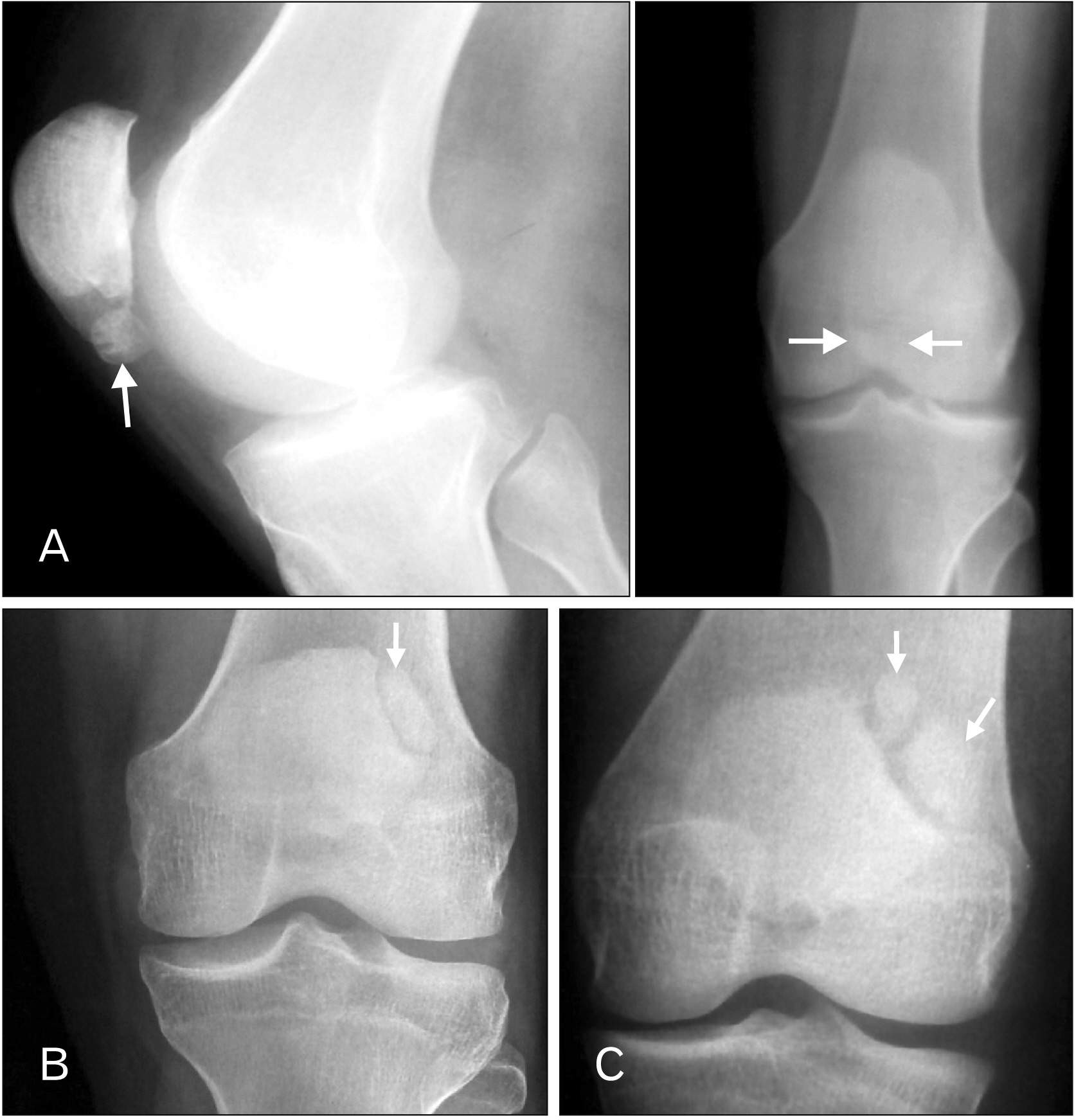
Fig. 2 (A) A 30-year-old male. Plain lateral and anteroposterior radiographs of the knee showing a type I (inferior margin) bipartite patella (arrows), according to the Saupe classification. (B) A 30-year-old male. Plain lateral and anteroposterior radiographs of the knee showing a type I (inferior margin) bipartite patella (arrow), according to the Saupe classification. (C) Plain anteroposterior radiograph of the knee showing a type III (superolateral margin) tripartite patella (arrows), according to the Saupe classification.
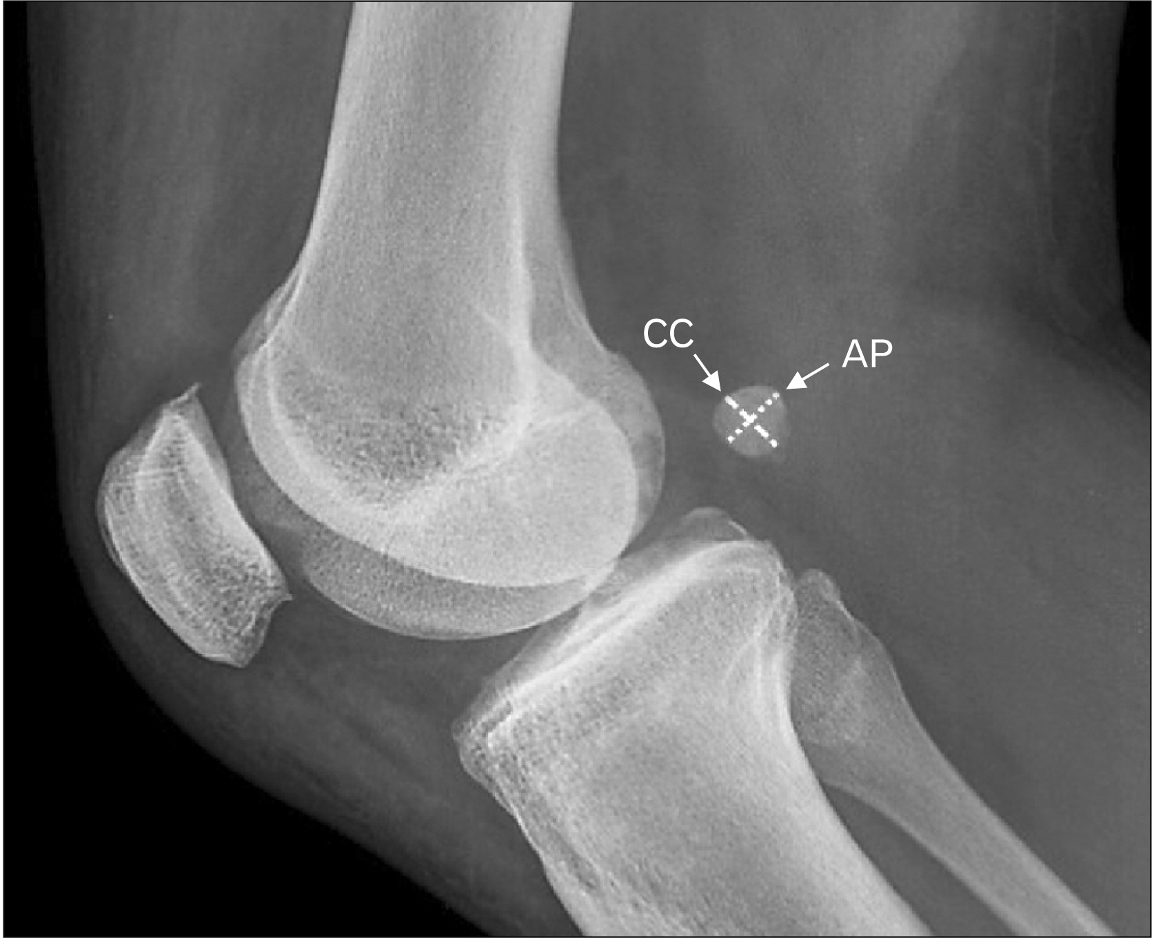
Fig. 3 A 41-year-old female. Plain lateral radiograph of the knee showing measurements of the CC and AP diameters of the fabella. AP, anteroposterior; CC, craniocaudal.

Fig. 4 A 41-year-old female. Plain lateral radiograph of the knee showing measurements of the FFD and FTD. FFD, fabello-femoral distance; FTD, fabello-tibial distance.
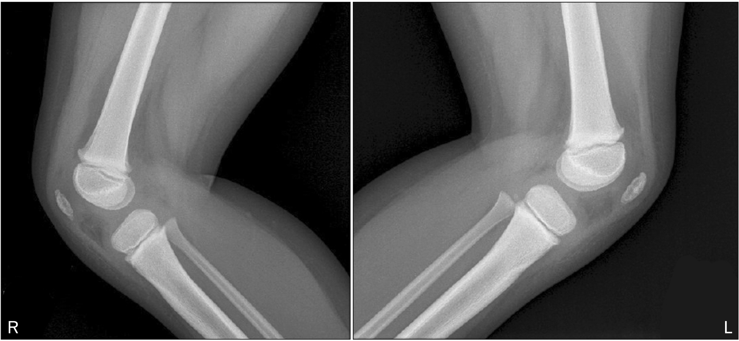
Fig. 5 A 4-year-old male. Plain lateral radiographs of both knees showing ‘just ossifying’ patellae.
Reference
-
References
1. Eyal S, Rubin S, Krief S, Levin L, Zelzer E. 2019; Common cellular origin and diverging developmental programs for different sesamoid bones. Development. 146:dev167452. DOI: 10.1242/dev.167452. PMID: 30745426. PMCID: PMC6919532.
Article2. Seguritan RE, Wolfe AR, Mena P, Bibawy J, Bianchi C, Solomon N, Kikkeri V. 2018; Bipartite patella separation and partial quadriceps tendon rupture in the setting of trauma. Radiol Case Rep. 14:526–9. DOI: 10.1016/j.radcr.2018.10.003. PMID: 30906491. PMCID: PMC6412164.
Article3. Sarkodieh J, Gobindpuri A. c2016. The age of patella ossification [Internet]. European Society of Radiology;Vienna: Available from: https://epos.myesr.org/esr/viewing/index.php?module=viewing_posteraction&task=downloadpdf&pi=135454. cited 2020 May 7.4. Zabierek S, Zabierek J, Kwapisz A, Domzalski ME. 2016; Bipartite patella in 35-year-old fitness instructor: a case report. Int J Sports Phys Ther. 11:777–83. PMID: 27757290. PMCID: PMC5046971.5. Gruber W. 1883; In Bildungsanomalie mit Bildungshemmung begründete Bipartition beider Patellae eines jungen Subjectes. Arch Pathol Anat. 94:358–361. German. DOI: 10.1007/BF01916049.6. Saupe E. 1921; Beitrag zur Patella bipartite. Fortschr Rontgenstr. 28:37–41.7. Oohashi Y, Koshino T, Oohashi Y. 2010; Clinical features and classification of bipartite or tripartite patella. Knee Surg Sports Traumatol Arthrosc. 18:1465–9. DOI: 10.1007/s00167-010-1047-y. PMID: 20111951.
Article8. Perdikakis E, Pothitakis C. 2013; Symptomatic multipartite patella: imaging findings and pain relief. A description of two cases. Cent Eur J Med. 8:172–175. DOI: 10.2478/s11536-012-0126-5.
Article9. Omar IM, Schweitzer M. 2004; Bipartite patella: MR imaging characteristics and frequency. Proc Intl Soc Mag Reson Med. 11:804.10. Dalip D, Iwanaga J, Oskouian RJ, Tubbs RS. 2018; A comprehensive review of the fabella bone. Cureus. 10:e2736. DOI: 10.7759/cureus.2736. PMID: 30087812. PMCID: PMC6075638.
Article11. Friedman AC, Naidich TP. 1978; The fabella sign: fabella displacement in synovial effusion and popliteal fossa masses. Normal and abnormal fabello-femoral and fabello-tibial distances. Radiology. 127:113–21. DOI: 10.1148/127.1.113. PMID: 635171.12. Minowa T, Murakami G, Kura H, Suzuki D, Han SH, Yamashita T. 2004; Does the fabella contribute to the reinforcement of the posterolateral corner of the knee by inducing the development of associated ligaments? J Orthop Sci. 9:59–65. DOI: 10.1007/s00776-003-0739-2. PMID: 14767706.
Article13. Hauser NH, Hoechel S, Toranelli M, Klaws J, Müller-Gerbl M. 2015; Functional and structural details about the fabella: what the important stabilizer looks like in the central European population. Biomed Res Int. 2015:343728. DOI: 10.1155/2015/343728. PMID: 26413516. PMCID: PMC4564579.
Article14. Chew CP, Lee KH, Koh JS, Howe TS. 2014; Incidence and radiological characteristics of fabellae in an Asian population. Singapore Med J. 55:198–201. DOI: 10.11622/smedj.2014052. PMID: 24763835. PMCID: PMC4291947.
Article15. Franceschi F, Longo UG, Ruzzini L, Leonardi F, Rojas M, Gualdi G, Denaro V. 2007; Dislocation of an enlarged fabella as uncommon cause of knee pain: a case report. Knee. 14:330–2. DOI: 10.1016/j.knee.2007.03.007. PMID: 17490883.16. Agathangelidis F, Vampertzis T, Gkouliopoulou E, Papastergiou S. 2016; Symptomatic enlarged fabella. BMJ Case Rep. 2016:bcr2016218085. DOI: 10.1136/bcr-2016-218085. PMID: 27807024. PMCID: PMC5128986.
Article17. Heideman GM, Baynes KE, Mautz AP, DuBois MS, Roberts JW. 2011; Fabella fracture with CT imaging: a case report. Emerg Radiol. 18:357–61. DOI: 10.1007/s10140-011-0941-z. PMID: 21305331.
Article18. Theodorou SJ, Theodorou DJ, Resnick D. 2005; Painful stress fractures of the fabella in patients with total knee arthroplasty. AJR Am J Roentgenol. 185:1141–4. DOI: 10.2214/AJR.04.1230. PMID: 16247123.
Article19. Driessen A, Balke M, Offerhaus C, White WJ, Shafizadeh S, Becher C, Bouillon B, Höher J. 2014; The fabella syndrome- a rare cause of posterolateral knee pain: a review of the literature and two case reports. BMC Musculoskelet Disord. 15:100. DOI: 10.1186/1471-2474-15-100. PMID: 24666711. PMCID: PMC3987160.
Article20. Phukubye P, Oyedele O. 2011; The incidence and structure of the fabella in a South African cadaver sample. Clin Anat. 24:84–90. DOI: 10.1002/ca.21049. PMID: 20830786.
Article21. Pritchett JW. 1984; The incidence of fabellae in osteoarthrosis of the knee. J Bone Joint Surg Am. 66:1379–80. DOI: 10.2106/00004623-198466090-00009. PMID: 6501334.
Article22. Takebe K, Kita K, Hirohata K. 1983; Radiological and anatomical observation on fabella. Orthop Surg. 34:1163–70.23. Sarin VK, Erickson GM, Giori NJ, Bergman AG, Carter DR. 1999; Coincident development of sesamoid bones and clues to their evolution. Anat Rec. 257:174–80. DOI: 10.1002/(SICI)1097-0185(19991015)257:5<174::AID-AR6>3.0.CO;2-O. PMID: 10597342.
Article24. Yu JS, Salonen DC, Hodler J, Haghighi P, Trudell D, Resnick D. 1996; Posterolateral aspect of the knee: improved MR imaging with a coronal oblique technique. Radiology. 198:199–204. DOI: 10.1148/radiology.198.1.8539378. PMID: 8539378.
Article25. Egerci OF, Kose O, Turan A, Kilicaslan OF, Sekerci R, Keles-Celik N. 2017; Prevalence and distribution of the fabella: a radiographic study in Turkish subjects. Folia Morphol (Warsz). 76:478–83. DOI: 10.5603/FM.a2016.0080. PMID: 28026849.
Article26. Er A, Murphy A. 2016. Knee (horizontal beam lateral view) [Internet]. Radiopaedia;Available from: https://radiopaedia.org/articles/knee-horizontal-beam-lateral-view-1. cited 2020 May 7.27. Zhou F, Zhang F, Deng G, Bi C, Wang J, Wang Q, Wang Q. 2017; Fabella fracture with radiological imaging: a case report. Trauma Case Rep. 12:19–23. DOI: 10.1016/j.tcr.2017.10.010. PMID: 29644278. PMCID: PMC5887092.
Article28. Hou W, Xu L, Wang J, Wang B, Liu L, Xu K, Cai Y, Guo H, Xu P. 2019; Fabellar prevalence, degeneration and association with knee osteoarthritis in the Chinese population. Sci Rep. 9:13046. DOI: 10.1038/s41598-019-49174-1. PMID: 31506455. PMCID: PMC6736872.
Article29. Pop TS, Pop AM, Olah P, Trâmbiţaş C. 2018; Prevalence of the fabella and its association with pain in the posterolateral corner of the knee: a cross-sectional study in a Romanian population. Medicine (Baltimore). 97:e13333. DOI: 10.1097/MD.0000000000013333. PMID: 30461651. PMCID: PMC6392660.30. Berthaume MA, Bull AMJ. 2020; Human biological variation in sesamoid bone prevalence: the curious case of the fabella. J Anat. 236:228–42. DOI: 10.1111/joa.13091. PMID: 31623020. PMCID: PMC6956444.
Article31. Zeng SX, Dong XL, Dang RS, Wu GS, Wang JF, Wang D, Huang HL, Guo XD. 2012; Anatomic study of fabella and its surrounding structures in a Chinese population. Surg Radiol Anat. 34:65–71. DOI: 10.1007/s00276-011-0828-4. PMID: 21626275.
Article32. Tabira Y, Saga T, Takahashi N, Watanabe K, Nakamura M, Yamaki K. 2013; Influence of a fabella in the gastrocnemius muscle on the common fibular nerve in Japanese subjects. Clin Anat. 26:893–902. DOI: 10.1002/ca.22153. PMID: 22933414.
Article33. Takebe K, Hirohata K. 1981; Peroneal nerve palsy due to fabella. Arch Orthop Trauma Surg. 99:91–5. DOI: 10.1007/BF00389743. PMID: 7316708.
Article34. Mangieri JV. 1973; Peroneal-nerve injury from an enlarged fabella. A case report. J Bone Joint Surg Am. 55:395–7. DOI: 10.2106/00004623-197355020-00019. PMID: 4696171.35. Hall FM. 1978; The fabella sign and radiologic assessment of knee joint effusion. Radiology. 129:541–2. DOI: 10.1148/129.2.541a. PMID: 704877.
Article36. Jerome JT, Varghese M, Sankaran B, Thomas S. 2008; Bilateral congenital absence of patella. Indian J Orthop. 42:228–30. DOI: 10.4103/0019-5413.40264. PMID: 19826534. PMCID: PMC2759612.
Article37. Sferopoulos NK. 2018; Congenital aplasia of the patella. Int J Radiol. 5:172–176. DOI: 10.17554/j.issn.2313-3406.2018.05.54. PMID: 30303847.
