Symptomatic Os Paracuneiforme: A Case Report
- Affiliations
-
- 1Department of Orthopedic Surgery, Pusan National University Yangsan Hospital, Pusan National University School of Medicine, Yangsan, Korea
- KMID: 2516649
- DOI: http://doi.org/10.14193/jkfas.2021.25.2.108
Abstract
- An extremely rare accessory bone of the foot located in the distal portion of the navicular that articulates with the medial cuneiform was observed. Os paracuneiforme is usually located medial to the medial cuneiform or the naviculocuneiform joint. Although os paracuneiforme is often referred to as an accessory bone around the foot, few cases of this type of accessory bone have been reported. This paper reports a patient with a painful Os paracuneiform who underwent surgical excision of the accessory bone.
Keyword
Figure
-
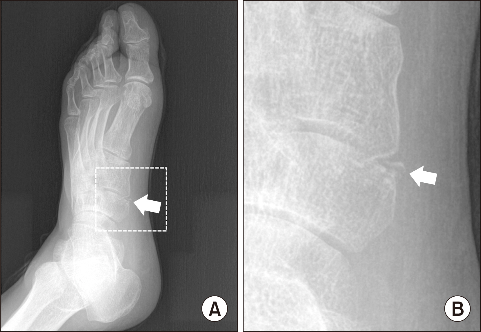
Figure. 1 Radiographs of the affected left foot revealing the wedge shaped accessory bone (arrows) between the navicular and medial cuneiform. (A) External oblique view, (B) enlarged marked portion of external oblique view.

Figure. 2 Sagittal view of computed tomography showing that the accessory bone was positioned between the navicular and medial cuneiform (arrow).
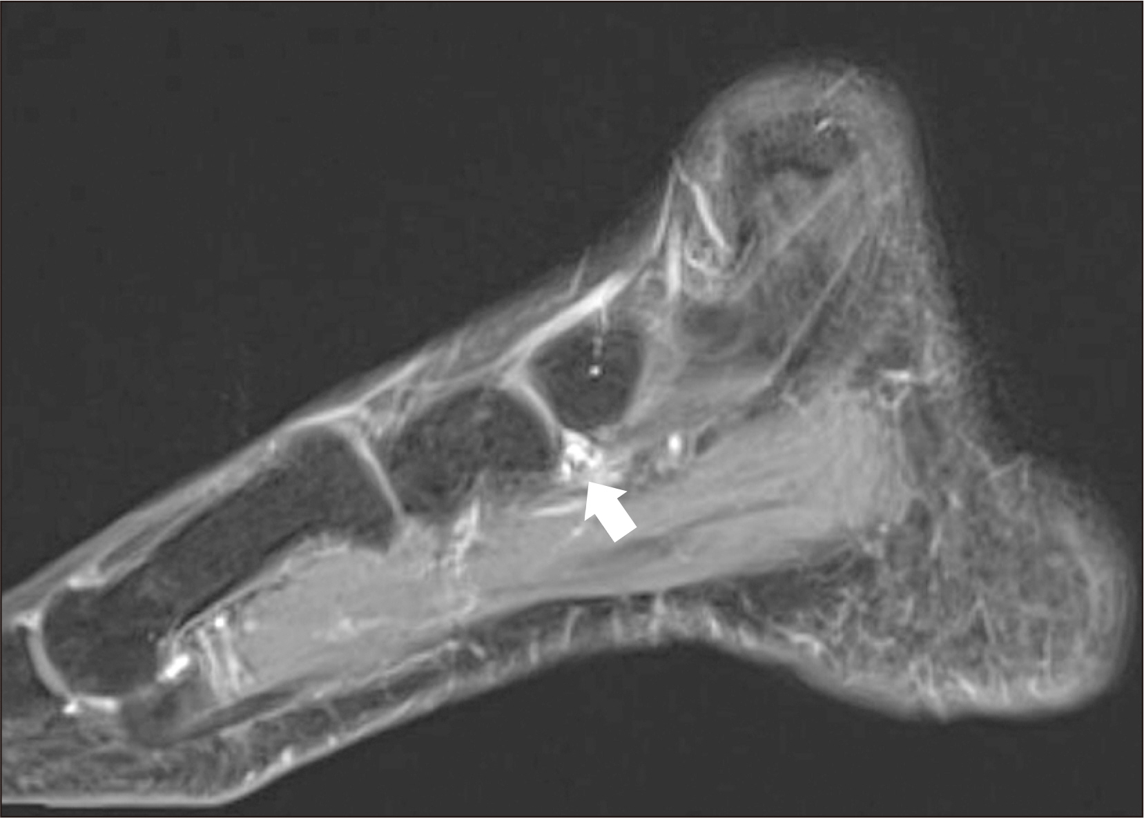
Figure. 3 Sagittal view of contrast-enhanced T1-weighted magnetic resonance images of the accessory bone. White arrow shows focal nodular enhancing lesion noted at inferior medial naviculocuneiform joint. About 5 mm sized accessary bone with adjacent joint osteophytes.

Figure. 4 Gross photo of specimen of the accessory bone: wedge shape mass about 4.0 mm×8.0 mm size.

Figure. 5 Microscopic finding of the os paracuneiforme, which shows small aggregates of chondrocytes surrounded by the hyaline matrix (H&E stain, ×100).
Reference
-
1. Coughlin MJ. Coughlin MJ, Saltzman CL, Anderson RB, editors. 2013. Sesamoids and accessory bones of the foot. Mann's surgery of the foot and ankle. 9th ed. Saunders/Elsevier;Philadelphia: p. 492–568.2. Keles-Celik N, Kose O, Sekerci R, Aytac G, Turan A, Güler F. 2017; Accessory ossicles of the foot and ankle: disorders and a review of the literature. Cureus. 9:e1881. doi: 10.7759/cureus.1881. DOI: 10.7759/cureus.1881. PMID: 29387510. PMCID: PMC5786346.
Article3. Kurashige T. 2020; Removal of painful os paracuneiforme: a case report. Foot Ankle Spec. 13:236–41. doi: 10.1177/1938640020905414. DOI: 10.1177/1938640020905414. PMID: 32059623.
Article4. Morrison AB. 1953; The os paracuneiforme; some observations on an example removed at operation. J Bone Joint Surg Br. 35:254–5. doi: 10.1302/0301-620X.35B2.254. DOI: 10.1302/0301-620X.35B2.254. PMID: 13061536.5. Kim JK, Roh KJ. 2013; Symptomatic os infranaviculare. Clin Orthop Surg. 5:152–4. doi: 10.4055/cios.2013.5.2.152. DOI: 10.4055/cios.2013.5.2.152. PMID: 23730481. PMCID: PMC3664676.
Article6. Coskun N, Yuksel M, Cevener M, Arican RY, Ozdemir H, Bircan O, et al. 2009; Incidence of accessory ossicles and sesamoid bones in the feet: a radiographic study of the Turkish subjects. Surg Radiol Anat. 31:19–24. doi: 10.1007/s00276-008-0383-9. DOI: 10.1007/s00276-008-0383-9. PMID: 18633564.
Article7. DiGiovanni CW, Patel A, Calfee R, Nickisch F. 2007; Osteonecrosis in the foot. J Am Acad Orthop Surg. 15:208–17. doi: 10.5435/00124635-200704000-00004. DOI: 10.5435/00124635-200704000-00004. PMID: 17426292.
Article8. Mair SD, Coogan AC, Speer KP, Hall RL. 1995; Gout as a source of sesamoid pain. Foot Ankle Int. 16:613–6. doi: 10.1177/107110079501601006. DOI: 10.1177/107110079501601006. PMID: 8574372.
Article9. Miller TT. 2002; Painful accessory bones of the foot. Semin Musculoskelet Radiol. 6:153–61. doi: 10.1055/s-2002-32361. DOI: 10.1055/s-2002-32361. PMID: 12077704.
Article
