Imaging Findings of CT and MRI of Os Supratalare: A Case Report
- Affiliations
-
- 1Department of Radiology, Haeundae Paik Hospital, Inje University College of Medicine, Busan, Korea. bluesingirl@paik.ac.kr
- KMID: 2041954
- DOI: http://doi.org/10.3348/jksr.2013.69.4.317
Abstract
- The os supratalare is quite a rare accessory ossicle of the ankle and the foot. We present imaging findings of a symptomatic os supratalare in a 21-year-old woman with a painful bump of the dorsal aspect on her hind foot. CT and MRI are helpful to distinguish this accessory ossicle from a fracture or an osteochondroma. Knowledge of imaging findings and clinical significances of os supratalare will be helpful for accurate diagnosis and appropriate management.
Figure
-
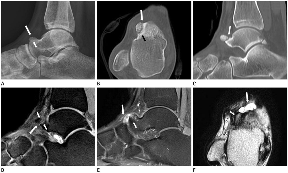
Fig. 1 A 21-year-old woman with os supratalare. A. Lateral radiograph of the ankle shows the os supratalare located dorsal aspect of the talus (long arrow). Subtle radiolucent line (short arrow) between the os supratalare and the talus is noted. B, C. Axial and sagittal CT images show the os supratalare close to the talar head. A narrow and mild irregular interface between the os and the talus is noted (long arrows in B and C). Small cysts and sclerosis along the interface and small bony productive changes are also noted (short arrow in B). D. Sagittal proton density image shows mild hyperintense signal intensity (long arrow) between the os supratalare and the talus with bone marrow edema (short arrows). E. Gadolinium-enhanced T1-weighted spectral pre-saturation with inversion recovery image shows bone marrow edema (short arrow) in the os supratalare and peripheral enhancing multiloculated cystic lesion in the anterior aspect of the os supratalare (long arrow). F. Axial T2-weighted image shows the os supratalare (short arrow) and multiloculated cystic lesion (long arrow).
Reference
-
1. Coughlin MJ. Sesamoids and accessory bones of the foot. In : Coughlin MJ, Mann RA, Saltzman CL, editors. Surgery of the Foot and Ankle. Philadelphia: Mosby;2007. p. 531–610.2. Coskun N, Yuksel M, Cevener M, Arican RY, Ozdemir H, Bircan O, et al. Incidence of accessory ossicles and sesamoid bones in the feet: a radiographic study of the Turkish subjects. Surg Radiol Anat. 2009; 31:19–24.3. Cilli F, Akçaoğlu M. [The incidence of accessory bones of the foot and their clinical significance]. Acta Orthop Traumatol Turc. 2005; 39:243–246.4. Tsuruta T, Shiokawa Y, Kato A, Matsumoto T, Yamazoe Y, Oike T, et al. [Radiological study of the accessory skeletal elements in the foot and ankle (author's transl)]. Nihon Seikeigeka Gakkai Zasshi. 1981; 55:357–370.5. Mellado JM, Ramos A, Salvadó E, Camins A, Danús M, Saurí A. Accessory ossicles and sesamoid bones of the ankle and foot: imaging findings, clinical significance and differential diagnosis. Eur Radiol. 2003; 13:Suppl 6. L164–L177.6. Mellado JM, Salvadó E, Camins A, Ramos A, Saurí A. Painful os sustentaculi: imaging findings of another symptomatic skeletal variant. Skeletal Radiol. 2002; 31:53–56.7. Karasick D, Schweitzer ME. The os trigonum syndrome: imaging features. AJR Am J Roentgenol. 1996; 166:125–129.8. Miller TT, Staron RB, Feldman F, Parisien M, Glucksman WJ, Gandolfo LH. The symptomatic accessory tarsal navicular bone: assessment with MR imaging. Radiology. 1995; 195:849–853.9. Osuji OU, McAdams TR. Dorsoulnar wrist ganglion associated with os ulnostyloideum: a case report. Am J Orthop (Belle Mead NJ). 2007; 36:E94–E96.10. Romanowski CA, Barrington NA. The accessory navicular--an important cause of medial foot pain. Clin Radiol. 1992; 46:261–264.
- Full Text Links
-
- Actions
-
Cited
- CITED
-
- Close
- Share
-
- Similar articles
-
- Imaging Findings of a Solitary Fibrous Tumor in Pancreas: A Case Report
- CT and MRI Findings of Low-Flow Mediastinal Vascular Malformation: A Case Report
- A Comparative Study of Survivor Outcomes between Preoperative Evaluation Using CT Alone and Combined CT and MRI in Patients with Pancreatic Ductal Adenocarcinoma
- US, CT and MR Imaging Findings of Leiomyoma of Urinary Bladder: Case Report
- Wallerian degeneration of brain: MRI and CT findings
