Neonatal Med.
2021 May;28(2):77-82. 10.5385/nm.2021.28.2.77.
A Neonate Diagnosed with Wolff-Parkinson-White Syndrome Presenting with Cardiogenic Shock
- Affiliations
-
- 1Department of Pediatrics, Soonchunhyang University Bucheon Hospital, Soonchunhyang University College of Medicine, Bucheon, Korea
- 2Department of Laboratory Medicine and Genetics, Soonchunhyang University Bucheon Hospital, Soonchunhyang University College of Medicine, Bucheon, Korea
- KMID: 2516489
- DOI: http://doi.org/10.5385/nm.2021.28.2.77
Abstract
- We present the case of a healthy 28-day-old female full-term neonate who was admitted to the neonatal intensive care unit for severe metabolic acidosis, hypoglycemia, and an initial sinus rhythm. The first diagnostic hypothesis was hypovolemic shock, and fluid resuscitation was started immediately. During fluid therapy, cardiovascular collapse occurred with supraventricular tachycardia. The latter was successfully treated with adenosine and beta-blockers. After 8 days, electrocardiography showed ventricular pre-excitation, and Wolff-Parkinson-White syndrome was diagnosed. A novel variant of the MYL2 gene that is related to hypertrophic cardiomyopathy and conduction defect was found after discharge. Cardiogenic shock should be considered, despite being a rare cause of shock in neonates.
Keyword
Figure
-
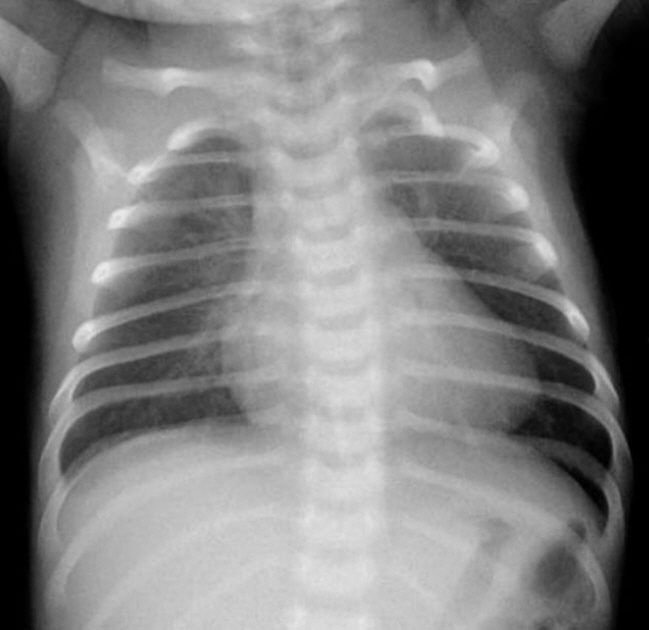
Figure 1. Chest radiograph of a 28-day-old female with cardiovascular collapse shows mild cardiomegaly (cardiothoracic ratio 56%) without pulmonary edema.

Figure 2. (A) Electrocardiography tracing of a 28-day-old female with cardiovascular collapse. Electrocardiography after the first episode of supraventricular tachycardia shows normal sinus rhythm (heart rate of 150 beats per minute). (B) Electrocardiography after the second episode of supraventricular tachycardia shows a short PR interval and wide QRS complex in all leads. This indicates ventricular pre-excitation (delta wave) resulting in the diagnosis of Wolff-Parkinson-White syndrome.
Reference
-
1. Chambers S, Jnah A, Newberry D. The pathophysiology, diagnosis, and management of Wolff-Parkinson-White syndrome in the neonate. Adv Neonatal Care. 2020; Aug. 19. [Epub]. https://doi.org/10.1097/ANC.0000000000000785.2. Hoeffler CD, Krenek ME, Brand MC. Wolff-Parkinson-White syndrome in a term infant presenting with cardiopulmonary arrest. Adv Neonatal Care. 2016; 16:44–51.3. Tsao S, Deal BJ. Management of symptomatic Wolff-Parkinson- White syndrome in childhood. Prog Pediatr Cardiol. 2013; 35:7–15.4. Dharnidharka VR, Lieh-Lai M, Sarnaik A, Clapp S. A child with cardiogenic shock and supraventricular tachycardia presenting in normal sinus rhythm. Pediatr Emerg Care. 1996; 12:420–1.5. Gikonyo BM, Dunnigan A, Benson DW Jr. Cardiovascular collapse in infants: association with paroxysmal atrial tachycardia. Pediatrics. 1985; 76:922–6.6. Viveiros E, Aveiro AC, Costa E, Nunes JL. Cardiogenic shock in a neonate. BMJ Case Rep. 2013; 2013:bcr2012008440.7. Al-Khatib SM, Pritchett EL. Clinical features of Wolff-Parkinson- White syndrome. Am Heart J. 1999; 138(3 Pt 1):403–13.8. Munger TM, Packer DL, Hammill SC, Feldman BJ, Bailey KR, Ballard DJ, et al. A population study of the natural history of Wolff-Parkinson-White syndrome in Olmsted County, Minnesota, 1953-1989. Circulation. 1993; 87:866–73.9. Richardson C, Silver ES. Management of supraventricular tachycardia in infants. Paediatr Drugs. 2017; 19:539–51.10. MacRae CA, Ghaisas N, Kass S, Donnelly S, Basson CT, Watkins HC, et al. Familial hypertrophic cardiomyopathy with Wolff-Parkinson-White syndrome maps to a locus on chromosome 7q3. J Clin Invest. 1995; 96:1216–20.11. van der Steld LP, Campuzano O, Perez-Serra A, Moura de Barros Zamorano M, Sousa Matos S, Brugada R. Wolff-Parkinson-White syndrome with ventricular hypertrophy in a Brazilian family. Am J Case Rep. 2017; 18:766–76.12. Gilljam T, Jaeggi E, Gow RM. Neonatal supraventricular tachycardia: outcomes over a 27-year period at a single institution. Acta Paediatr. 2008; 97:1035–9.13. Riggs TW, Byrd JA, Weinhouse E. Recurrence risk of supraventricular tachycardia in pediatric patients. Cardiology. 1999; 91:25–30.14. National Center for Biotechnology Information, U.S. National Library of Medicine. MYL2 myosin light chain 2 [homo sapiens (human)] [Internet]. Bethesda: NCBI Gene;2020. [cited 2021 May 12]. Available from: http://www.ncbi.nlm.nih.gov/gene/4633.15. Andersen PS, Havndrup O, Hougs L, Sorensen KM, Jensen M, Larsen LA, et al. Diagnostic yield, interpretation, and clinical utility of mutation screening of sarcomere encoding genes in Danish hypertrophic cardiomyopathy patients and relatives. Hum Mutat. 2009; 30:363–70.16. Claes GR, van Tienen FH, Lindsey P, Krapels IP, Helderman-van den Enden AT, Hoos MB, et al. Hypertrophic remodelling in cardiac regulatory myosin light chain (MYL2) founder mutation carriers. Eur Heart J. 2016; 37:1815–22.17. Ho CY, Day SM, Colan SD, Russe ll MW, Towbin JA, Sherrid MV, et al. The burden of early phenotypes and the influence of wall thickness in hypertrophic cardiomyopathy mutation carriers: findings from the HCMNet study. JAMA Cardiol. 2017; 2:419–28.
- Full Text Links
-
- Actions
-
Cited
- CITED
-
- Close
- Share
-
- Similar articles
-
- One Case of Cerebral Embolism Associated with Paroxysmal Tachycardia in Wolff-Parkinson-White Syndrome
- Two Cases of Wolff-Parkinson-White Syndrome in a Family
- Wolff-Parkinson-White Syndrome Treated with Radiofrequency Ablation in Father and His Son
- Atrial fibrillation in patient with Wolff-Parkinson-White syndrome mimicking ventricular tachycardia
- A Case of Early Onset MELAS Patient with Wolff-Parkinson-White Syndrome
