Current Approaches to the Treatment of Post-Stroke Aphasia
- Affiliations
-
- 1Department of Communication Sciences and Disorders, University of South Carolina, Columbia, SC, USA
- 2Department of Neurology, Johns Hopkins University School of Medicine, Baltimore, MD, USA
- KMID: 2516408
- DOI: http://doi.org/10.5853/jos.2020.05015
Abstract
- Aphasia, impairment of language after stroke or other neurological insult, is a common and often devastating condition that affects nearly every social activity and interaction. Behavioral speech and language therapy is the mainstay of treatment, although other interventions have been introduced to augment the effects of the behavioral therapy. In this narrative review, we discuss advances in aphasia therapy in the last 5 years and focus primarily on properly powered, randomized, controlled trials of both behavioral therapies and interventions to augment therapy for post-stroke aphasia. These trials include evaluation of behavioral therapies and computer-delivered language therapies. We also discuss outcome prediction trials as well as interventional trials that have employed noninvasive brain stimulation, or medications to augment language therapy. Supported by evidence from Phase III trials and large meta-analyses, it is now generally accepted that aphasia therapy can improve language processing for many patients. Not all patients respond similarly to aphasia therapy with the most severe patients being the least likely responders. Nevertheless, it is imperative that all patients, regardless of severity, receive aphasia management focused on direct therapy of language deficits, counseling, or both. Emerging evidence from Phase II trials suggests transcranial brain stimulation is a promising method to boost aphasia therapy outcomes.
Keyword
Figure
-

Figure 1. The relationship between improvement in naming (y-axis) and aphasia severity (x-axis). Improvement in correct naming was measured using the Philadelphia Naming Test (PNT) whereas aphasia severity was qualified as the overall severity score, aphasia quotient on the Western Aphasia Battery-Revised Aphasia Quotient (WAB-R AQ). (A) The graph shows raw change in correct naming whereas (B) the graph shows change in naming as proportion of potential room for improvement. Note the discrepancy in the graphs at the higher end of the WAB AQ spectrum, which is caused by the mildest cases having relatively limited room for improvement at baseline compared to their counterparts with more severe forms of aphasia.
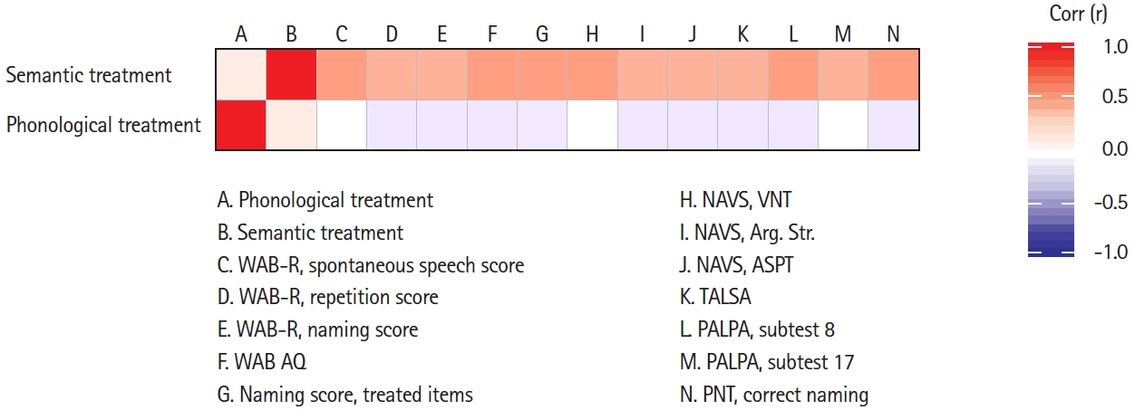
Figure 2. Correlation matrix showing improvements in correct naming following semantic therapy (top row) and phonological therapy (bottom row) in relation to several different language tests. AQ, aphasia quotient; NAVS, Northwestern Assessment of Verbs and Sentences; VNT, verb naming test; Arg. Str., argument structure; ASPT, argument structure production test; TALSA, Temple Assessment of Language and Short-term Memory in Aphasia; PALPA, Psycholinguistic Assessment of Language Processing in Aphasia (Subtest 8: Nonword repetition; Subtest 17: Phonological segmentation of final sounds in words); PNT, Philadelphia Naming Test.
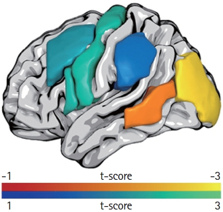
Figure 3. Overall improvement in correct naming on the Philadelphia Naming Test (PNT) was predicted by less damage to the middle occipital gyrus and posterior middle temporal gyrus (red-yellow scale) whereas greater PNT improvement following the phonological treatment phase was predicted by greater damage to anterior regions and the supramarginal gyrus (blue-green scale). The color scales indicate t-scores.
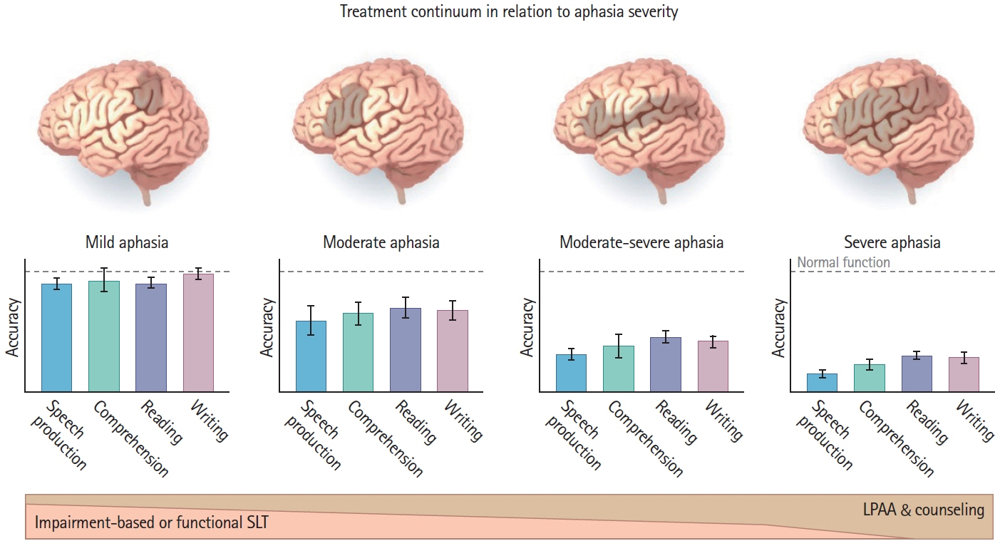
Figure 4. A simple schema suggesting how clinicians might determine how much of their effort in treatment should be devoted to speech and language therapy (SLT) versus Life Participation Approaches to Aphasia (LPAA) based on aphasia severity
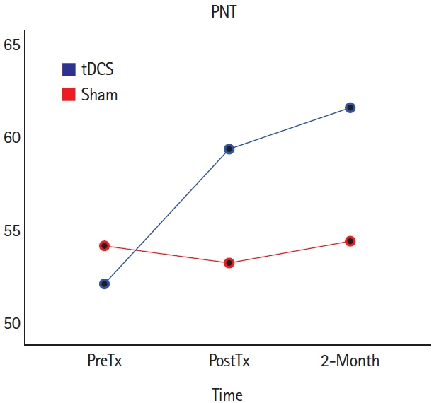
Figure 5. Mean accuracy in naming untrained words on the Philadelphia Naming Test (PNT) with right cerebellar transcranial direct current stimulation (tDCS) versus sham conditions, at pre-treatment (PreTx), post-treatment (PostTx), and 2 months PostTx. This figure illustrates data from reference 133 but combines data for anodal and cathodal right cerebellar tDCS, which did not have significantly different effects. Both tDCS conditions resulted in greater improvements than sham from PreTx to PostTx, and sustained 2 months later. To see results reported separately for anodal and cathodal tDCS, see reference 111.
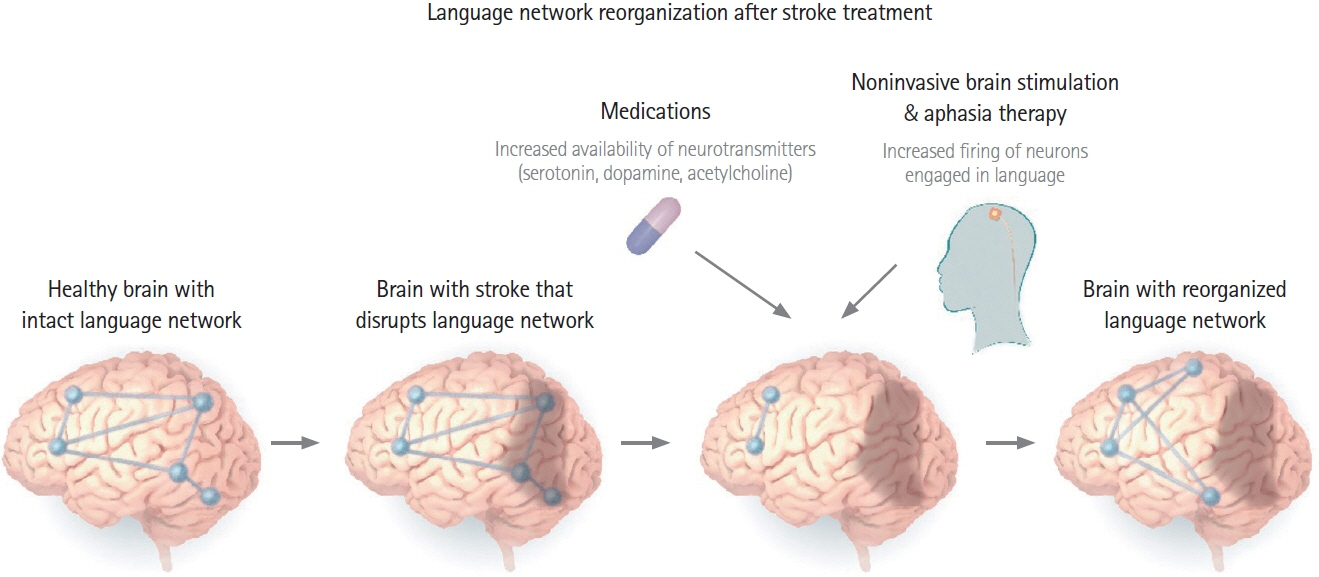
Figure 6. Combined behavioral, noninvasive brain stimulation, and pharmaceutical intervention promote reorganization of the language network to support improved language processing, as demonstrated through a variety of functional imaging studies of aphasia recovery with treatment.[3-6, 112]
Cited by 1 articles
-
Predictors of Therapy Response in Chronic Aphasia: Building a Foundation for Personalized Aphasia Therapy
Sigfus Kristinsson, Dirk B. den Ouden, Chris Rorden, Roger Newman-Norlund, Jean Neils-Strunjas, Julius Fridriksson
J Stroke. 2022;24(2):189-206. doi: 10.5853/jos.2022.01102.
Reference
-
References
1. Nardo D, Holland R, Leff AP, Price CJ, Crinion JT. Less is more: neural mechanisms underlying anomia treatment in chronic aphasic patients. Brain. 2017; 140:3039–3054.
Article2. Abel S, Lambon Ralph MA. Cognitive neuroscience of aphasia recovery and therapy. Aphasiology. 2018; 32:739–741.
Article3. Crinion JT, Leff AP. Using functional imaging to understand therapeutic effects in poststroke aphasia. Curr Opin Neurol. 2015; 28:330–337.
Article4. Fridriksson J, Smith K. Neuroplasticity associated with treated aphasia recovery. In: Hickok G, Small SL. Neurobiology of Language. Amsterdam, NL: Elsevier;2016. p. 1007–1013.5. Kiran S, Meier EL, Johnson JP. Neuroplasticity in aphasia: a proposed framework of language recovery. J Speech Lang Hear Res. 2019; 62:3973–3985.
Article6. Hartwigsen G, Saur D. Neuroimaging of stroke recovery from aphasia: insights into plasticity of the human language network. Neuroimage. 2019; 190:14–31.7. Berker EA, Berker AH, Smith A. Translation of Broca’s 1865 report: localization of speech in the third left frontal convolution. Arch Neurol. 1986; 43:1065–1072.8. Fama ME, Turkeltaub PE. Treatment of poststroke aphasia: current practice and new directions. Semin Neurol. 2014; 34:504–513.
Article9. Boyle M. Semantic feature analysis treatment for aphasic word retrieval impairments: what’s in a name? Top Stroke Rehabil. 2010; 17:411–422.
Article10. Edmonds LA, Mammino K, Ojeda J. Effect of Verb Network Strengthening Treatment (VNeST) in persons with aphasia: extension and replication of previous findings. Am J Speech Lang Pathol. 2014; 23:S312–S329.
Article11. Kendall DL, Rosenbek JC, Heilman KM, Conway T, Klenberg K, Gonzalez Rothi LJ, et al. Phoneme-based rehabilitation of anomia in aphasia. Brain Lang. 2008; 105:1–17.
Article12. Martin N, Thompson CK, Worrall L. Aphasia Rehabilitation: The Impairment and Its Consequences. San Diego, CA: Plural Publishing;2007.13. Berthier ML, Green C, Lara JP, Higueras C, Barbancho MA, Dávila G, et al. Memantine and constraint-induced aphasia therapy in chronic poststroke aphasia. Ann Neurol. 2009; 65:577–585.
Article14. Kertesz A, McCabe P. Recovery patterns and prognosis in aphasia. Brain. 1977; 100 Pt 1:1–18.
Article15. Smania N, Gandolfi M, Aglioti SM, Girardi P, Fiaschi A, Girardi F. How long is the recovery of global aphasia?: twenty-five years of follow-up in a patient with left hemisphere stroke. Neurorehabil Neural Repair. 2010; 24:871–875.
Article16. Brady MC, Kelly H, Godwin J, Enderby P. Speech and language therapy for aphasia following stroke. Cochrane Database Syst Rev . 2012; (5):CD000425.
Article17. Brady MC, Kelly H, Godwin J, Enderby P, Campbell P. Speech and language therapy for aphasia following stroke. Cochrane Database Syst Rev. 2016; 6:CD000425.
Article18. Breitenstein C, Grewe T, Flöel A, Ziegler W, Springer L, Martus P, et al. Intensive speech and language therapy in patients with chronic aphasia after stroke: a randomised, open-label, blinded-endpoint, controlled trial in a health-care setting. Lancet. 2017; 389:1528–1538.19. Linebaugh CW, Baron CR, Corcoran KJ. Assessing treatment efficacy in acute aphasia: paradoxes, presumptions, problems and principles. Aphasiology. 1998; 12:519–536.20. Robey RR. A meta-analysis of clinical outcomes in the treatment of aphasia. J Speech Lang Hear Res. 1998; 41:172–187.
Article21. Robey RR. The efficacy of treatment for aphasic persons: a meta-analysis. Brain Lang. 1994; 47:582–608.
Article22. Blomert L, Kean ML, Koster C, Schokker J. Amsterdam—Nijmegen everyday language test: construction, reliability and validity. Aphasiology. 1994; 8:381–407.23. Doesborgh SJ, van de Sandt-Koenderman MW, Dippel DW, van Harskamp F, Koudstaal PJ, Visch-Brink EG. Effects of semantic treatment on verbal communication and linguistic processing in aphasia after stroke: a randomized controlled trial. Stroke. 2004; 35:141–146.24. de Jong-Hagelstein M, van de Sandt-Koenderman WM, Prins ND, Dippel DW, Koudstaal PJ, Visch-Brink EG. Efficacy of early cognitive-linguistic treatment and communicative treatment in aphasia after stroke: a randomised controlled trial (RATS-2). J Neurol Neurosurg Psychiatry. 2011; 82:399–404.
Article25. Lincoln NB, McGuirk E, Mulley GP, Lendrem W, Jones AC, Mitchell JR. Effectiveness of speech therapy for aphasic stroke patients: a randomized controlled trial. Lancet. 1984; 1:1197–1200.26. de Courten-Myers GM, Kleinholz M, Wagner KR, Xi G, Myers RE. Efficacious experimental stroke treatment with high-dose methylprednisolone. Stroke. 1994; 25:487–493.
Article27. Nouwens F, Visch-Brink EG, Van de Sandt-Koenderman MM, Dippel DW, Koudstaal PJ, de Lau LM. Optimal timing of speech and language therapy for aphasia after stroke: more evidence needed. Expert Rev Neurother. 2015; 15:885–893.
Article28. Warren RL, Gabriel C, Johnston A, Gaddie A. Efficacy during acute rehabilitation. Clinical Aphasiology Conference. 1987. May 31-Jun 4. Lake of the Ozarks, MO: Jackson, WY: BRK Publishers; 1-11.29. Nouwens F, de Lau LM, Visch-Brink EG, van de Sandt-Koenderman WM, Lingsma HF, Goosen S, et al. Efficacy of early cognitive-linguistic treatment for aphasia due to stroke: a randomised controlled trial (Rotterdam Aphasia Therapy Study-3). Eur Stroke J. 2017; 2:126–136.
Article30. Godecke E, Armstrong E, Rai T, Ciccone N, Rose ML, Middleton S, et al. A randomized control trial of intensive aphasia therapy after acute stroke: the Very Early Rehabilitation for SpEech (VERSE) study. Int J Stroke . 2020. Oct. 6. G[Epub]. https://doi.org/10.1177/1747493020961926.
Article31. Fridriksson J. Preservation and modulation of specific left hemisphere regions is vital for treated recovery from anomia in stroke. J Neurosci. 2010; 30:11558–11564.
Article32. Johnson L, Basilakos A, Yourganov G, Cai B, Bonilha L, Rorden C, et al. Progression of aphasia severity in the chronic stages of stroke. Am J Speech Lang Pathol. 2019; 28:639–649.
Article33. Holland A, Fromm D, Forbes M, MacWhinney B. Long-term recovery in stroke accompanied by aphasia: a reconsideration. Aphasiology. 2017; 31:152–165.
Article34. Hope TM, Leff AP, Prejawa S, Bruce R, Haigh Z, Lim L, et al. Right hemisphere structural adaptation and changing language skills years after left hemisphere stroke. Brain. 2017; 140:1718–1728.
Article35. Basilakos A, Stark BC, Johnson L, Rorden C, Yourganov G, Bonilha L, et al. Leukoaraiosis is associated with a decline in language abilities in chronic aphasia. Neurorehabil Neural Repair. 2019; 33:718–729.
Article36. Wilmskoetter J, Marebwa B, Basilakos A, Fridriksson J, Rorden C, Stark BC, et al. Long-range fibre damage in small vessel brain disease affects aphasia severity. Brain. 2019; 142:3190–3201.
Article37. Levine DA, Galecki AT, Langa KM, Unverzagt FW, Kabeto MU, Giordani B, et al. Trajectory of cognitive decline after incident stroke. JAMA. 2015; 314:41–51.
Article38. Darley FL. The efficacy of language rehabilitation in aphasia. J Speech Hear Disord. 1972; 37:3–21.
Article39. Borod JC, Carper JM, Naeser M. Long-term language recovery in left-handed aphasic patients. Aphasiology. 1990; 4:561–572.
Article40. Laska AC, Hellblom A, Murray V, Kahan T, Von Arbin M. Aphasia in acute stroke and relation to outcome. J Intern Med. 2001; 249:413–422.
Article41. Pedersen PM, Vinter K, Olsen TS. Aphasia after stroke: type, severity and prognosis: the Copenhagen aphasia study. Cerebrovasc Dis. 2004; 17:35–43.42. Engelter ST, Gostynski M, Papa S, Frei M, Born C, Ajdacic-Gross V, et al. Epidemiology of aphasia attributable to first ischemic stroke: incidence, severity, fluency, etiology, and thrombolysis. Stroke. 2006; 37:1379–1384.43. Seniów J, Litwin M, Leśniak M. The relationship between non-linguistic cognitive deficits and language recovery in patients with aphasia. J Neurol Sci. 2009; 283:91–94.
Article44. Hope TM, Seghier ML, Leff AP, Price CJ. Predicting outcome and recovery after stroke with lesions extracted from MRI images. Neuroimage Clin. 2013; 2:424–433.
Article45. Selnes OA, Knopman DS, Niccum N, Rubens AB, Larson D. Computed tomographic scan correlates of auditory comprehension deficits in aphasia: a prospective recovery study. Ann Neurol. 1983; 13:558–566.
Article46. Hillis AE, Beh YY, Sebastian R, Breining B, Tippett DC, Wright A, et al. Predicting recovery in acute poststroke aphasia. Ann Neurol. 2018; 83:612–622.
Article47. Marchina S, Zhu LL, Norton A, Zipse L, Wan CY, Schlaug G. Impairment of speech production predicted by lesion load of the left arcuate fasciculus. Stroke. 2011; 42:2251–2256.
Article48. Plowman E, Hentz B, Ellis C Jr. Post-stroke aphasia prognosis: a review of patient-related and stroke-related factors. J Eval Clin Pract. 2012; 18:689–694.
Article49. Cheng BB, Worrall LE, Copland DA, Wallace SJ. Prognostication in post-stroke aphasia: how do speech pathologists formulate and deliver information about recovery? Int J Lang Commun Disord. 2020; 55:520–536.
Article50. Spell LA, Richardson JD, Basilakos A, Stark BC, Teklehaimanot A, Hillis AE, et al. Developing, implementing, and improving assessment and treatment fidelity in clinical aphasia research. Am J Speech Lang Pathol. 2020; 29:286–298.
Article51. Roach A, Schwartz MF, Martin N, Grewal RS, Brecher A. The Philadelphia naming test: scoring and rationale. Clin Aphasiol. 1996; 24:121–133.52. Dignam J, Copland D, O’Brien K, Burfein P, Khan A, Rodriguez AD. Influence of cognitive ability on therapy outcomes for anomia in adults with chronic poststroke aphasia. J Speech Lang Hear Res. 2017; 60:406–421.
Article53. Gilmore N, Meier EL, Johnson JP, Kiran S. Nonlinguistic cognitive factors predict treatment-induced recovery in chronic poststroke aphasia. Arch Phys Med Rehabil. 2019; 100:1251–1258.
Article54. Kristinsson S, Basilakos A, Elm J, Spell LA, Bonilha L, Rorden C, et al. Individualized response to different types of aphasia rehabilitation. Brain Commun. 2021. In Press.55. Fridriksson J, Yourganov G, Bonilha L, Basilakos A, Den Ouden DB, Rorden C. Revealing the dual streams of speech processing. Proc Natl Acad Sci U S A. 2016; 113:15108–15113.
Article56. Conroy P, Sotiropoulou Drosopoulou C, Humphreys GF, Halai AD, Lambon Ralph MA. Time for a quick word?: the striking benefits of training speed and accuracy of word retrieval in post-stroke aphasia. Brain. 2018; 141:1815–1827.
Article57. Bonilha L, Gleichgerrcht E, Nesland T, Rorden C, Fridriksson J. Success of anomia treatment in aphasia is associated with preserved architecture of global and left temporal lobe structural networks. Neurorehabil Neural Repair. 2016; 30:266–279.
Article58. Toth E, Gersner R, Wilf-Yarkoni A, Raizel H, Dar DE, Richter-Levin G, et al. Age-dependent effects of chronic stress on brain plasticity and depressive behavior. J Neurochem. 2008; 107:522–532.
Article59. deToledo-Morrell L, Geinisman Y, Morrell F. Age-dependent alterations in hippocampal synaptic plasticity: relation to memory disorders. Neurobiol Aging. 1988; 9:581–590.
Article60. Vara H, Muñoz-Cuevas J, Colino A. Age-dependent alterations of long-term synaptic plasticity in thyroid-deficient rats. Hippocampus. 2003; 13:816–825.
Article61. Harvey S, Carragher M, Dickey MW, Pierce JE, Rose ML. Dose effects in behavioural treatment of post-stroke aphasia: a systematic review and meta-analysis. Disabil Rehabil. 2020. Nov. 9. [Epub]. https://doi.org/10.1080/09638288.2020.1843079.
Article62. Duchan JF, Byng S. Challenging Aphasia Therapies: Broadening the Discourse and Extending the Boundaries. Hove, UK: Psychology Press;2004.63. Carpenter J, Cherney LR. Increasing aphasia treatment intensity in an acute inpatient rehabilitation program: a feasibility study. Aphasiology. 2016; 30:542–565.64. Dignam J, Copland D, McKinnon E, Burfein P, O’Brien K, Farrell A, et al. Intensive versus distributed aphasia therapy: a nonrandomized, parallel-group, dosage-controlled study. Stroke. 2015; 46:2206–2211.65. Katz RC, Hallowell B, Code C, Armstrong E, Roberts P, Pound C, et al. A multinational comparison of aphasia management practices. Int J Lang Commun Disord. 2000; 35:303–314.
Article66. Hoover EL, Caplan DN, Waters GS, Carney A. Communication and quality of life outcomes from an interprofessional intensive, comprehensive, aphasia program (ICAP). Top Stroke Rehabil. 2017; 24:82–90.
Article67. Lanyon L, Worrall L, Rose M. Exploring participant perspectives of community aphasia group participation: from “I know where I belong now” to “some people didn’t really fit in”. Aphasiology. 2018; 32:139–163.
Article68. Ribeiro Lima R, Rose ML, Lima HD, Guarinello AC, Santos RS, Massi GA. Socio-demographic factors associated with quality of life after a multicomponent aphasia group therapy in people with sub-acute and chronic post-stroke aphasia. Aphasiology. 2020. Feb 24 [Epub]. https://doi.org/10.1080/02687038.2020.1727710.
Article69. Simmons-Mackie N. Aphasia in North America. Moorestown, NJ: Aphasia Access;2018.70. Øra HP, Kirmess M, Brady MC, Partee I, Hognestad RB, Johannessen BB, et al. The effect of augmented speech-language therapy delivered by telerehabilitation on poststroke aphasia: a pilot randomized controlled trial. Clin Rehabil. 2020; 34:369–381.71. Woolf C, Caute A, Haigh Z, Galliers J, Wilson S, Kessie A, et al. A comparison of remote therapy, face to face therapy and an attention control intervention for people with aphasia: a quasi-randomised controlled feasibility study. Clin Rehabil. 2016; 30:359–373.
Article72. Meltzer JA, Baird AJ, Steele RD, Harvey SJ. Computer-based treatment of poststroke language disorders: a non-inferiority study of telerehabilitation compared to in-person service delivery. Aphasiology. 2018; 32:290–311.
Article73. Steele RD. Lingraphic methods in interface design. In: Proceedings of the International Workshop on Human Interface Technology IWHIT 95; 1995 Oct 12-13; Aizu, JP: 53-58.74. Pitt R, Theodoros D, Hill AJ, Russell T. The impact of the telerehabilitation group aphasia intervention and networking programme on communication, participation, and quality of life in people with aphasia. Int J Speech Lang Pathol. 2019; 21:513–523.
Article75. Kurland J, Liu A, Stokes P. Effects of a tablet-based home practice program with telepractice on treatment outcomes in chronic aphasia. J Speech Lang Hear Res. 2018; 61:1140–1156.
Article76. Palmer R, Dimairo M, Cooper C, Enderby P, Brady M, Bowen A, et al. Self-managed, computerised speech and language therapy for patients with chronic aphasia post-stroke compared with usual care or attention control (Big CACTUS): a multicentre, single-blinded, randomised controlled trial. Lancet Neurol. 2019; 18:821–833.
Article77. Latimer NR, Bhadhuri A, Alshreef A, Palmer R, Cross E, Dimairo M, et al. Self-managed, computerised word finding therapy as an add-on to usual care for chronic aphasia post-stroke: an economic evaluation. Clin Rehabil. 2021; 35:703–717.
Article78. Fleming V, Brownsett S, Krason A, Maegli MA, Coley-Fisher H, Ong YH, et al. Efficacy of spoken word comprehension therapy in patients with chronic aphasia: a cross-over randomised controlled trial with structural imaging. J Neurol Neurosurg Psychiatry. 2021; 92:418–424.
Article79. Howard D, Swinburn K, Porter G. Putting the CAT out: what the Comprehensive Aphasia Test has to offer. Aphasiology. 2010; 24:56–74.
Article80. Stark BC, Warburton EA. Improved language in chronic aphasia after self-delivered iPad speech therapy. Neuropsychol Rehabil. 2018; 28:818–831.
Article81. Thompson CK, Shapiro LP. Treating agrammatic aphasia within a linguistic framework: treatment of underlying forms. Aphasiology. 2005; 19:1021–1036.
Article82. Murray L, Timberlake A, Eberle R. Treatment of underlying forms in a discourse context. Aphasiology. 2007; 21:139–163.
Article83. Barbieri E, Mack J, Chiappetta B, Europa E, Thompson CK. Recovery of offline and online sentence processing in aphasia: language and domain-general network neuroplasticity. Cortex. 2019; 120:394–418.
Article84. Thompson CK. Treatment of syntactic and morphologic deficits in agrammatic aphasia: Treatment of underlying forms. In: Chapey R. Language Intervention Strategies in Aphasia and Related Neurogenic Communication Disorders. 5th. Philadelphia, PA: Wolters Kluwer Health;2012. p. 735–755.85. Zhang J, Yu J, Bao Y, Xie Q, Xu Y, Zhang J, et al. Constraint-induced aphasia therapy in post-stroke aphasia rehabilitation: a systematic review and meta-analysis of randomized controlled trials. PLoS One. 2017; 12:e0183349.
Article86. Pulvermüller F, Neininger B, Elbert T, Mohr B, Rockstroh B, Koebbel P, et al. Constraint-induced therapy of chronic aphasia after stroke. Stroke. 2001; 32:1621–1626.
Article87. Szaflarski JP, Ball AL, Vannest J, Dietz AR, Allendorfer JB, Martin AN, et al. Constraint-induced aphasia therapy for treatment of chronic post-stroke aphasia: a randomized, blinded, controlled pilot trial. Med Sci Monit. 2015; 21:2861–2869.
Article88. Sickert A, Anders LC, Münte TF, Sailer M. Constraint-induced aphasia therapy following sub-acute stroke: a single-blind, randomised clinical trial of a modified therapy schedule. J Neurol Neurosurg Psychiatry. 2014; 85:51–55.
Article89. Ciccone N, West D, Cream A, Cartwright J, Rai T, Granger A, et al. Constraint-induced aphasia therapy (CIAT): a randomised controlled trial in very early stroke rehabilitation. Aphasiology. 2016; 30:566–584.
Article90. Rose ML, Copland D, Nickels L, Togher L, Meinzer M, Rai T, et al. Constraint-induced or multi-modal personalized aphasia rehabilitation (COMPARE): a randomized controlled trial for stroke-related chronic aphasia. Int J Stroke. 2019; 14:972–976.
Article91. Kagan A, Simmons-Mackie N, Victor JC. The impact of exposure with no training: implications for future partner training research. J Speech Lang Hear Res. 2018; 61:2347–2352.
Article92. Kagan A, Winckel J, Black S, Duchan JF, Simmons-Mackie N, Square P. A set of observational measures for rating support and participation in conversation between adults with aphasia and their conversation partners. Top Stroke Rehabil. 2004; 11:67–83.
Article93. Simmons-Mackie N, Savage MC, Worrall L. Conversation therapy for aphasia: a qualitative review of the literature. Int J Lang Commun Disord. 2014; 49:511–526.
Article94. Holland A, Forbes M, Fromm D, MacWhinney B. Communicative strengths in severe aphasia: the famous people protocol and its value in planning treatment. Am J Speech Lang Pathol. 2019; 28:1010–1018.
Article95. Simmons-Mackie N, Holland AL. Aphasia centers in North America: a survey. Semin Speech Lang. 2011; 32:203–215.
Article96. Attard MC, Loupis Y, Togher L, Rose ML. Staff experiences of an interprofessional community aphasia group. Aphasiology. 2020; 34:1–18.
Article97. Elman RJ. Aphasia centers and the life participation approach to aphasia. Top Lang Disord. 2016; 36:154–167.
Article98. Holland AL. Pragmatic aspects of intervention in aphasia. J Neurolinguist. 1991; 6:197–211.
Article99. Purdie H, Baldwin S. Music therapy: challenging low self-esteem in people with a stroke. J Br Music Ther. 1994; 8:19–24.
Article100. Nicholas M, Elliott S. C-Speak Aphasia: A Communication System for Adults with Aphasia. Solana Beach, CA: Mayer-Johnson Co;1998.101. Nicholas M, Sinotte M, Helm-Estabrooks N. Using a computer to communicate: effect of executive function impairments in people with severe aphasia. Aphasiology. 2005; 19:1052–1065.
Article102. National Institute of Neurological Disorders and Stroke RT-PA Stroke Study Group. Tissue plasminogen activator for acute ischemic stroke. N Engl J Med. 1995; 333:1581–1587.103. Berkhemer OA, Fransen PS, Beumer D, van den Berg LA, Lingsma HF, Yoo AJ, et al. A randomized trial of intraarterial treatment for acute ischemic stroke. N Engl J Med. 2015; 372:11–20.104. Campbell BC, Mitchell PJ, Kleinig TJ, Dewey HM, Churilov L, Yassi N, et al. Endovascular therapy for ischemic stroke with perfusion-imaging selection. N Engl J Med. 2015; 372:1009–1018.
Article105. Hillis AE. Pharmacological, surgical, and neurovascular interventions to augment acute aphasia recovery. Am J Phys Med Rehabil. 2007; 86:426–434.
Article106. Hillis AE, Ulatowski JA, Barker PB, Torbey M, Ziai W, Beauchamp NJ, et al. A pilot randomized trial of induced blood pressure elevation: effects on function and focal perfusion in acute and subacute stroke. Cerebrovasc Dis. 2003; 16:236–246.
Article107. Berthier ML. Ten key reasons for continuing research on pharmacotherapy for post-stroke aphasia. Aphasiology. 2020. May. 31. [Epub]. https://doi.org/10.1080/02687038.2020.1769987.
Article108. Crosson B, Rodriguez AD, Copland D, Fridriksson J, Krishnamurthy LC, Meinzer M, et al. Neuroplasticity and aphasia treatments: new approaches for an old problem. J Neurol Neurosurg Psychiatry. 2019; 90:1147–1155.
Article109. Stefaniak JD, Halai AD, Lambon Ralph MA. The neural and neurocomputational bases of recovery from post-stroke aphasia. Nat Rev Neurol. 2020; 16:43–55.
Article110. Brzosko Z, Mierau SB, Paulsen O. Neuromodulation of spike-timing-dependent plasticity: past, present, and future. Neuron. 2019; 103:563–581.
Article111. Kirkwood A, Rozas C, Kirkwood J, Perez F, Bear MF. Modulation of long-term synaptic depression in visual cortex by acetylcholine and norepinephrine. J Neurosci. 1999; 19:1599–1609.
Article112. Kilgard MP, Merzenich MM. Cortical map reorganization enabled by nucleus basalis activity. Science. 1998; 279:1714–1718.
Article113. Llano DA, Small SL. Pharmacotherapy for aphasia. In: Hickok G, Small SL. Neurobiology of Language. Amsterdam, NL: Academic Press;2015. p. 1067–1083.114. Walker-Batson D, Curtis S, Natarajan R, Ford J, Dronkers N, Salmeron E, et al. A double-blind, placebo-controlled study of the use of amphetamine in the treatment of aphasia. Stroke. 2001; 32:2093–2098.
Article115. Seniów J, Litwin M, Litwin T, Leśniak M, Członkowska A. New approach to the rehabilitation of post-stroke focal cognitive syndrome: effect of levodopa combined with speech and language therapy on functional recovery from aphasia. J Neurol Sci. 2009; 283:214–218.
Article116. Ashtary F, Janghorbani M, Chitsaz A, Reisi M, Bahrami A. A randomized, double-blind trial of bromocriptine efficacy in nonfluent aphasia after stroke. Neurology. 2006; 66:914–916.
Article117. Breitenstein C, Korsukewitz C, Baumgärtner A, Flöel A, Zwitserlood P, Dobel C, et al. L-dopa does not add to the success of high-intensity language training in aphasia. Restor Neurol Neurosci. 2015; 33:115–120.
Article118. Berthier ML, Green C, Higueras C, Fernández I, Hinojosa J, Martín MC. A randomized, placebo-controlled study of donepezil in poststroke aphasia. Neurology. 2006; 67:1687–1689.
Article119. Haixia YE, Shilin LI. A clinical randomized case-control study of donepezil in the treatment of aphasia after stroke. Chin J Behav Med Brain Sci. 2014; 23:225–227.120. Lipton SA. Paradigm shift in neuroprotection by NMDA receptor blockade: memantine and beyond. Nat Rev Drug Discov. 2006; 5:160–170.
Article121. Chollet F, Tardy J, Albucher JF, Thalamas C, Berard E, Lamy C, et al. Fluoxetine for motor recovery after acute ischaemic stroke (FLAME): a randomised placebo-controlled trial. Lancet Neurol. 2011; 10:123–130.
Article122. Jorge RE, Acion L, Moser D, Adams HP Jr, Robinson RG. Escitalopram and enhancement of cognitive recovery following stroke. Arch Gen Psychiatry. 2010; 67:187–196.
Article123. FOCUS Trial Collaboration. Effects of fluoxetine on functional outcomes after acute stroke (FOCUS): a pragmatic, double-blind, randomised, controlled trial. Lancet. 2019; 393:265–274.124. Kraglund KL, Mortensen JK, Damsbo AG, Modrau B, Simonsen SA, Iversen HK, et al. Neuroregeneration and vascular protection by citalopram in acute ischemic stroke (TALOS). Stroke. 2018; 49:2568–2576.
Article125. Bindman LJ, Lippold OC, Redfearn JW. The action of brief polarizing currents on the cerebral cortex of the rat (1) during current flow and (2) in the production of long-lasting after-effects. J Physiol. 1964; 172:369–382.
Article126. Fritsch B, Reis J, Martinowich K, Schambra HM, Ji Y, Cohen LG, et al. Direct current stimulation promotes BDNF-dependent synaptic plasticity: potential implications for motor learning. Neuron. 2010; 66:198–204.
Article127. Fridriksson J, Elm J, Stark BC, Basilakos A, Rorden C, Sen S, et al. BDNF genotype and tDCS interaction in aphasia treatment. Brain Stimul. 2018; 11:1276–1281.
Article128. Holland R, Leff AP, Josephs O, Galea JM, Desikan M, Price CJ, et al. Speech facilitation by left inferior frontal cortex stimulation. Curr Biol. 2011; 21:1403–1407.
Article129. Crinion J. Speech facilitation by left inferior frontal cortex stimulation. In : Hillis AE, editor. The Handbook of Adult Language Disorders. New York, NY: Psychology Press;2015. 458–475140.130. Biou E, Cassoudesalle H, Cogné M, Sibon I, De Gabory I, Dehail P, et al. Transcranial direct current stimulation in post-stroke aphasia rehabilitation: a systematic review. Ann Phys Rehabil Med. 2019; 62:104–121.
Article131. Elsner B, Kugler J, Pohl M, Mehrholz J. Transcranial direct current stimulation (tDCS) for improving aphasia in adults with aphasia after stroke. Cochrane Database Syst Rev. 2019; 5:CD009760.
Article132. Campana S, Caltagirone C, Marangolo P. Combining voxel-based lesion-symptom mapping (VLSM) with A-tDCS language treatment: predicting outcome of recovery in nonfluent chronic aphasia. Brain Stimul. 2015; 8:769–776.
Article133. Darkow R, Martin A, Würtz A, Flöel A, Meinzer M. Transcranial direct current stimulation effects on neural processing in post-stroke aphasia. Hum Brain Mapp. 2017; 38:1518–1531.
Article134. Meinzer M, Darkow R, Lindenberg R, Flöel A. Electrical stimulation of the motor cortex enhances treatment outcome in post-stroke aphasia. Brain. 2016; 139(Pt 4):1152–1163.
Article135. Fridriksson J, Rorden C, Elm J, Sen S, George MS, Bonilha L. Transcranial direct current stimulation vs sham stimulation to treat aphasia after stroke: a randomized clinical trial. JAMA Neurol. 2018; 75:1470–1476.136. Silva FR, Mac-Kay AP, Chao JC, Santos MD, Gagliadi RJ. Transcranial direct current stimulation: a study on naming performance in aphasic individuals. Codas. 2018; 30:e20170242.137. Naeser MA, Martin PI, Treglia E, Ho M, Kaplan E, Bashir S, et al. Research with rTMS in the treatment of aphasia. Restor Neurol Neurosci. 2010; 28:511–529.
Article138. Sebastian R, Saxena S, Tsapkini K, Faria AV, Long C, Wright A, et al. Cerebellar tDCS: a novel approach to augment language treatment post-stroke. Front Hum Neurosci. 2017; 10:695.
Article139. Sebastian R, Kim JH, Brenowitz R, Tippett DC, Desmond JE, Celnik PA, et al. Cerebellar neuromodulation improves naming in post-stroke aphasia. Brain Commun. 2020; 2:fcaa179.
Article140. Dvorak E, DeMarco A, Stoodley C, Turkeltaub P. A pilot study of right cerebellar tDCS as a therapeutic adjuvant in chronic aphasia after left-hemisphere stroke (P5.6-006). Neurology. 2019; 92(15 Suppl):P5.6–006.141. You DS, Kim DY, Chun MH, Jung SE, Park SJ. Cathodal transcranial direct current stimulation of the right Wernicke’s area improves comprehension in subacute stroke patients. Brain Lang. 2011; 119:1–5.
Article142. Spielmann K, van de Sandt-Koenderman WM, Heijenbrok-Kal MH, Ribbers GM. Transcranial direct current stimulation does not improve language outcome in subacute poststroke aphasia. Stroke. 2018; 49:1018–1020.
Article143. Rubi-Fessen I, Hartmann A, Huber W, Fimm B, Rommel T, Thiel A, et al. Add-on effects of repetitive transcranial magnetic stimulation on subacute aphasia therapy: enhanced improvement of functional communication and basic linguistic skills: a randomized controlled study. Arch Phys Med Rehabil. 2015; 96:1935–1944.
Article144. Khedr EM, Abo El-Fetoh N, Ali AM, El-Hammady DH, Khalifa H, Atta H, et al. Dual-hemisphere repetitive transcranial magnetic stimulation for rehabilitation of poststroke aphasia: a randomized, double-blind clinical trial. Neurorehabil Neural Repair. 2014; 28:740–750.145. Haghighi M, Mazdeh M, Ranjbar N, Seifrabie MA. Further evidence of the positive influence of repetitive transcranial magnetic stimulation on speech and language in patients with aphasia after stroke: results from a double-blind intervention with sham condition. Neuropsychobiology. 2017; 75:185–192.
Article146. Tsai PY, Wang CP, Ko JS, Chung YM, Chang YW, Wang JX. The persistent and broadly modulating effect of inhibitory rTMS in nonfluent aphasic patients: a sham-controlled, double-blind study. Neurorehabil Neural Repair. 2014; 28:779–787.147. Ren CL, Zhang GF, Xia N, Jin CH, Zhang XH, Hao JF, et al. Effect of low-frequency rTMS on aphasia in stroke patients: a meta-analysis of randomized controlled trials. PLoS One. 2014; 9:e102557.
Article148. Hara T, Abo M, Kakita K, Mori Y, Yoshida M, Sasaki N. The effect of selective transcranial magnetic stimulation with functional near-infrared spectroscopy and intensive speech therapy on individuals with post-stroke aphasia. Eur Neurol. 2017; 77:186–194.
Article149. Wang CP, Hsieh CY, Tsai PY, Wang CT, Lin FG, Chan RC. Efficacy of synchronous verbal training during repetitive transcranial magnetic stimulation in patients with chronic aphasia. Stroke. 2014; 45:3656–3662.
Article150. Lam JM, Wodchis WP. The relationship of 60 disease diagnoses and 15 conditions to preference-based health-related quality of life in Ontario hospital-based long-term care residents. Med Care. 2010; 48:380–387.
Article
- Full Text Links
-
- Actions
-
Cited
- CITED
-
- Close
- Share
-
- Similar articles
-
- Current Evidence for Post Stroke Aphasia Treatment
- Characteristics of Cognitive Impairment in Patients With Post-stroke Aphasia
- Diagnosis of Post-Stroke Aphasia: A Review of Language Tests in Korea
- Subcortical Aphasia in Stroke Patients
- Prolonged Ictal Aphasia Presenting as Clinical-Diffusion Mismatch in a Patient with Acute Ischemic Stroke
