Unusually Early Recurrence of Mitral Valve Myxoma in a Child
- Affiliations
-
- 1Department of Pediatrics, Seoul St. Mary's Hospital, College of Medicine, The Catholic University of Korea, Seoul, Korea
- 2Department of Thoracic and Cardiovascular Surgery, Seoul St. Mary's Hospital, College of Medicine, The Catholic University of Korea, Seoul, Korea
- KMID: 2516220
- DOI: http://doi.org/10.4070/kcj.2021.0112
Figure
-
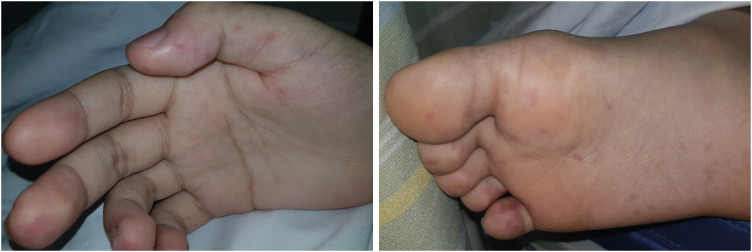
Figure 1 The patient had swellings of fingers and toes, and painful, red, and raised lesions (Osler's nodes), and non-tender, small erythematous or hemorrhagic macular lesions (Janeway lesions) on the palms and soles.
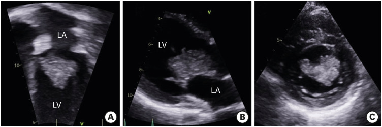
Figure 2 Echocardiography showing a huge, irregular shaped, and heterogeneous mass attached to the anterior leaflet of mitral valve at an apical 4-chamber (A), a parasternal long-axis (B), and a parasternal short-axis (C) views.LA = left atrium; LV = left ventricle.
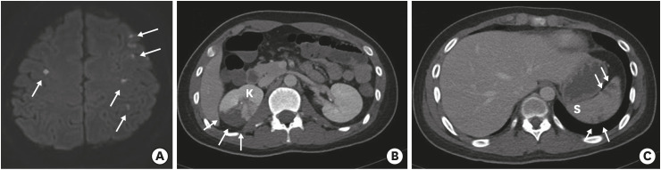
Figure 3 Brain magnetic resonance imaging and abdominal computed tomography showing multiple embolic infarct lesions (white arrows) in the brain (A), kidney (B), and spleen (C).K = right kidney; S = spleen.
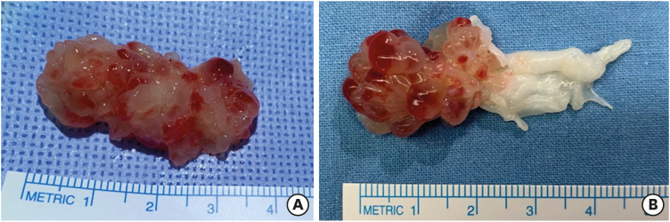
Figure 4 (A) A gelatinous and polypoid mass excised from mitral valve at the first operation. (B) An en bloc mass with the recurred tumor and mitral valve tissues excised at the second operation.
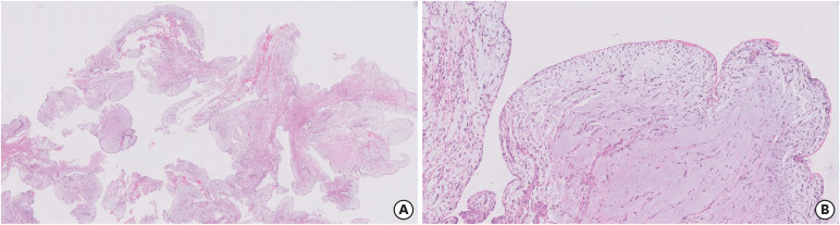
Figure 5 Histology of the mitral valve myxoma showing a lobulated, irregular, and frond-like surfaces, which is prone to embolization (A), and an abundant myxoid stroma with single spindle and stellate cells (B).
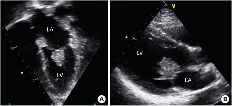
Figure 6 At a month after the first operation, a new tumor was identified at the ventricular side of mitral valve at the apical 4-chamber (A) and parasternal long-axis (B) views.LA = left atrium; LV = left ventricle.
Cited by 2 articles
-
Systemic Inflammation in the Setting of Cardiac Myxomas: an Overview of Clinical and Practical Considerations
Kenan Yalta, Cihan Ozturk, Tülin Yalta, Ertan Yetkin
Korean Circ J. 2021;51(9):784-786. doi: 10.4070/kcj.2021.0241.Author's Reply to Systemic Inflammation in the Setting of Cardiac Myxomas: an Overview of Clinical and Practical Considerations
Ji Hong Yoon, Ju Ae Shin, Yeon U Choi, Jae Young Lee
Korean Circ J. 2021;51(9):787-788. doi: 10.4070/kcj.2021.2411.
Reference
-
1. Yuan SM. Mitral valve myxoma: clinical features, current diagnostic approaches, and surgical management. Cardiol J. 2012; 19:105–109. PMID: 22298179.
Article2. Tasoglu I, Tutun U, Lafci G, et al. Primary cardiac myxomas: clinical experience and surgical results in 67 patients. J Card Surg. 2009; 24:256–259. PMID: 19438777.
Article
- Full Text Links
-
- Actions
-
Cited
- CITED
-
- Close
- Share
-
- Similar articles
-
- Cardiac Myxoma Originating from the Anterior Mitral Valve Leaflet
- A Case of Microangiopathic Hemolytic Anemia after Myxoma Excision and Mitral Valve Repair Presenting as Hemolytic Uremic Syndrome
- Left Atrial Myxoma Associated with Mitral Regurgitation and Coronary Artery Disease
- A case of myxoma attached to both interatrial septum and anterior mital leaflet
- Pregnancy, Paroxysmal Nocturnal Dyspnea, Presyncope, and Plop: A Case of Left Atrial Myxoma Causing Mitral Valve Obstruction in Pregnancy
