Healthc Inform Res.
2021 Apr;27(2):146-152. 10.4258/hir.2021.27.2.146.
Histoscope: A Web-Based Microscopy Tool for Oral Histology Education
- Affiliations
-
- 1School of Dentistry, Faculty of Medicine and Dentistry, University of Alberta, Edmonton, Canada
- KMID: 2515917
- DOI: http://doi.org/10.4258/hir.2021.27.2.146
Abstract
Objectives
Histology, the study of tissue structure under a microscope, is one of the most essential yet least engaging topics for health professional students. Understanding tissue microanatomy is crucial for students to be able to recognize cellular structures and follow disease pathogenesis. Traditional histology teaching labs rely on light microscopes and a limited array of slides, which inhibits simultaneous observation by multiple learners, and prevents in-class discussions. We have developed an interactive web-based microscopy tool called “Histoscope” for oral histology in this context.
Methods
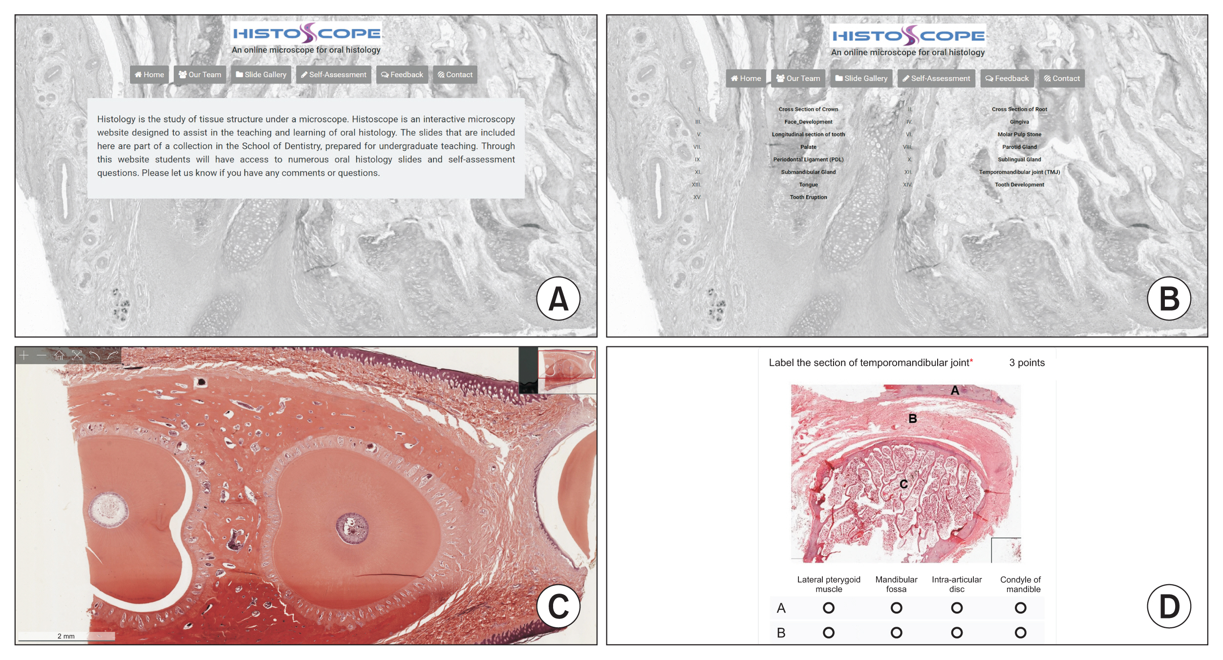
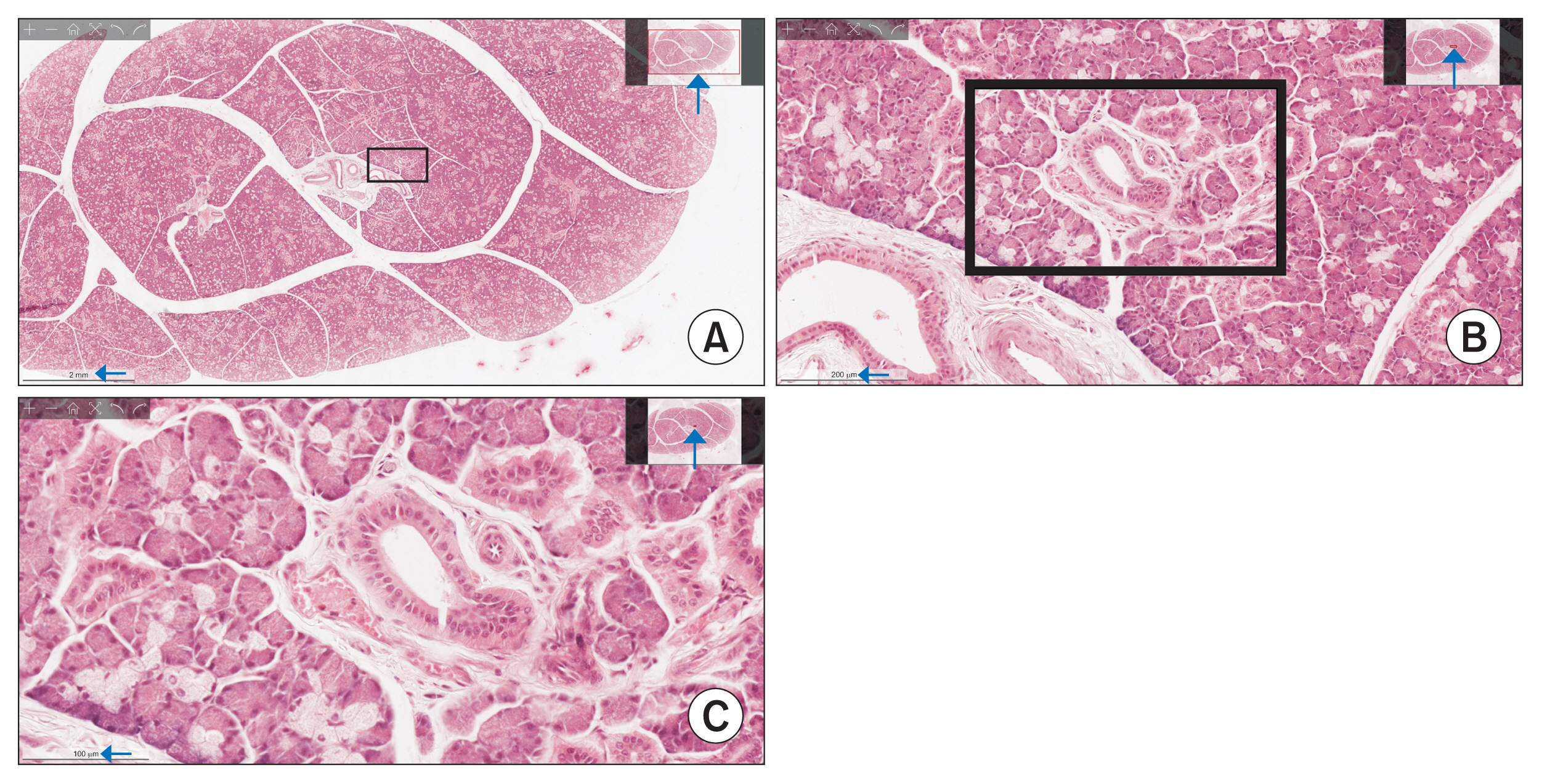
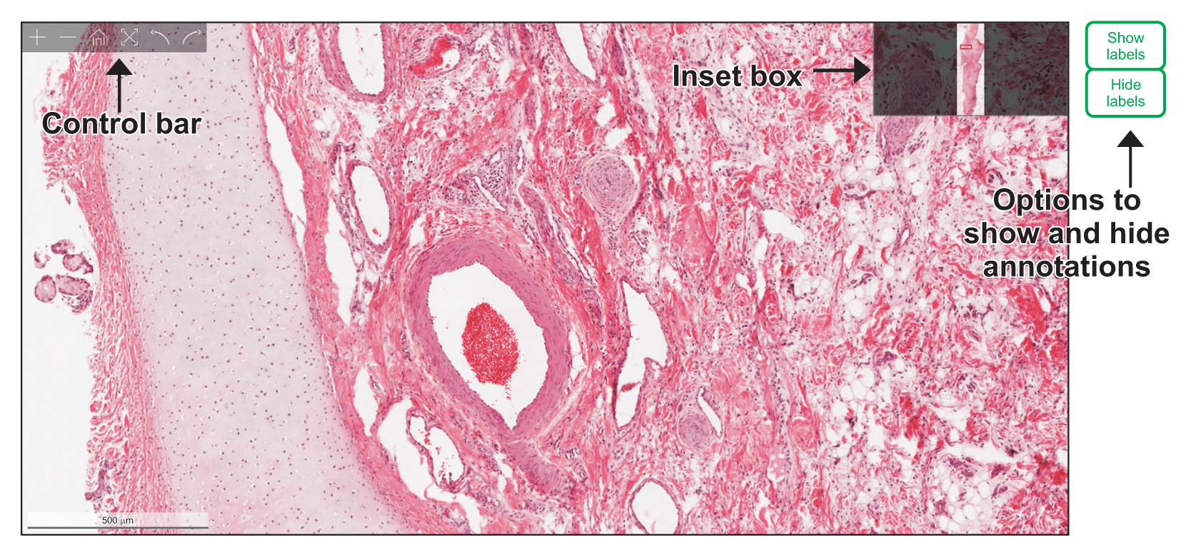
Good quality microscope slides were selected for digital scanning. The slides were scanned with multiple layers of z-stacking, a method of taking multiple images at different focal distances. The digital images were checked for quality and were archived on Histoscope. The slides were annotated, and self-assessment questions were prepared for the website. Interactive components were programmed on the website to mimic the experience of using a real light microscope.
Results
This web-based tool allows users to interact with histology slides, replicating the experience of observing and manipulating a slide under a real microscope. Through this website, learners can access a broad array of digital oral histology slides and self-assessment questions.
Conclusions
Incorporation of Histoscope in a course can shift traditional teacher-centered histology learning to a collaborative and student-centered learning environment. This platform can also provide students the flexibility to study histology at their own pace.
Figure
Reference
-
References
1. Garcia M, Victory N, Navarro-Sempere A, Segovia Y. Students’ views on difficulties in learning histology. Anat Sci Educ. 2019; 12(5):541–9.
Article2. Capela e Silva F, Rato LM, Lopes OS. Learning and teaching histology: traditional and computational methods. In : Proceedings of International Conference “Learning and Teaching in Higher Education”; 2010 Apr 15–16; Evora, Portugal.3. Blake CA, Lavoie HA, Millette CF. Teaching medical histology at the University of South Carolina School of Medicine: transition to virtual slides and virtual microscopes. Anat Rec B New Anat. 2003; 275(1):196–206.
Article4. Pantanowitz L, Szymas J, Yagi Y, Wilbur D. Whole slide imaging for educational purposes. J Pathol Inform. 2012; 3:46.
Article5. Kumar RK, Velan GM, Korell SO, Kandara M, Dee FR, Wakefield D. Virtual microscopy for learning and assessment in pathology. J Pathol. 2004; 204(5):613–8.
Article6. Helle L, Nivala M, Kronqvist P. More technology, better learning resources, better learning? Lessons from adopting virtual microscopy in undergraduate medical education. Anat Sci Educ. 2013; 6(2):73–80.
Article7. Hortsch M. The SecondLook [Internet]. Ann Arbor (MI): University of Michigan;c2019. [cited at 2021 Apr 5]. Available from: https://secondlook.med.umich.edu/histology .8. MacPherson BR, Tieman JG. Oral histology: a digital laboratory and atlas [Internet]. Lexington (KY): University of Kentucky;n.d.. [cited at 2021 Apr 5]. Available from: http://www.uky.edu/~brmacp/oralhist/index.htm .9. Rinaldi VD, Lorr NA, Williams K. Evaluating a technology supported interactive response system during the laboratory section of a histology course. Anat Sci Educ. 2017; 10(4):328–38.
Article10. Van Es SL, Pryor WM, Belinson Z, Salisbury EL, Velan GM. Cytopathology whole slide images and virtual microscopy adaptive tutorials: a software pilot. J Pathol Inform. 2015; 6:54.
Article11. Ray SF. Applied photographic optics: lenses and optical systems for photography, film, video, electronic imaging and digital imaging. 3rd ed. Oxford, UK: Focal Press;2002.12. Bloodgood RA, Ogilvie RW. Trends in histology laboratory teaching in United States medical schools. Anat Rec B New Anat. 2006; 289(5):169–75.
Article13. Cotter JR. Laboratory instruction in histology at the University at Buffalo: recent replacement of microscope exercises with computer applications. Anat Rec. 2001; 265(5):212–21.
Article14. McBride JM, Drake RL. National survey on anatomical sciences in medical education. Anat Sci Educ. 2018; 11(1):7–14.
Article15. Vella J. On teaching and learning: putting the principles and practices of dialogue education into action. San Francisco (CA): Jossey-Bass;2008.16. Karge BD, Phillips KM, Jessee T, McCabe M. Effective strategies for engaging adult learners. J Coll Teach Learn. 2011; 8(12):53–6.
Article17. Mayer RE. Multimedia learning. 2nd ed. Cambridge, UK: Cambridge University Press;2009.
- Full Text Links
- Actions
-
Cited
- CITED
-
- Close
- Share
- Similar articles
-
- Development and Evaluation of a Web-based Ostomy Self-care Education Program
- Survey of Oral Health Education Effects in Twenties
- Application of Evaluation Criteria for Web sites to Sexuality Education
- Development and Evaluation of a Web-based Education Program to Prevent Secondary Stroke
- How Can We Develop and Make Use of the Quality Assessment Tool of Web-Based Instruction(WBI) for Nutrition Education?


