Midfacial soft tissue changes after maxillary expansion using micro-implant-supported maxillary skeletal expanders in young adults: A retrospective study
- Affiliations
-
- 1Department of Orthodontics, Institute of Oral Health Science, Ajou University School of Medicine, Suwon, Korea
- 2Department of Orthodontics, Center for Advanced Dental Education, Saint Louis University, Saint Louis, MO, USA
- 3Department of Orthodontics, The Institute of Oral Health Science, Samsung Medical Center, Sungkyunkwan University School of Medicine, Seoul, Korea
- KMID: 2515818
- DOI: http://doi.org/10.4041/kjod.2021.51.3.145
Abstract
Objective
The aim of this retrospective study was to assess the midfacial soft tissue changes following maxillary expansion using micro-implantsupported maxillary skeletal expanders (MSEs) in young adults by cone-beam computerized tomography (CBCT) and to evaluate the correlations between hard and soft tissue changes after MSE usage.
Methods
Twenty patients (mean age, 22.4 years; range, 17.6–27.1) with maxillary transverse deficiency treated with MSEs were selected. Mean expansion amount was 6.5 mm. CBCT images taken before and after expansion were superimposed to measure the changes in soft and hard tissue landmarks. Statistical analyses were performed using paired t-test and Pearson’s correlation analysis on the basis of the normality of data.
Results
Average lateral movement of the cheek points was 1.35 mm (right) and 1.08 mm (left), and that of the alar curvature points was 1.03 mm (right) and 1.02 mm (left). Average forward displacement of the cheek points was 0.59 mm (right) and 0.44 mm (left), and that of the alar curvature points was 0.61 mm (right) and 0.77 mm (left) (p < 0.05). Anterior nasal spine (ANS), posterior nasal spine (PNS), and alveolar bone width showed significant increments (p < 0.05). Changes in the cheek and alar curvature points on both sides significantly correlated with hard tissue changes (p < 0.05).
Conclusions
Maxillary expansion using MSEs resulted in significant lateral and forward movements of the soft tissues of cheek and alar curvature points on both sides in young adults and correlated with the maxillary suture opening at the ANS and PNS.
Keyword
Figure
-
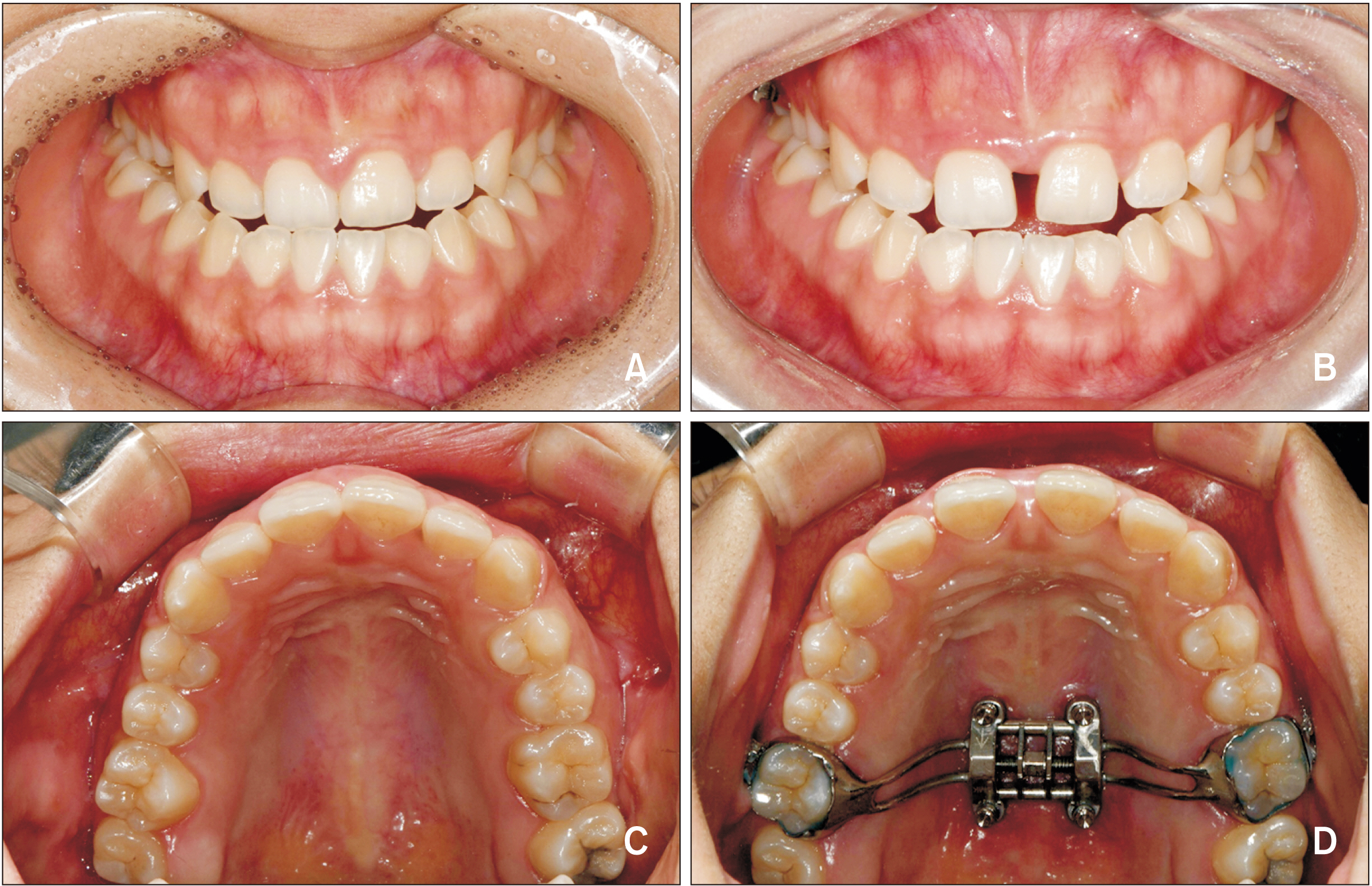
Figure 1 Intraoral photographs of a maxillary skeletal expander (MSE). A, C, Before activation of the MSE. B, Diastema following MSE use. D, After activation of the MSE.

Figure 2 OnDemand 3D (CyberMed Inc, Seoul, Korea) voxel-based superimposition on the cranial base. The yellow box was used to determine the area of the cranial base which was the reference for superimposition.
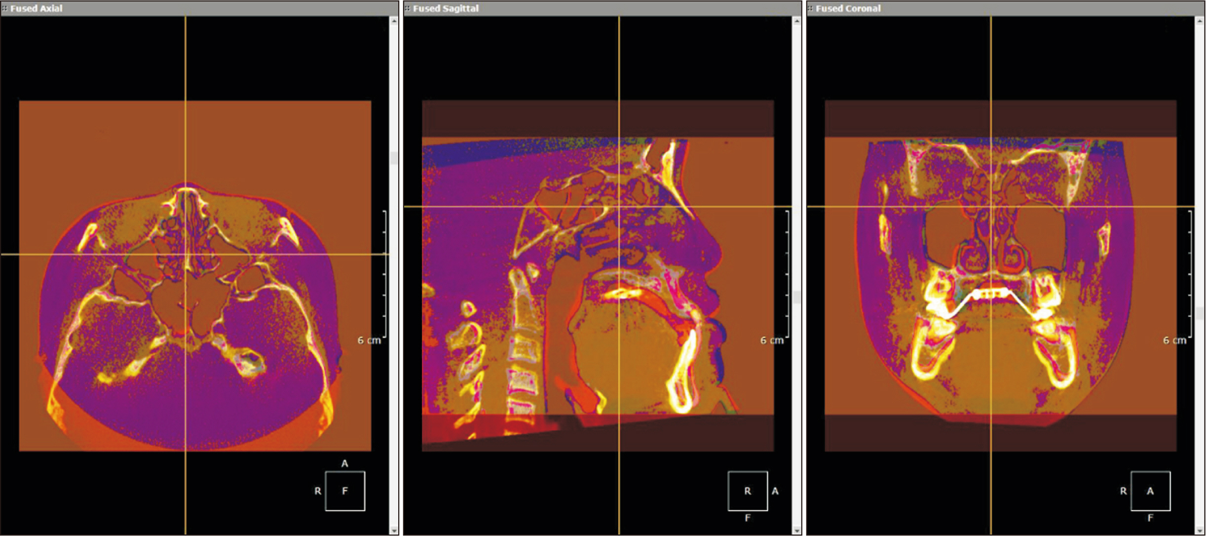
Figure 3 Superimposed pre- and post-expansion cone-beam computerized tomography (CBCT) scans following maxillary skeletal expander use (primary CBCT, violet; secondary CBCT, red).
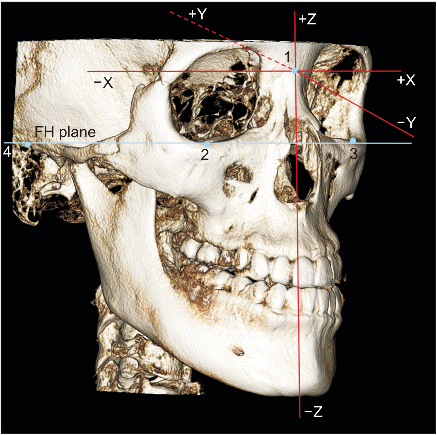
Figure 4 The coordinate system consists of three axes (x, y, z) with their origin (0, 0, 0) registered at nasion. 1, nasion; 2 and 3, orbitale (right and left); 4, porion (right). Positive values are to the left, posterior, and superior to the nasion point. FH, Frankfort horizontal.
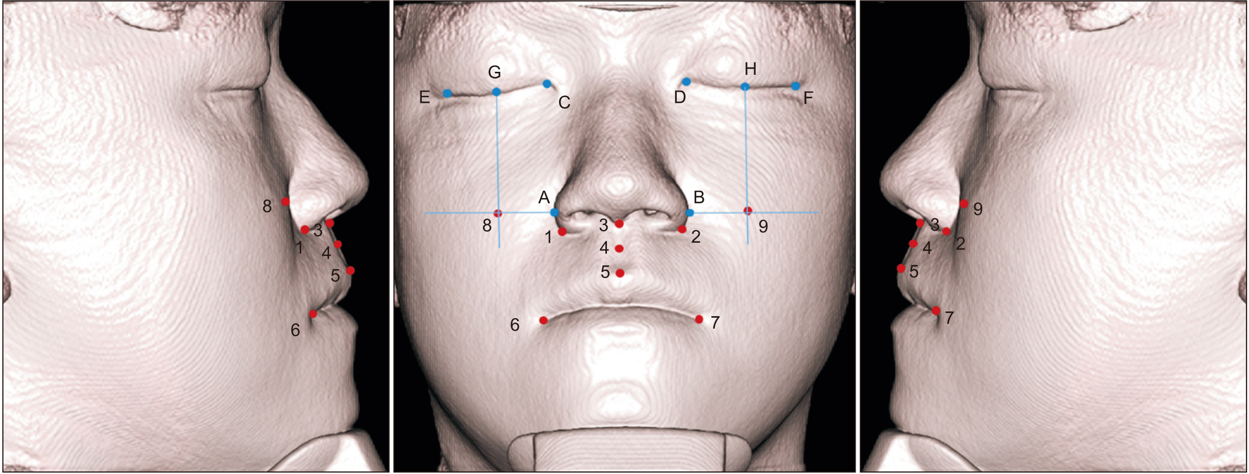
Figure 5 Soft tissue landmarks used in this study. 1 and 2, alar curvature points (right and left); 3, subnasale; 4, soft tissue A point; 5, labrale superius; 6 and 7, cheilion (right and left); 8 and 9, cheek points (right and left; the intersection point of the vertical and horizontal blue lines. The vertical blue line passes through the mid-canthus parallel to the z-axis. The horizontal blue line is perpendicular to the vertical line passing through the alare); A and B, alare (right and left; the most lateral point on each alar contour); C and D, endocanthus (right and left); E and F, exocanthus (right and left); G and H, mid-canthus (right and left).

Figure 6 Hard tissue landmarks used in this study. 1 and 2, A point (right and left); 3 and 4, prosthion (right and left); 5 and 6, ectocanine (right and left); 7 and 8, ectomolare (right and left); and 9 and 10, processus zygomaticus (right and left).
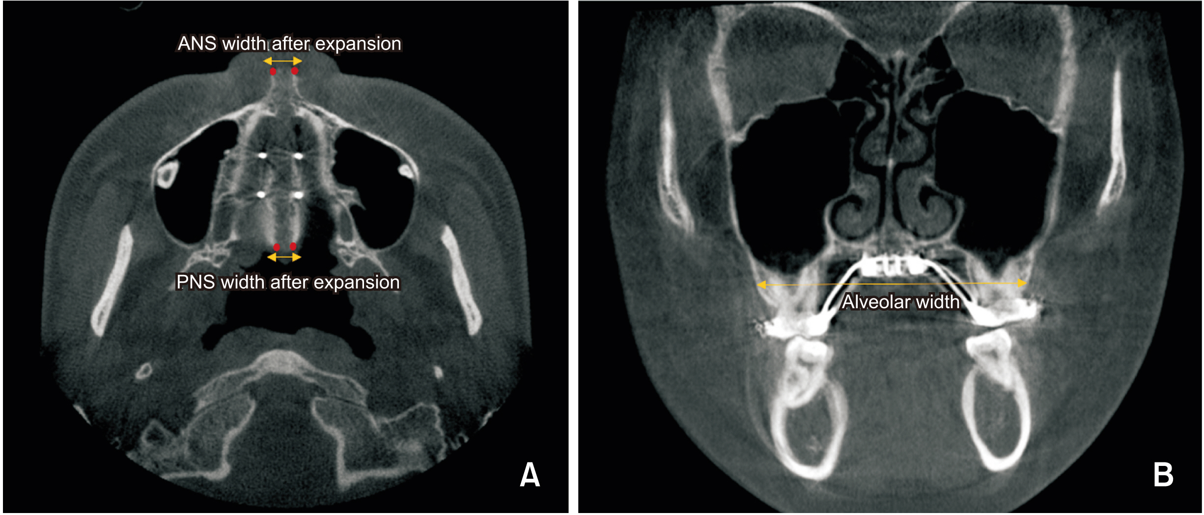
Figure 7 A, Anterior nasal spine (ANS) and posterior nasal spine (PNS) width after expansion. B, Alveolar width after expansion.
Cited by 1 articles
-
Effectiveness of miniscrew assisted rapid palatal expansion using cone beam computed tomography: A systematic review and meta-analysis
Patchaya Siddhisaributr, Kornkanok Khlongwanitchakul, Niwat Anuwongnukroh, Somchai Manopatanakul, Nita Viwattanatipa
Korean J Orthod. 2022;52(3):182-200. doi: 10.4041/kjod21.256.
Reference
-
1. McNamara JA. 2000; Maxillary transverse deficiency. Am J Orthod Dentofacial Orthop. 117:567–70. DOI: 10.1016/S0889-5406(00)70202-2. PMID: 18249297.2. Cross DL, McDonald JP. 2000; Effect of rapid maxillary expansion on skeletal, dental, and nasal structures: a postero-anterior cephalometric study. Eur J Orthod. 22:519–28. DOI: 10.1093/ejo/22.5.519. PMID: 11105408.
Article3. Haas AJ. 1970; Palatal expansion: just the beginning of dentofacial orthopedics. Am J Orthod. 57:219–55. DOI: 10.1016/0002-9416(70)90241-1. PMID: 5263785.
Article4. Adkins MD, Nanda RS, Currier GF. 1990; Arch perimeter changes on rapid palatal expansion. Am J Orthod Dentofacial Orthop. 97:194–9. DOI: 10.1016/S0889-5406(05)80051-4. PMID: 2178393.
Article5. Geran RG, McNamara JA Jr, Baccetti T, Franchi L, Shapiro LM. 2006; A prospective long-term study on the effects of rapid maxillary expansion in the early mixed dentition. Am J Orthod Dentofacial Orthop. 129:631–40. DOI: 10.1016/j.ajodo.2005.01.020. PMID: 16679203.
Article6. Gurel HG, Memili B, Erkan M, Sukurica Y. 2010; Long-term effects of rapid maxillary expansion followed by fixed appliances. Angle Orthod. 80:5–9. DOI: 10.2319/011209-22.1. PMID: 19852633.
Article7. MacGinnis M, Chu H, Youssef G, Wu KW, Machado AW, Moon W. 2014; The effects of micro-implant assisted rapid palatal expansion (MARPE) on the nasomaxillary complex--a finite element method (FEM) analysis. Prog Orthod. 15:52. DOI: 10.1186/s40510-014-0052-y. PMID: 25242527. PMCID: PMC4148550.
Article8. Lagravère MO, Carey J, Heo G, Toogood RW, Major PW. 2010; Transverse, vertical, and anteroposterior changes from bone-anchored maxillary expansion vs traditional rapid maxillary expansion: a randomized clinical trial. Am J Orthod Dentofacial Orthop. 137:304.e1–12. discussion 304–5. DOI: 10.1016/j.ajodo.2009.09.016. PMID: 20197161.
Article9. Lee KJ, Park YC, Park JY, Hwang WS. 2010; Miniscrew-assisted nonsurgical palatal expansion before orthognathic surgery for a patient with severe mandibular prognathism. Am J Orthod Dentofacial Orthop. 137:830–9. DOI: 10.1016/j.ajodo.2007.10.065. PMID: 20685540.
Article10. Carlson C, Sung J, McComb RW, Machado AW, Moon W. 2016; Microimplant-assisted rapid palatal expansion appliance to orthopedically correct transverse maxillary deficiency in an adult. Am J Orthod Dentofacial Orthop. 149:716–28. DOI: 10.1016/j.ajodo.2015.04.043. PMID: 27131254.
Article11. Abedini S, Elkenawy I, Kim E, Moon W. 2018; Three-dimensional soft tissue analysis of the face following micro-implant-supported maxillary skeletal expansion. Prog Orthod. 19:46. DOI: 10.1186/s40510-018-0243-z. PMID: 30450504. PMCID: PMC6240556.
Article12. Lee SR, Lee JW, Chung DH, Lee SM. 2020; Short-term impact of microimplant-assisted rapid palatal expansion on the nasal soft tissues in adults: a three-dimensional stereophotogrammetry study. Korean J Orthod. 50:75–85. DOI: 10.4041/kjod.2020.50.2.75. PMID: 32257933. PMCID: PMC7093666.
Article13. Staderini E, Patini R, De Luca M, Gallenzi P. 2018; Three-dimensional stereophotogrammetric analysis of nasolabial soft tissue effects of rapid maxillary expansion: a systematic review of clinical trials. Acta Otorhinolaryngol Ital. 38:399–408. DOI: 10.14639/0392-100X-2059. PMID: 30498268. PMCID: PMC6265666.
Article14. Kim KB, Adams D, Araújo EA, Behrents RG. 2012; Evaluation of immediate soft tissue changes after rapid maxillary expansion. Dental Press J Orthod. 17:157–64. DOI: 10.1590/S2176-94512012000500022.
Article15. Moss JP. 2006; The use of three-dimensional imaging in orthodontics. Eur J Orthod. 28:416–25. DOI: 10.1093/ejo/cjl025. PMID: 16957057.
Article16. Vanarsdall RL Jr. 1999; Transverse dimension and long-term stability. Semin Orthod. 5:171–80. DOI: 10.1016/S1073-8746(99)80008-5. PMID: 10860069.
Article17. Oh H, Park J, Lagravere-Vich MO. 2019; Comparison of traditional RPE with two types of micro-implant assisted RPE: CBCT study. Semin Orthod. 25:60–8. DOI: 10.1053/j.sodo.2019.02.007.
Article18. Cevidanes LH, Heymann G, Cornelis MA, DeClerck HJ, Tulloch JF. 2009; Superimposition of 3-dimensional cone-beam computed tomography models of growing patients. Am J Orthod Dentofacial Orthop. 136:94–9. DOI: 10.1016/j.ajodo.2009.01.018. PMID: 19577154. PMCID: PMC2750893.
Article19. Bazina M, Cevidanes L, Ruellas A, Valiathan M, Quereshy F, Syed A, et al. 2018; Precision and reliability of Dolphin 3-dimensional voxel-based superimposition. Am J Orthod Dentofacial Orthop. 153:599–606. DOI: 10.1016/j.ajodo.2017.07.025. PMID: 29602352.
Article20. Cantarella D, Dominguez-Mompell R, Mallya SM, Moschik C, Pan HC, Miller J, et al. 2017; Changes in the midpalatal and pterygopalatine sutures induced by micro-implant-supported skeletal expander, analyzed with a novel 3D method based on CBCT imaging. Prog Orthod. 18:34. DOI: 10.1186/s40510-017-0188-7. PMID: 29090368. PMCID: PMC5663987.
Article21. Lim HM, Park YC, Lee KJ, Kim KH, Choi YJ. 2017; Stability of dental, alveolar, and skeletal changes after miniscrew-assisted rapid palatal expansion. Korean J Orthod. 47:313–22. DOI: 10.4041/kjod.2017.47.5.313. PMID: 28861393. PMCID: PMC5548712.
Article22. Sarver DM. 2015; Interactions of hard tissues, soft tissues, and growth over time, and their impact on orthodontic diagnosis and treatment planning. Am J Orthod Dentofacial Orthop. 148:380–6. DOI: 10.1016/j.ajodo.2015.04.030. PMID: 26321335.
Article23. Cevidanes LH, Motta A, Proffit WR, Ackerman JL, Styner M. 2010; Cranial base superimposition for 3-dimensional evaluation of soft-tissue changes. Am J Orthod Dentofacial Orthop. 137(4 Suppl):S120–9. DOI: 10.1016/j.ajodo.2009.04.021. PMID: 20381752. PMCID: PMC2859472.
Article24. Nur RB, Çakan DG, Arun T. 2016; Evaluation of facial hard and soft tissue asymmetry using cone-beam computed tomography. Am J Orthod Dentofacial Orthop. 149:225–37. DOI: 10.1016/j.ajodo.2015.07.038. PMID: 26827979.
Article25. Lee TY, Kim KH, Yu HS, Kim KD, Jung YS, Baik HS. 2014; Correlation analysis of three-dimensional changes of hard and soft tissues in class III orthognathic surgery patients using cone-beam computed tomography. J Craniofac Surg. 25:1530–40. DOI: 10.1097/SCS.0000000000000620. PMID: 25006925.
Article26. Torun GS. 2017; Soft tissue changes in the orofacial region after rapid maxillary expansion: a cone beam computed tomography study. J Orofac Orthop. 78:193–200. DOI: 10.1007/s00056-016-0074-9. PMID: 27957595.27. Nada RM, van Loon B, Maal TJ, Bergé SJ, Mostafa YA, Kuijpers-Jagtman AM, et al. 2013; Three-dimensional evaluation of soft tissue changes in the orofacial region after tooth-borne and bone-borne surgically assisted rapid maxillary expansion. Clin Oral Investig. 17:2017–24. DOI: 10.1007/s00784-013-0927-1. PMID: 23377777.
Article28. Magnusson A, Bjerklin K, Kim H, Nilsson P, Marcusson A. 2013; Three-dimensional computed tomographic analysis of changes to the external features of the nose after surgically assisted rapid maxillary expansion and orthodontic treatment: a prospective longitudinal study. Am J Orthod Dentofacial Orthop. 144:404–13. DOI: 10.1016/j.ajodo.2013.04.013. PMID: 23992813.29. Lima SM Jr, de Moraes M, Asprino L. 2011; Photoelastic analysis of stress distribution of surgically assisted rapid maxillary expansion with and without separation of the pterygomaxillary suture. J Oral Maxillofac Surg. 69:1771–5. DOI: 10.1016/j.joms.2010.07.035. PMID: 21292367.30. Cantarella D, Dominguez-Mompell R, Moschik C, Sfogliano L, Elkenawy I, Pan HC, et al. 2018; Zygomaticomaxillary modifications in the horizontal plane induced by micro-implant-supported skeletal expander, analyzed with CBCT images. Prog Orthod. 19:41. DOI: 10.1186/s40510-018-0240-2. PMID: 30345476. PMCID: PMC6196147.
Article31. Cantarella D, Dominguez-Mompell R, Moschik C, Mallya SM, Pan HC, Alkahtani MR, et al. 2018; Midfacial changes in the coronal plane induced by microimplant-supported skeletal expander, studied with cone-beam computed tomography images. Am J Orthod Dentofacial Orthop. 154:337–45. DOI: 10.1016/j.ajodo.2017.11.033. PMID: 30173836.
Article32. Pangrazio-Kulbersh V, Wine P, Haughey M, Pajtas B, Kaczynski R. 2012; Cone beam computed tomography evaluation of changes in the naso-maxillary complex associated with two types of maxillary expanders. Angle Orthod. 82:448–57. DOI: 10.2319/072211-464.1. PMID: 22032536.
Article33. Abouei E, Lee S, Ford NL. 2015; Quantitative performance characterization of image quality and radiation dose for a CS 9300 dental cone beam computed tomography machine. J Med Imaging (Bellingham). 2:044002. DOI: 10.1117/1.JMI.2.4.044002. PMID: 26587550. PMCID: PMC4650966.
Article34. Molen AD. 2010; Considerations in the use of cone-beam computed tomography for buccal bone measurements. Am J Orthod Dentofacial Orthop. 137(4 Suppl):S130–5. DOI: 10.1016/j.ajodo.2010.01.015. PMID: 20381753.
Article
- Full Text Links
-
- Actions
-
Cited
- CITED
-
- Close
- Share
-
- Similar articles
-
- Differences in dentoskeletal and soft tissue changes due to rapid maxillary expansion using a tooth-borne expander between adolescents and adults: A retrospective observational study
- Does mini-implant-supported rapid maxillary expansion cause less root resorption than traditional approaches? A micro-computed tomography study
- Soft tissue change of the midface in skeletal class III orthognathic surgery patients
- Alterations of the soft tissue dimensions around implant-supported single-tooth replacements in the maxillary anterior region: A retrospective longitudinal study
- Relationship between midfacial fractures and maxillary sinus pathology
