Ann Clin Neurophysiol.
2021 Apr;23(1):56-60. 10.14253/acn.2021.23.1.56.
Tectal glioma presenting with adult-onset epileptic seizures
- Affiliations
-
- 1Department of Neurology, Samsung Medical Center, Sungkyunkwan University School of Medicine, Seoul, Korea
- 2Department of Neurosurgery, Samsung Medical Center, Sungkyunkwan University School of Medicine, Seoul, Korea
- KMID: 2515609
- DOI: http://doi.org/10.14253/acn.2021.23.1.56
Abstract
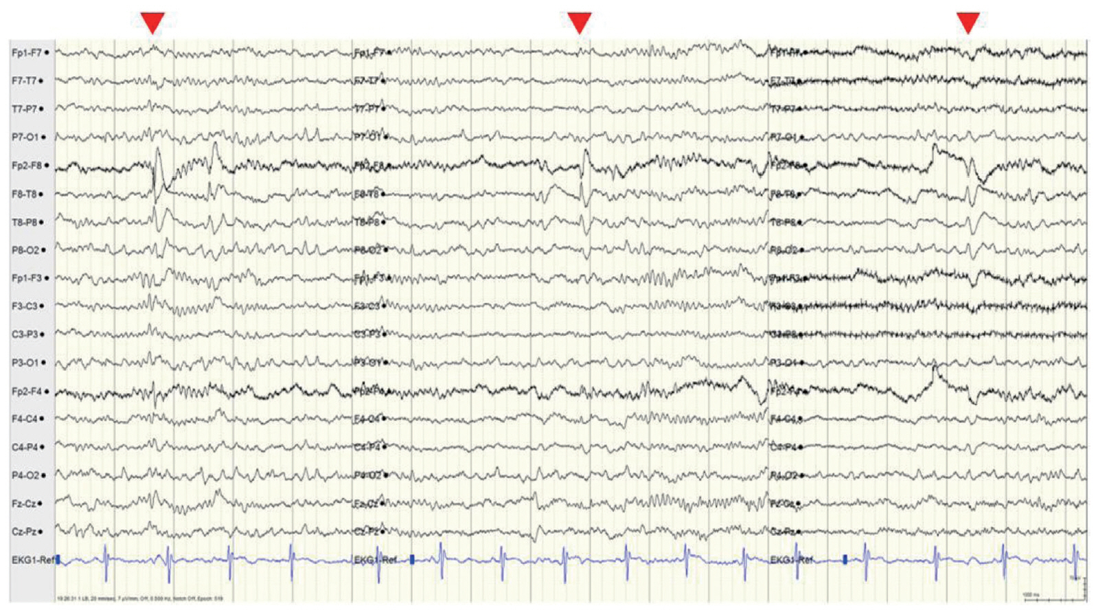
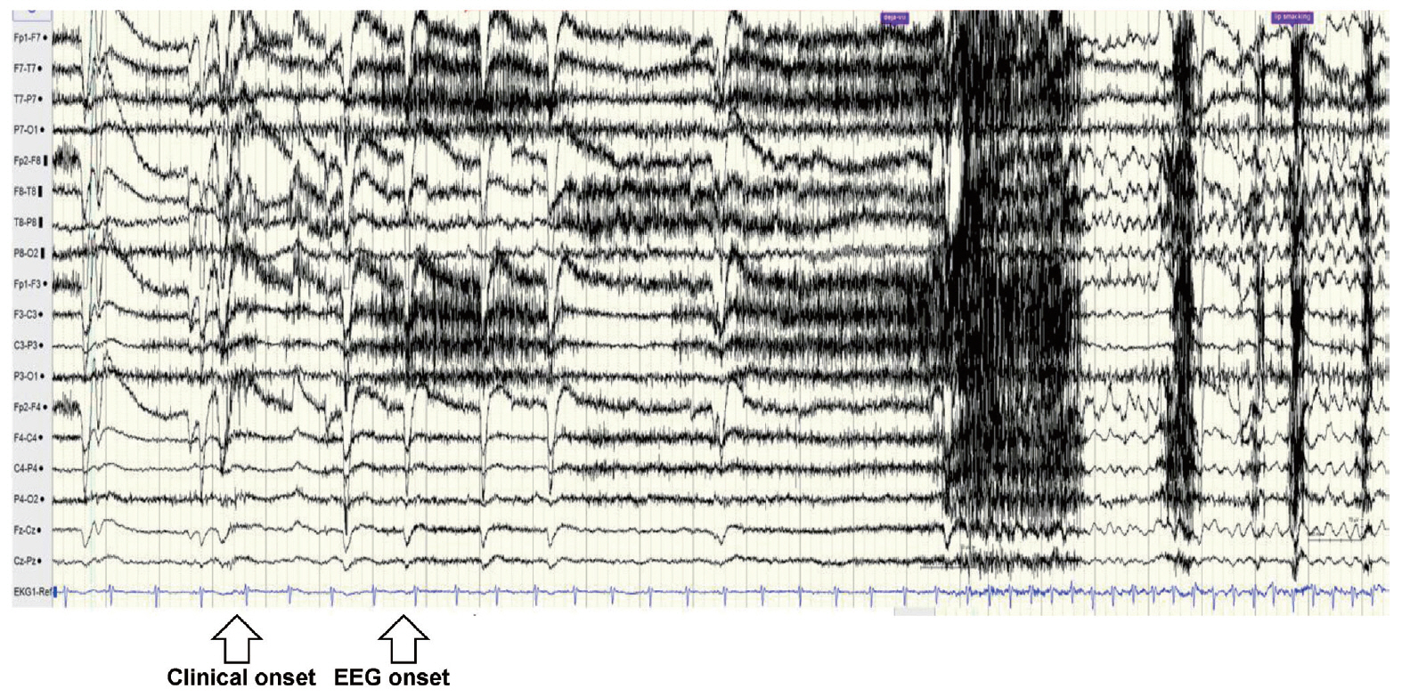
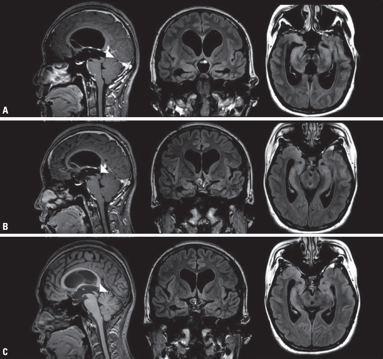
- Tectal glioma is an indolent and benign tumor that occurs predominantly in the pediatric population. It arises in the tectum of the midbrain and, due to its location, contributes to the development of obstructive hydrocephalus, typically presenting with increased intracranial pressure (IICP) symptoms or signs. Here we report a rare case of tectal glioma that presented as adult-onset epileptic seizures without IICP symptoms and was treated with endoscopic third ventriculostomy and antiepileptic drugs.
Keyword
Figure
Reference
-
1. Hosking GP. Fits in hydrocephalic children. Arch Dis Child. 1974; 49:633–635.
Article2. Phi JH, Kim SK, Kang TH, Wang KC. Hydrocephalic fits: forgotten but not gone. Childs Nerv Syst. 2012; 28:1863–1868.
Article3. Dabscheck G, Prabhu SP, Manley PE, Goumnerova L, Ullrich NJ. Risk of seizures in children with tectal gliomas. Epilepsia. 2015; 56:e139–e142.
Article4. Igboechi C, Vaddiparti A, Sorenson EP, Rozzelle CJ, Tubbs RS, Loukas M. Tectal plate gliomas: a review. Childs Nerv Syst. 2013; 29:1827–1833.
Article5. Reyes-Botero G, Mokhtari K, Martin-Duverneuil N, Delattre JY, Laigle-Donadey F. Adult brainstem gliomas. Oncologist. 2012; 17:388–397.
Article6. Liu APY, Harreld JH, Jacola LM, Gero M, Acharya S, Ghazwani Y, et al. Tectal glioma as a distinct diagnostic entity: a comprehensive clinical, imaging, histologic and molecular analysis. Acta Neuropathol Commun. 2018; 6:101.
Article7. Lapras C, Bognar L, Turjman F, Villanyi E, Mottolese C, Fischer C, et al. Tectal plate gliomas. Part I: microsurgery of the tectal plate gliomas. Acta Neurochir (Wien). 1994; 126:76–83.
Article8. Li KW, Roonprapunt C, Lawson HC, Abbott IR, Wisoff J, Epstein F, et al. Endoscopic third ventriculostomy for hydrocephalus associated with tectal gliomas. Neurosurg Focus. 2005; 18:E2.
Article9. Heo K. Sleep and epilepsy. J Korean Sleep Res Soc. 2009; 6:69–73.
Article
- Full Text Links
- Actions
-
Cited
- CITED
-
- Close
- Share
- Similar articles
-
- Comparison of 3 and 7 Tesla Magnetic Resonance Imaging of Obstructive Hydrocephalus Caused by Tectal Glioma
- Non-epileptic paroxysmal events during sleep: Differentiation from epileptic seizures
- Seizure Induced or Aggravated by Carbamazepine
- Seizure Disorders Mimicking Epilepsy
- Case Reports of Tectal Plate Gliomas Showing Indolent Course


