Epidural hematoma treated by aspiration after transforaminal epidural steroid injection - A case report -
- Affiliations
-
- 1Department of Anesthesiology and Pain Medicine, Kangdong Sacred Heart Hospital, College of Medicine, Hallym University, Seoul, Korea
- KMID: 2515545
- DOI: http://doi.org/10.17085/apm.20085
Abstract
- Background
Spinal epidural hematoma is rare condition that can rapidly develop into severe neurologic deficits. The pathophysiology of this development remains unclear. There are several case reports of emergency hematoma evacuations after epidural steroid injection. Case: We report on two patients who developed acute, large amounts of epidural hematoma without neurological deficits after transforaminal epidural steroid injection. After fluoroscopy guided aspiration for epidural hematoma was performed, neurological defects did not progress and the hematoma was shown to be absorbed on magnetic resonance imaging.
Conclusions
These reports are believed to be the first of treating epidural hematoma occurring after transforaminal epidural steroid injection through non-surgical hematoma aspiration. If large amounts of epidural hematoma are not causing neurological issues, it can be aspirated until it is absorbed.
Keyword
Figure
-
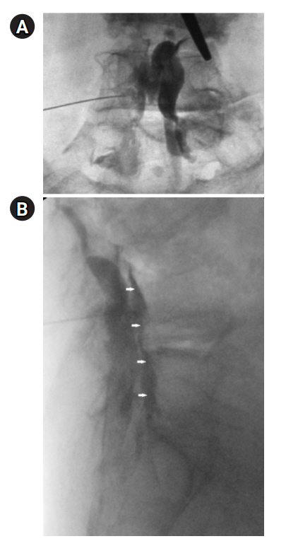
Fig. 1. (A, B) The contrast was injected and spread well in the anterior epidural space (white arrows).

Fig. 2. (A–D) T1- and T2-weighted sagittal and axial MRI images showing the 14-centimeter epidural hematoma (white arrows) at the T11-L4 level. MRI: magnetic resonance imaging.
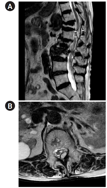
Fig. 3. (A, B) T2-weighted sagittal and axial MRI images showing a decrease in the epidural hematoma (white arrows) and nerve compression. MRI: magnetic resonance imaging.
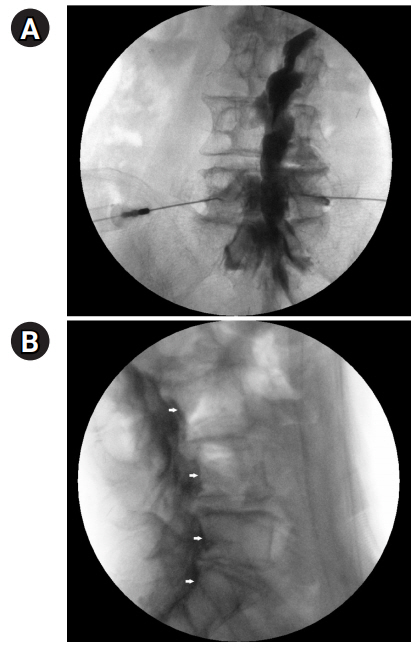
Fig. 4. (A, B) AP/lat. fluoroscopy image showing the anterior epidural space spreading (white arrows).
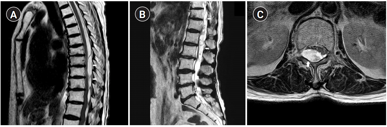
Fig. 5. (A, B) T2-weighted sagittal MRI image showing a large amount of epidural hematoma (white arrows) at the lower T-L spine and clumping of cauda equina at the L1-2 level. (C) T2-weighted axial MRI image at the L1 level showing epidural hematoma and nerve compression (white arrow). MRI: magnetic resonance imaging.
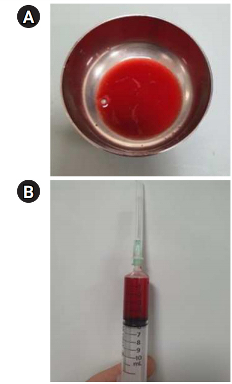
Fig. 6. (A, B) A total of 5 ml of diluted blood aspirated through an epidural catheter.
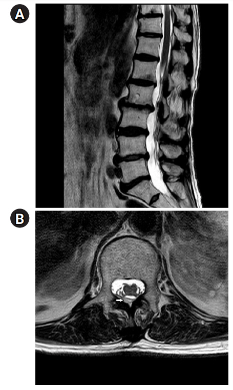
Fig. 7. (A, B) T2-weighted sagittal and axial MRI images showing decreased epidural hematoma (white arrow) and nerve compression. MRI: magnetic resonance imaging.
Reference
-
1. Wong AY, Karppinen J, Samartzis D. Low back pain in older adults: risk factors, management options and future directions. Scoliosis Spinal Disord. 2017; 12:14.2. Kim SI, Lee DH, Kim SH, Cho YH. Spinal epidural hematoma occurring at a distance from the transforaminal epidural injection site: a case report. Medicine (Baltimore). 2019; 98:e16654.3. Epstein NE. The risks of epidural and transforaminal steroid injections in the spine: commentary and a comprehensive review of the literature. Surg Neurol Int. 2013; 4(Suppl 2):S74–93.4. Mandell JC, Czuczman GJ, Gaviola GC, Ghazikhanian V, Cho CH. The lumbar neural foramen and transforaminal epidural steroid injections: an anatomic review with key safety considerations in planning the percutaneous approach. AJR Am J Roentgenol. 2017; 209:W26–35.5. Davis N, Hourigan P, Clarke A. Transforaminal epidural steroid injection in lumbar spinal stenosis: an observational study with two-year follow-up. Br J Neurosurg. 2017; 31:205–8.6. Manchikanti L, Singh V, Pampati V, Falco FJ, Hirsch JA. Comparison of the efficacy of caudal, interlaminar, and transforaminal epidural injections in managing lumbar disc herniation: is one method superior to the other? Korean J Pain. 2015; 28:11–21.7. Al-Mutair A, Bednar DA. Spinal epidural hematoma. J Am Acad Orthop Surg. 2010; 18:494–502.8. Xu R, Bydon M, Gokaslan ZL, Wolinsky JP, Witham TF, Bydon A. Epidural steroid injection resulting in epidural hematoma in a patient despite strict adherence to anticoagulation guidelines. J Neurosurg Spine. 2009; 11:358–64.9. Lefranc F, David P, Brotchi J, De Witte O. Traumatic epidural hematoma of the cervical spine: magnetic resonance imaging diagnosis and spontaneous resolution: case report. Neurosurgery. 1999; 44:408–10.10. Jang JW, Lee JK, Seo BR, Kim JH, Kim SH. Spontaneous resolution of a traumatic cervicothoracic epidural hematoma presenting with transient paraplegia: a case report. Spine (Phila Pa 1976). 2010; 35:E564–7.11. Gungor S, Aiyer R. Epidural hematoma development contralateral to dura after lumbar transforaminal epidural steroid injection. Pain Manag. 2017; 7:367–75.12. Choi JJ, Chang YJ, Jung WS, Lee KC, Kim JH, Jo YY. Discordant lumbar epidural hematoma after caudal steroid injection: a case report (CARE-compliant). Medicine (Baltimore). 2017; 96:e7127.13. Shanthanna H, Park J. Acute epidural haematoma following epidural steroid injection in a patient with spinal stenosis. Anaesthesia. 2011; 66:837–9.14. Narawong D, Gibbons VP, McLaughlin JR, Bouhasin JD, Kotagal S. Conservative management of spinal epidural hematoma in hemophilia. Pediatr Neurol. 1988; 4:169–71.
- Full Text Links
-
- Actions
-
Cited
- CITED
-
- Close
- Share
-
- Similar articles
-
- Acute Cervical Subdural Hematoma with Quadriparesis after Cervical Transforaminal Epidural Block
- Comparison of Transforaminal Epidural Steroid Injection and Lumbar/Caudal Epidural Steroid Injection for the Treatment of Lumbosacral Radiculopathy
- Epidural Steroid Injection
- Oblique interlaminar lumbar epidural steroid injection for management of low back pain with lumbosacral radicular pain: A case report
- Selective Epidural Steroid Injection in a Patient with Refractory Radicular Leg Pain: A case report
