Radioactive Parathyroid Adenomas on Sestamibi Scans: Low Parathyroid Hormone Secretory Potential and Large Volume
- Affiliations
-
- 1Department of Internal Medicine, Seoul National University Hospital, Seoul National University College of Medicine, Seoul, Korea
- 2Department of Internal Medicine, Seoul National University Bundang Hospital, Seongnam, Korea
- 3Department of Internal Medicine, Seoul Metropolitan Government Seoul National University Boramae Medical Center, Seoul, Korea
- KMID: 2515461
- DOI: http://doi.org/10.3803/EnM.2020.823
Abstract
- Background
We investigated the clinical characteristics of parathyroid adenomas according to radioactivity on 99mTc-methoxyisobutylisonitrile (99mTc-MIBI) single-photon emission computed tomography/computed tomography (SPECT/CT) in primary hyperparathyroidism (PHPT) patients.
Methods
The study included 217 patients diagnosed with PHPT from 2000 to 2019 at Seoul National University Hospital who underwent 99mTc-MIBI SPECT/CT scans. On SPECT/CT, the radioactivity of parathyroid adenomas was measured as the ratio of the mean radioactivity count of the parathyroid adenoma to that of the contralateral thyroid.
Results
Tumors were localized by MIBI scans in 190 patients (MIBI [+] group) and by ultrasound or parathyroid four-dimensional CT in 27 patients (MIBI [–] group). The mean age was 55 years, and mean body mass index was 23.4 kg/m2. Patients in the MIBI (+) group had higher parathyroid hormone (iPTH) and lower 25-hydroxy vitamin D levels than those in the MIBI (–) group (168.0 pg/mL [interquartile range, IQR, 111.0 to 250.7] vs. 134.7 pg/mL [IQR, 98.2 to 191.2], P=0.049; 15.4 ng/mL [IQR, 11.1 to 20.8] vs. 21.2 ng/mL [IQR, 13.9 to 24.8], P=0.012, respectively). Patients in the MIBI (+) group had larger tumor volumes, but lower iPTH/volume ratios than those in the MIBI (–) group (1,216.66 [IQR, 513.40 to 2,663.02], 499.82 mm3 [IQR, 167.77 to 1,229.80], P=0.002; 0.18 [IQR, 0.08 to 0.46], 0.40 pg/mL/mm3 [IQR, 0.16 to 1.29], P=0.016, respectively). Adenoma radioactivity was positively correlated with calcium, iPTH, and volume (r=0.180, P=0.020; r=0.208, P=0.006; r=0.288, P<0.001, respectively), but not with iPTH/volume.
Conclusion
Parathyroid adenomas with positive MIBI scans had larger volumes and higher iPTH than adenomas with negative scans, but lower iPTH per unit volume.
Keyword
Figure
-
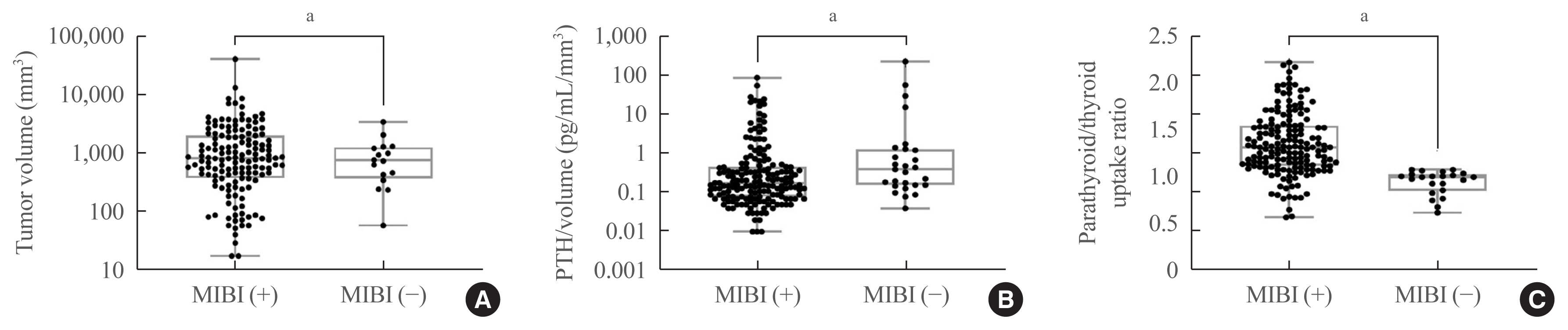
Fig. 1 (A) Tumor volume, (B) parathyroid hormone (PTH)/volume, and (C) radioactivity of parathyroid adenomas according to methoxyisobutylisonitrile (MIBI) scan uptake. Radioactivity of parathyroid adenomas refers to the parathyroid/thyroid uptake ratio.
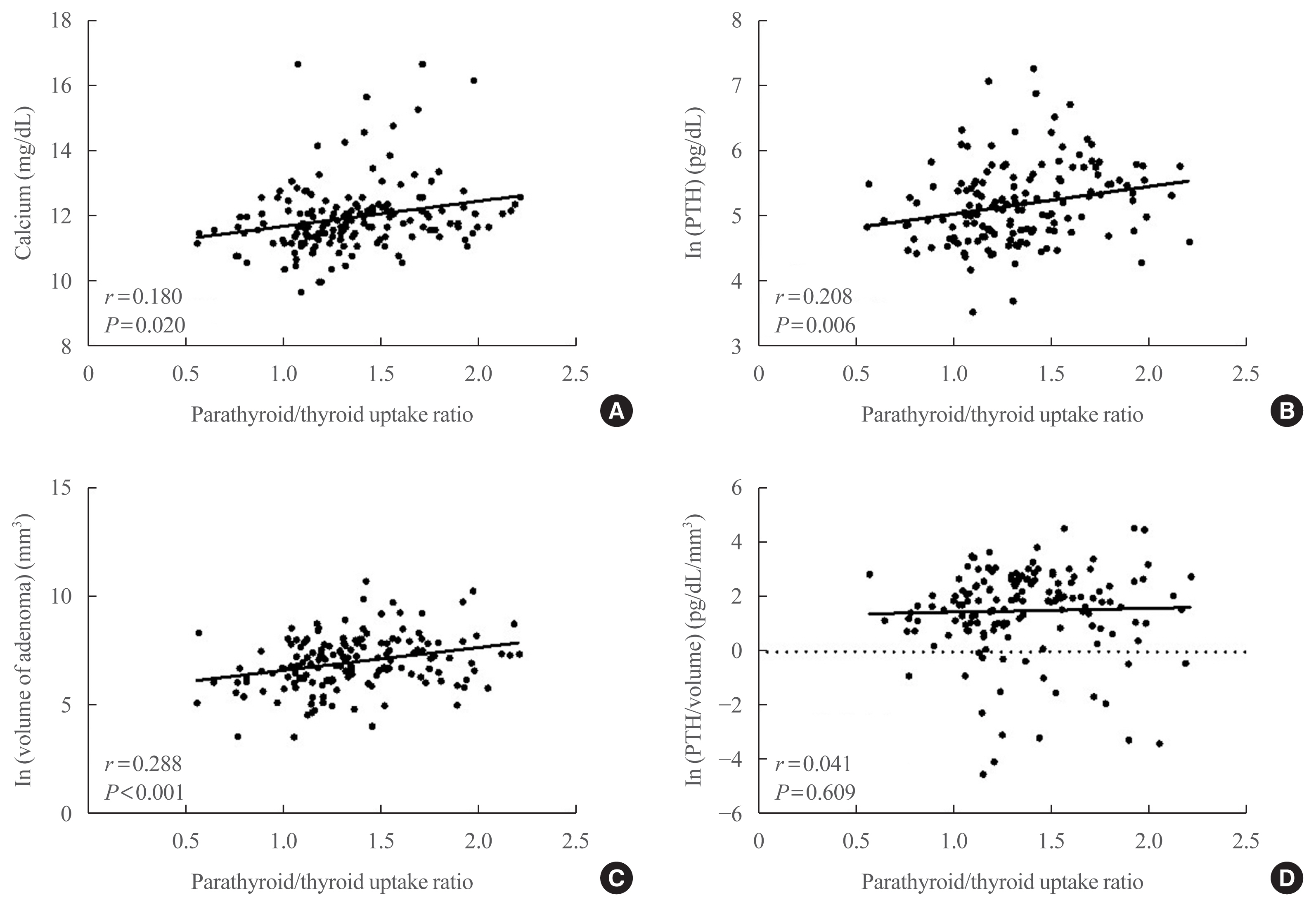
Fig. 2 Correlations between the radioactivity of parathyroid adenomas and (A) calcium and (B) parathyroid hormone (PTH) levels, (C) volume of adenomas, and (D) the PTH/volume ratio. Radioactivity of parathyroid adenomas refers to the parathyroid/thyroid uptake ratio. ln, natural logarithm.
Cited by 2 articles
-
Atypical parathyroid tumor: clinical and parathyroid hormone response to surgical treatment
Antonio Giulio Napolitano, Massimo Monacelli, Valeria Liparulo, Eleonora Coviello, Domenico Pourmolkara, Stefano Avenia, Andrea Polistena
Ann Surg Treat Res. 2023;105(2):76-81. doi: 10.4174/astr.2023.105.2.76.Protein Signatures of Parathyroid Adenoma according to Tumor Volume and Functionality
Sung Hye Kong, Jeong Mo Bae, Jung Hee Kim, Sang Wan Kim, Dohyun Han, Chan Soo Shin
Endocrinol Metab. 2024;39(2):375-386. doi: 10.3803/EnM.2023.1827.
Reference
-
1. Khan A, Bilezikian J. Primary hyperparathyroidism: pathophysiology and impact on bone. CMAJ. 2000; 163:184–7.2. Silverberg SJ, Lewiecki EM, Mosekilde L, Peacock M, Rubin MR. Presentation of asymptomatic primary hyperparathyroidism: proceedings of the third international workshop. J Clin Endocrinol Metab. 2009; 94:351–65.
Article3. Rubin MR, Bilezikian JP, McMahon DJ, Jacobs T, Shane E, Siris E, et al. The natural history of primary hyperparathyroidism with or without parathyroid surgery after 15 years. J Clin Endocrinol Metab. 2008; 93:3462–70.
Article4. Mihai R, Simon D, Hellman P. Imaging for primary hyperparathyroidism: an evidence-based analysis. Langenbecks Arch Surg. 2009; 394:765–84.
Article5. Kim YI, Jung YH, Hwang KT, Lee HY. Efficacy of 99mTc-sestamibi SPECT/CT for minimally invasive parathyroidectomy: comparative study with 99mTc-sestamibi scintigraphy, SPECT, US and CT. Ann Nucl Med. 2012; 26:804–10.
Article6. Chiu ML, Kronauge JF, Piwnica-Worms D. Effect of mitochondrial and plasma membrane potentials on accumulation of hexakis (2-methoxyisobutylisonitrile) technetium(I) in cultured mouse fibroblasts. J Nucl Med. 1990; 31:1646–53.7. Melloul M, Paz A, Koren R, Cytron S, Feinmesser R, Gal R. 99mTc-MIBI scintigraphy of parathyroid adenomas and its relation to tumour size and oxyphil cell abundance. Eur J Nucl Med. 2001; 28:209–13.
Article8. Mshelia DS, Hatutale AN, Mokgoro NP, Nchabaleng ME, Buscombe JR, Sathekge MM. Correlation between serum calcium levels and dual-phase (99m)Tc-sestamibi parathyroid scintigraphy in primary hyperparathyroidism. Clin Physiol Funct Imaging. 2012; 32:19–24.
Article9. Biertho LD, Kim C, Wu HS, Unger P, Inabnet WB. Relationship between sestamibi uptake, parathyroid hormone assay, and nuclear morphology in primary hyperparathyroidism. J Am Coll Surg. 2004; 199:229–33.10. Carpentier A, Jeannotte S, Verreault J, Lefebvre B, Bisson G, Mongeau CJ, et al. Preoperative localization of parathyroid lesions in hyperparathyroidism: relationship between technetium-99m-MIBI uptake and oxyphil cell content. J Nucl Med. 1998; 39:1441–4.11. Akin M, Atasever T, Kurukahvecioglu O, Dogan M, Gokaslan D, Poyraz A, et al. Preoperative detection of parathyroid adenomas with Tc-99m MIBI and Tc-99m pertechnetate scintigraphy: histopathological and biochemical correlation with Tc-99m MIBI uptake. Bratisl Lek Listy. 2009; 110:166–9.12. Chen CC, Skarulis MC, Fraker DL, Alexander R, Marx SJ, Spiegel AM. Technetium-99m-sestamibi imaging before reoperation for primary hyperparathyroidism. J Nucl Med. 1995; 36:2186–91.13. Leslie WD, Riese KT, Dupont JO, Peterdy AE. Parathyroid adenomas without sestamibi retention. Clin Nucl Med. 1995; 20:699–702.
Article14. Hiromatsu Y, Ishibashi M, Nishida H, Okuda S, Miyake I. Technetium-99m tetrofosmin parathyroid imaging in patients with primary hyperparathyroidism. Intern Med. 2000; 39:101–6.
Article15. Piga M, Bolasco P, Satta L, Altieri P, Loi G, Nicolosi A, et al. Double phase parathyroid technetium-99m-MIBI scintigraphy to identify functional autonomy in secondary hyperparathyroidism. J Nucl Med. 1996; 37:565–9.16. Bhatnagar A, Vezza PR, Bryan JA, Atkins FB, Ziessman HA. Technetium-99m-sestamibi parathyroid scintigraphy: effect of P-glycoprotein, histology and tumor size on detectability. J Nucl Med. 1998; 39:1617–20.17. Williams JG, Wheeler MH, Aston JP, Brown RC, Woodhead JS. The relationship between adenoma weight and intact (1–84) parathyroid hormone level in primary hyperparathyroidism. Am J Surg. 1992; 163:301–4.
Article18. Cantley LK, Ontjes DA, Cooper CW, Thomas CG, Leight GS, Wells SA Jr. Parathyroid hormone secretion from dispersed human hyperparathyroid cells: increased secretion in cells from hyperplastic glands versus adenomas. J Clin Endocrinol Metab. 1985; 60:1032–7.
Article19. Randhawa PS, Mace AD, Nouraei SA, Stearns MP. Primary hyperparathyroidism: do perioperative biochemical variables correlate with parathyroid adenoma weight or volume? Clin Otolaryngol. 2007; 32:179–84.
Article20. Kamani F, Najafi A, Mohammadi SS, Tavassoli S, Shojaei SP. Correlation of biochemical markers of primary hyperparathyroidism with single adenoma weight and volume. Indian J Surg. 2013; 75:102–5.
Article21. Harris L, Yoo J, Driedger A, Fung K, Franklin J, Gray D, et al. Accuracy of technetium-99m SPECT-CT hybrid images in predicting the precise intraoperative anatomical location of parathyroid adenomas. Head Neck. 2008; 30:509–17.
Article22. Oksuz MO, Dittmann H, Wicke C, Mussig K, Bares R, Pfannenberg C, et al. Accuracy of parathyroid imaging: a comparison of planar scintigraphy, SPECT, SPECT-CT, and C-11 methionine PET for the detection of parathyroid adenomas and glandular hyperplasia. Diagn Interv Radiol. 2011; 17:297–307.23. Cheung K, Wang TS, Farrokhyar F, Roman SA, Sosa JA. A meta-analysis of preoperative localization techniques for patients with primary hyperparathyroidism. Ann Surg Oncol. 2012; 19:577–83.
Article24. Boi F, Lombardo C, Cocco MC, Piga M, Serra A, Lai ML, et al. Thyroid diseases cause mismatch between MIBI scan and neck ultrasound in the diagnosis of hyperfunctioning parathyroids: usefulness of FNA-PTH assay. Eur J Endocrinol. 2012; 168:49–58.
Article25. Khan AA, Hanley DA, Rizzoli R, Bollerslev J, Young JE, Rejnmark L, et al. Primary hyperparathyroidism: review and recommendations on evaluation, diagnosis, and management: a Canadian and international consensus. Osteoporos Int. 2017; 28:1–19.
Article26. Sinha TK, Miller S, Feming J, Khairi R, Edmondson J, Johnston CC Jr, et al. Demonstration of a diurnal variation in serum parathyroid hormone in primary and secondary hyperparathyroidism. J Clin Endocrinol Metab. 1975; 41:1009–13.
Article27. Moosgaard B, Vestergaard P, Heickendorff L, Melsen F, Christiansen P, Mosekilde L. Vitamin D status, seasonal variations, parathyroid adenoma weight and bone mineral density in primary hyperparathyroidism. Clin Endocrinol (Oxf). 2005; 63:506–13.
Article28. Nevo-Shor A, Kogan S, Joshua BZ, Bahat-Dinur A, Novack V, Fraenkel M. Seasonal changes in serum calcium, PTH and vitamin D levels in patients with primary hyperparathyroidism. Bone. 2016; 89:59–63.
Article
- Full Text Links
-
- Actions
-
Cited
- CITED
-
- Close
- Share
-
- Similar articles
-
- Parathyroid Adenoma without Hyperparathyroidism Presenting as a Large Neck Mass
- Supernumerary Ectopic Mediastinal Parathyroid Adenoma Combined With Parathyroid Hyperplasia
- Association of the Parathyroid Adenoma Volume and the Biochemical Parameters in Primary Hyperparathyroidism
- False-Positive Parathyroid Sestamibi in Minimally Invasive Radioguided Parathyroidectomy
- Giant Parathyroid Adenomas: Differential Aspects Compared to Atypical Parathyroid Adenomas
