Relationships Between Relative Ankle Muscle Ratios, Severity of Symptoms, and Radiologic Parameters in Adolescent Patients With Symptomatic Flexible Flat Feet
- Affiliations
-
- 1Department of Rehabilitation Medicine, Chungnam National University Hospital, Chungnam National University College of Medicine, Daejeon, Korea
- KMID: 2515393
- DOI: http://doi.org/10.5535/arm.20174
Abstract
Objective
To investigate differences in the relative sizes of the ankle-stabilizing muscles in individuals with versus without flexible flat feet and to determine predictors of symptom severity.
Methods
This cross-sectional study included 30 patients with symptomatic flexible flat feet and 24 normal controls. The following were evaluated: foot posture index, resting calcaneal stance position angle, radiographic findings (calcaneal pitch, Meary’s angle, talocalcaneal angle, talonavicular coverage angle [TNCA]), foot function index (FFI), and cross-sectional areas (CSA) of the tibialis anterior (TA), tibialis posterior (TP), and peroneus longus (PL) upon ultrasonographic examination. To address morphometric differences among participants, individual muscle measurements were normalized to proportions of total muscle CSA. Between-group differences were evaluated with independent t-tests. Correlations between muscle ratios, radiographic parameters, and FFI scores were investigated. Logistic regression analysis was performed to determine which parameters predicted severe symptoms.
Results
The relative size of the TP was significantly greater and those of the TA and PL were significantly smaller in patients with flat feet than in normal controls. Correlations were found among relative muscle CSA ratios, radiographic parameters, and FFI score. Linear regression analysis confirmed that the TNCA and the relative CSA of the PL were independent predictors of symptom severity.
Conclusion
This study found significant differences in the relative CSAs of the ankle muscles in patients with flexible flat feet versus individuals without flat feet; these differences were significantly correlated with anatomic abnormalities. Symptoms were more severe in patients with relatively greater forefoot abduction and relatively smaller PL.
Keyword
Figure
-
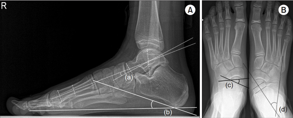
Fig. 1. (A, B) Radiographic parameters: (a) Meary’s angle, (b) calcaneal pitch, (c) talonavicular coverage angle, and (d) talocalcaneal angle.
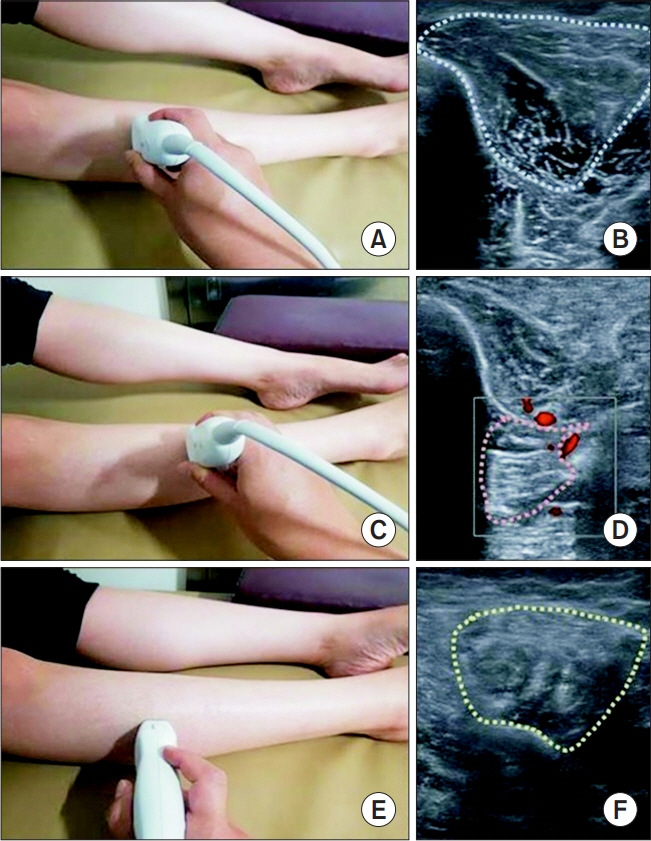
Fig. 2. Scanned structures, probe position, and corresponding sample images. (A) TA probe position. (B) CSA of TA. (C) TP probe position. (D) CSA of TP. (E) PL probe position. (F) CSA of PL. TA, tibialis anterior; TP, tibialis posterior; PL, peroneus longus; CSA, cross-sectional area.
Cited by 2 articles
-
Biomechanical Evidence From Ultrasonography Supports Rigid Foot Orthoses in Children With Flatfoot
Joon-Ho Shin
Ann Rehabil Med. 2021;45(6):411-412. doi: 10.5535/arm.21189.Effect of Foot Orthoses in Children With Symptomatic Flexible Flatfoot Based on Ultrasonography of the Ankle Invertor and Evertor Muscles
Dong Joon Cho, So Young Ahn, Soo-Kyung Bok
Ann Rehabil Med. 2021;45(6):459-470. doi: 10.5535/arm.21137.
Reference
-
1. Harris EJ, Vanore JV, Thomas JL, Kravitz SR, Mendelson SA, Mendicino RW, et al. Diagnosis and treatment of pediatric flatfoot. J Foot Ankle Surg. 2004; 43:341–73.
Article2. Garcia-Rodriguez A, Martin-Jimenez F, Carnero-Varo M, Gomez-Gracia E, Gomez-Aracena J, Fernandez-Crehuet J. Flexible flat feet in children: a real problem? Pediatrics. 1999; 103:e84.3. Evans AM, Rome K. A Cochrane review of the evidence for non-surgical interventions for flexible pediatric flat feet. Eur J Phys Rehabil Med. 2011; 47:69–89.4. Banwell HA, Paris ME, Mackintosh S, Williams CM. Paediatric flexible flat foot: how are we measuring it and are we getting it right?: a systematic review. J Foot Ankle Res. 2018; 11:21.
Article5. Buldt AK, Levinger P, Murley GS, Menz HB, Nester CJ, Landorf KB. Foot posture is associated with kinematics of the foot during gait: a comparison of normal, planus and cavus feet. Gait Posture. 2015; 42:42–8.
Article6. Hunt AE, Smith RM. Mechanics and control of the flat versus normal foot during the stance phase of walking. Clin Biomech (Bristol, Avon). 2004; 19:391–7.
Article7. Murley GS, Menz HB, Landorf KB. Foot posture influences the electromyographic activity of selected lower limb muscles during gait. J Foot Ankle Res. 2009; 2:35.
Article8. Murley GS, Tan JM, Edwards RM, De Luca J, Munteanu SE, Cook JL. Foot posture is associated with morphometry of the peroneus longus muscle, tibialis anterior tendon, and Achilles tendon. Scand J Med Sci Sports. 2014; 24:535–41.
Article9. Angin S, Crofts G, Mickle KJ, Nester CJ. Ultrasound evaluation of foot muscles and plantar fascia in pes planus. Gait Posture. 2014; 40:48–52.
Article10. Pontaga I. Ankle joint evertor-invertor muscle torque ratio decrease due to recurrent lateral ligament sprains. Clin Biomech (Bristol, Avon). 2004; 19:760–2.
Article11. Schon LC, Weinfeld SB, Horton GA, Resch S. Radiographic and clinical classification of acquired midtarsus deformities. Foot Ankle Int. 1998; 19:394–404.
Article12. Youn KJ, Ahn SY, Kim BO, Park IS, Bok SK. Long-term effect of rigid foot orthosis in children older than six years with flexible flat foot. Ann Rehabil Med. 2019; 43:224–9.
Article13. Budiman-Mak E, Conrad KJ, Roach KE. The Foot Function Index: a measure of foot pain and disability. J Clin Epidemiol. 1991; 44:561–70.
Article14. Lee DY, Kim YM, Lee JH, Kim J, Kim JB, Kim BS, et al. Validation of electronic foot function index in patients with foot and ankle disease: a randomized, prospective multicenter study. J Korean Foot Ankle Soc. 2019; 23:24–30.
Article15. Ishida H, Watanabe S. Influence of inward pressure of the transducer on lateral abdominal muscle thickness during ultrasound imaging. J Orthop Sports Phys Ther. 2012; 42:815–8.
Article16. Hintermann B. Tibialis posterior dysfunction: a review of the problem and personal experience. Foot Ankle Surg. 1997; 3:61–70.
Article17. Ross MH, Smith MD, Mellor R, Vicenzino B. Exercise for posterior tibial tendon dysfunction: a systematic review of randomised clinical trials and clinical guidelines. BMJ Open Sport Exerc Med. 2018; 4:e000430.
Article18. Pomeroy GC, Pike RH, Beals TC, Manoli A 2nd. Acquired flatfoot in adults due to dysfunction of the posterior tibial tendon. J Bone Joint Surg Am. 1999; 81:1173–82.
Article19. Kerr CM, Stebbins J, Theologis T, Zavatsky AB. Static postural differences between neutral and flat feet in children with and without symptoms. Clin Biomech (Bristol, Avon). 2015; 30:314–7.
Article20. Kerr CM, Zavatsky AB, Theologis T, Stebbins J. Kinematic differences between neutral and flat feet with and without symptoms as measured by the Oxford foot model. Gait Posture. 2019; 67:213–8.
Article21. Dars S, Uden H, Banwell HA, Kumar S. The effectiveness of non-surgical intervention (Foot Orthoses) for paediatric flexible pes planus: a systematic review: update. PLoS One. 2018; 13:e0193060.
Article22. Unver B, Erdem EU, Akbas E. Effects of short-foot exercises on foot posture, pain, disability, and plantar pressure in pes planus. J Sport Rehabil. 2019; 29:436–40.
Article
- Full Text Links
-
- Actions
-
Cited
- CITED
-
- Close
- Share
-
- Similar articles
-
- Long Term Effect of Custom-Molded Foot Orthoses on Foot Pain and Balance in Children with Symptomatic Flexible Flat Feet
- Effect of Foot Orthoses in Children With Symptomatic Flexible Flatfoot Based on Ultrasonography of the Ankle Invertor and Evertor Muscles
- Flat Foot and Postural Harmony in 6-Year-Old Caucasians: What is Their Relationship?
- Effect of Custom-Molded Foot Orthoses on Foot Pain and Balance in Children With Symptomatic Flexible Flat Feet
- The Treatment of Failed Kidner Procedure for Adolescent Prehallux: A Case Report
