Increased Expression of S100B and RAGE in a Mouse Model of Bile Duct Ligation-induced Liver Fibrosis
- Park JW
 1,2,3
1,2,3 - Kim MJ1,4
- Kim SE2,3
- Kim HJ4
- Jeon YC4
- Shin HY4
- Park SJ4
- Jang MK3,5
- Kim DJ3,6
- Park CK2,3
- Choi EK1,4
- Affiliations
-
- 1Department of Biomedical Gerontology, Graduate School of Hallym University, Chuncheon, Korea
- 2Department of Internal Medicine, Hallym University Medical Center, Anyang, Korea
- 3Institute for Liver and Digestive Diseases, Hallym University, Chuncheon, Korea
- 4Ilsong Institute of Life Science, Hallym University, Anyang, Korea
- 5Department of Internal Medicine, Kangdong Sacred Heart Hospital of Hallym University Medical Center, Seoul, Korea
- 6Department of Internal Medicine, Chuncheon Sacred Heart Hospital of Hallym University Medical Center, Chuncheon, Korea
- KMID: 2514901
- DOI: http://doi.org/10.3346/jkms.2021.36.e90
Abstract
- Background
Liver fibrosis is defined as the accumulation of the extracellular matrix and scar formation. The receptor for advanced glycation end products (RAGE) has been demonstrated to participate in fibrogenesis. S100B is a ligand of RAGE and exerts extracellular functions by inducing a series of signal transduction cascades. However, the involvement of S100B and RAGE in cholestasis-induced liver fibrosis remains unclear. In this study, we investigated S100B and RAGE expression during liver fibrosis in mice that underwent common bile duct ligation (BDL).
Methods
BDL was performed in 10-week-old male C57BL/6J mice with sham control (n = 26) and BDL (n = 26) groups. Expression levels of S100B, RAGE and fibrotic markers in the livers from both groups at week 1 and 3 after BDL were examined by western blot and quantitative real-time reverse transcription polymerase chain reaction analysis. Liver fibrotic changes were examined by histological and ultrastructural analysis.
Results
Histological staining with Sirius Red and the evaluation of the messenger RNA expression of fibrotic markers showed noticeable periportal fibrosis and bile duct proliferation. S100B was mainly present in bile duct epithelial cells, and its expression was upregulated in proportion to the ductular reaction during fibrogenesis by BDL. RAGE expression was also increased, and interestingly, triple immunofluorescence staining and transmission electron microscopy showed that both S100B and RAGE were expressed in proliferating bile duct epithelial cells and activated hepatic stellate cells (HSCs) of the BDL livers. In addition, in rat HSCs (HSC-T6), treatment with recombinant S100B protein significantly increased fibrotic markers in a dose-dependent manner, and RAGE small interfering RNA (siRNA) suppressed S100B-stimulated upregulation of fibrotic markers compared with cells treated with scramble siRNA and S100B.
Conclusion
These findings suggest that the increased expression of S100B and RAGE and the interaction between S100B and RAGE may play an important role in ductular reaction and liver fibrosis induced by BDL.
Figure
-
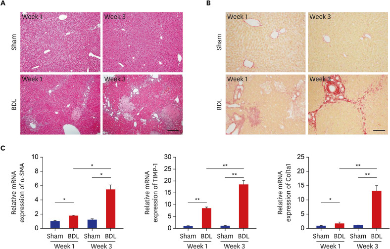
Fig. 1 Development of liver fibrosis after BDL. (A, B) Liver sections from sham control (upper panels) and BDL (lower panels) mice were examined by hematoxylin and eosin staining (A) and Sirius Red staining (B) at weeks 1 and 3 after BDL. (C) The mRNA levels of fibrotic marker genes at weeks 1 and 3 after BDL were determined by quantitative real-time reverse transcription polymerase chain reaction and normalized to β-actin (n = 9). The data represent the mean ± standard error of mean of three independent experiments. Scale bars, 25 μm.BDL = bile duct ligation, mRNA = messenger RNA, α-SMA = α-smooth muscle actin, TIMP-1 = tissue inhibitor of metalloproteinases-1, Col1a1 = collagen type I.*P < 0.05, **P < 0.01.
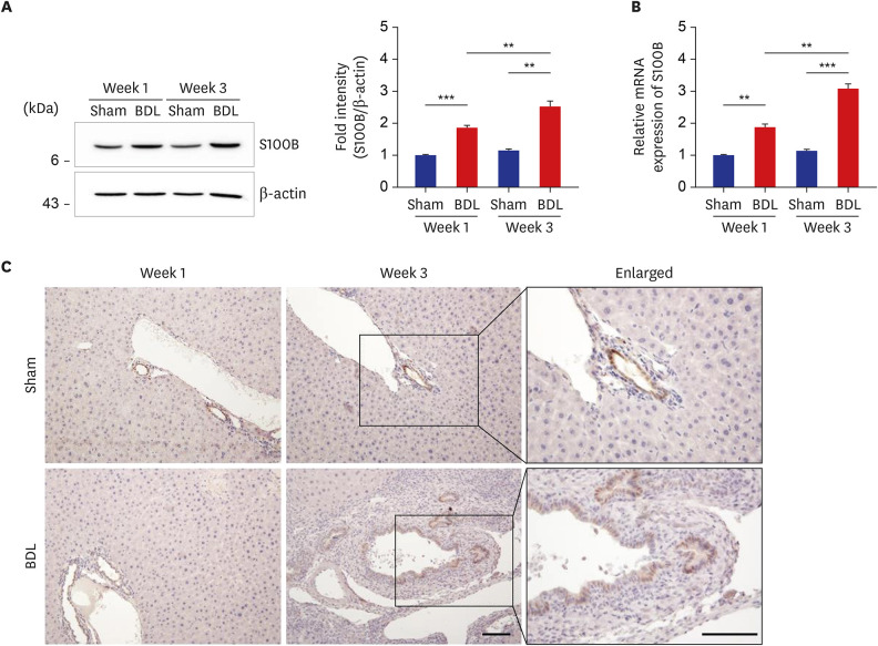
Fig. 2 Upregulated S100B expression in the livers of BDL mice. (A, B) Expression levels of S100B protein (A) and mRNA (B) in the livers from sham control and BDL mice was determined by Western blot analysis (A) and quantitative real-time reverse transcription polymerase chain reaction (B) and normalized to β-actin (n = 9) at each time point. The data represent the mean ± standard error of mean of three independent experiments. (C) Immunohistochemical staining of S100B in liver tissues from sham control and BDL mice. Note that an increased number of S100B-positive cells was found in the bile duct of BDL liver. Scale bars, 25 μm.BDL = bile duct ligation, mRNA = messenger RNA.**P < 0.01, ***P < 0.001.

Fig. 3 Subcellular localization of S100B and RAGE in the livers of BDL mice. (A) Expression of RAGE protein in the livers of sham control and BDL mice at 1 week and 3 weeks after operation was analyzed by Western blot analysis (left panel) and normalized to β-actin (right panel) (n = 9). The data represent the mean ± standard error of mean of three independent experiments. (B, C) Triple immunofluorescence staining of S100B, RAGE, and α-SMA (B) or CK19 (C) were performed in the liver sections of sham control and BDL mice at 3 weeks after operation. Note the colocalization of S100B and RAGE in α-SMA-positive activated HSCs (B) as well as CK19-positive bile duct epithelial cells (C) in the BDL livers. Green, S100B; red, α-SMA, and CK19; white, RAGE. Scale bars, 20 μm.RAGE = receptor for advanced glycation end products, BDL = bile duct ligation, α-SMA = α-smooth muscle actin, CK19 = cytokeratin19.*P < 0.05, **P < 0.01, ***P < 0.001.

Fig. 4 Double immunogold labeling of S100B and RAGE in the livers of BDL mice. The liver sections from sham control and BDL mice at week 3 were incubated with rabbit monoclonal anti-S100B (1:100) and mouse monoclonal anti-RAGE (1:100), followed by incubation with goat anti-rabbit IgG-conjugated 15 nm gold particles for S100B and goat anti-mouse IgG-conjugated 25 nm gold particles for RAGE. Note that immunogold particles indicating S100B (arrows) and RAGE (arrowhead) are present in the cytoplasm and microvilli of bile duct epithelial cells (upper panels), and in the HSCs, both particles were found in the nucleus and its environs (lower panels). Scale bars, 2 μm.RAGE = receptor for advanced glycation end products, BDL = bile duct ligation, HSC = hepatic stellate cell, IgG = immunoglobulin G, N = nuclei, M = mitochondria, L = lumen, LD = lipid droplet.

Fig. 5 S100B regulates HSC activation via RAGE. (A, B) HSC-T6 cells were treated with recombinant mouse S100B protein in a dose-dependent manner. After 12 hours, the cells were lysed, and HSC activation-related protein and gene expression levels were detected by Western blotting (A) and qRT-PCR (B), respectively. (C, D) HSC-T6 cells were transiently transfected with either SCR or si-RAGE in a dose-dependent manner, then incubated with 25 ng of recombinant S100B protein for 12 hours and analyzed by Western blotting (C) and qRT-PCR (D). The data represent the mean ± standard error of mean of three independent experiments.RAGE = receptor for advanced glycation end products, Col1a1 = collagen type I, α-SMA = α-smooth muscle actin, GAPDH = glyceraldehyde 3-phosphate dehydrogenase, mRNA = messenger RNA, PBS = phosphate-buffered saline, SCR = scrambled, si-RAGE = small interfering RNA-mediated knockdown of endogenous RAGE, qRT-PCR = quantitative real-time reverse transcription polymerase chain reaction, HSC = hepatic stellate cell.*P < 0.05, **P < 0.01, ***P < 0.001.
Reference
-
1. Tacke F, Trautwein C. Mechanisms of liver fibrosis resolution. J Hepatol. 2015; 63(4):1038–1039. PMID: 26232376.
Article2. Pinzani M. Pathophysiology of liver fibrosis. Dig Dis. 2015; 33(4):492–497. PMID: 26159264.
Article3. Lemoinne S, Cadoret A, El Mourabit H, Thabut D, Housset C. Origins and functions of liver myofibroblasts. Biochim Biophys Acta. 2013; 1832(7):948–954. PMID: 23470555.
Article4. Xia JR, Liu NF, Zhu NX. Specific siRNA targeting the receptor for advanced glycation end products inhibits experimental hepatic fibrosis in rats. Int J Mol Sci. 2008; 9(4):638–661. PMID: 19325776.
Article5. Yan SF, Yan SD, Ramasamy R, Schmidt AM. Tempering the wrath of RAGE: an emerging therapeutic strategy against diabetic complications, neurodegeneration, and inflammation. Ann Med. 2009; 41(6):408–422. PMID: 19322705.
Article6. Bierhaus A, Stern DM, Nawroth PP. RAGE in inflammation: a new therapeutic target? Curr Opin Investig Drugs. 2006; 7(11):985–991.7. Neeper M, Schmidt AM, Brett J, Yan SD, Wang F, Pan YC, et al. Cloning and expression of a cell surface receptor for advanced glycosylation end products of proteins. J Biol Chem. 1992; 267(21):14998–15004. PMID: 1378843.
Article8. Stern D, Yan SD, Yan SF, Schmidt AM. Receptor for advanced glycation endproducts: a multiligand receptor magnifying cell stress in diverse pathologic settings. Adv Drug Deliv Rev. 2002; 54(12):1615–1625. PMID: 12453678.
Article9. Lohwasser C, Neureiter D, Popov Y, Bauer M, Schuppan D. Role of the receptor for advanced glycation end products in hepatic fibrosis. World J Gastroenterol. 2009; 15(46):5789–5798. PMID: 19998499.10. Basta G, Navarra T, De Simone P, Del Turco S, Gastaldelli A, Filipponi F. What is the role of the receptor for advanced glycation end products-ligand axis in liver injury? Liver Transpl. 2011; 17(6):633–640. PMID: 21438128.
Article11. Ekong U, Zeng S, Dun H, Feirt N, Guo J, Ippagunta N, et al. Blockade of the receptor for advanced glycation end products attenuates acetaminophen-induced hepatotoxicity in mice. J Gastroenterol Hepatol. 2006; 21(4):682–688. PMID: 16677153.
Article12. Donato R. S100: a multigenic family of calcium-modulated proteins of the EF-hand type with intracellular and extracellular functional roles. Int J Biochem Cell Biol. 2001; 33(7):637–668. PMID: 11390274.
Article13. Gonçalves CA, Leite MC, Nardin P. Biological and methodological features of the measurement of S100B, a putative marker of brain injury. Clin Biochem. 2008; 41(10-11):755–763. PMID: 18454941.
Article14. Donato R, Sorci G, Riuzzi F, Arcuri C, Bianchi R, Brozzi F, et al. S100B's double life: intracellular regulator and extracellular signal. Biochim Biophys Acta. 2009; 1793(6):1008–1022. PMID: 19110011.
Article15. Leclerc E, Fritz G, Vetter SW, Heizmann CW. Binding of S100 proteins to RAGE: an update. Biochim Biophys Acta. 2009; 1793(6):993–1007. PMID: 19121341.
Article16. Pelinka LE, Toegel E, Mauritz W, Redl H. Serum S 100 B: a marker of brain damage in traumatic brain injury with and without multiple trauma. Shock. 2003; 19(3):195–200. PMID: 12630517.
Article17. Raabe A, Kopetsch O, Woszczyk A, Lang J, Gerlach R, Zimmermann M, et al. Serum S-100B protein as a molecular marker in severe traumatic brain injury. Restor Neurol Neurosci. 2003; 21(3-4):159–169. PMID: 14530578.18. Steiner J, Bernstein HG, Bogerts B, Gos T, Richter-Landsberg C, Wunderlich MT, et al. S100B is expressed in, and released from, OLN-93 oligodendrocytes: Influence of serum and glucose deprivation. Neuroscience. 2008; 154(2):496–503. PMID: 18472341.
Article19. Netto CB, Conte S, Leite MC, Pires C, Martins TL, Vidal P, et al. Serum S100B protein is increased in fasting rats. Arch Med Res. 2006; 37(5):683–686. PMID: 16740441.
Article20. Pelinka LE, Harada N, Szalay L, Jafarmadar M, Redl H, Bahrami S. Release of S100B differs during ischemia and reperfusion of the liver, the gut, and the kidney in rats. Shock. 2004; 21(1):72–76. PMID: 14676687.
Article21. Starkel P, Leclercq IA. Animal models for the study of hepatic fibrosis. Best Pract Res Clin Gastroenterol. 2011; 25(2):319–333. PMID: 21497748.
Article22. Vogel S, Piantedosi R, Frank J, Lalazar A, Rockey DC, Friedman SL, et al. An immortalized rat liver stellate cell line (HSC-T6): a new cell model for the study of retinoid metabolism in vitro. J Lipid Res. 2000; 41(6):882–893. PMID: 10828080.
Article23. Van Eldik LJ, Staecker JL, Winningham-Major F. Synthesis and expression of a gene coding for the calcium-modulated protein S100 beta and designed for cassette-based, site-directed mutagenesis. J Biol Chem. 1988; 263(16):7830–7837. PMID: 3372506.
Article24. Mely Y, Gérard D. Large-scale, one-step purification of oxidized and reduced forms of bovine brain S100b protein by HPLC. J Neurochem. 1988; 50(3):739–744. PMID: 3339349.
Article25. Junqueira LC, Bignolas G, Brentani RR. Picrosirius staining plus polarization microscopy, a specific method for collagen detection in tissue sections. Histochem J. 1979; 11(4):447–455. PMID: 91593.
Article26. Kim SE, Park JW, Kim MJ, Jang B, Jeon YC, Kim HJ, et al. Accumulation of citrullinated glial fibrillary acidic protein in a mouse model of bile duct ligation-induced hepatic fibrosis. PLoS One. 2018; 13(8):e0201744. PMID: 30071078.
Article27. Fehrenbach H, Weiskirchen R, Kasper M, Gressner AM. Up-regulated expression of the receptor for advanced glycation end products in cultured rat hepatic stellate cells during transdifferentiation to myofibroblasts. Hepatology. 2001; 34(5):943–952. PMID: 11679965.
Article28. Cataldegirmen G, Zeng S, Feirt N, Ippagunta N, Dun H, Qu W, et al. RAGE limits regeneration after massive liver injury by coordinated suppression of TNF-alpha and NF-kappaB. J Exp Med. 2005; 201(3):473–484. PMID: 15699076.29. Yamagishi S, Matsui T. Role of receptor for advanced glycation end products (RAGE) in liver disease. Eur J Med Res. 2015; 20(1):15. PMID: 25888859.
Article30. Roskams T, van den Oord JJ, De Vos R, Desmet VJ. Neuroendocrine features of reactive bile ductules in cholestatic liver disease. Am J Pathol. 1990; 137(5):1019–1025. PMID: 1700614.31. Desmet V, Roskams T, Van Eyken P. Ductular reaction in the liver. Pathol Res Pract. 1995; 191(6):513–524. PMID: 7479372.32. Alvaro D, Mancino MG, Glaser S, Gaudio E, Marzioni M, Francis H, et al. Proliferating cholangiocytes: a neuroendocrine compartment in the diseased liver. Gastroenterology. 2007; 132(1):415–431. PMID: 17241889.
Article33. Franchitto A, Onori P, Renzi A, Carpino G, Mancinelli R, Alvaro D, et al. Recent advances on the mechanisms regulating cholangiocyte proliferation and the significance of the neuroendocrine regulation of cholangiocyte pathophysiology. Ann Transl Med. 2013; 1(3):27. PMID: 25332971.34. Jensen K, Marzioni M, Munshi K, Afroze S, Alpini G, Glaser S. Autocrine regulation of biliary pathology by activated cholangiocytes. Am J Physiol Gastrointest Liver Physiol. 2012; 302(5):G473–83. PMID: 22194419.
Article35. Marzioni M, Francis H, Benedetti A, Ueno Y, Fava G, Venter J, et al. Ca2+-dependent cytoprotective effects of ursodeoxycholic and tauroursodeoxycholic acid on the biliary epithelium in a rat model of cholestasis and loss of bile ducts. Am J Pathol. 2006; 168(2):398–409. PMID: 16436655.
Article36. Heizmann CW, Fritz G, Schäfer BW. S100 proteins: structure, functions and pathology. Front Biosci. 2002; 7:d1356–68. PMID: 11991838.
Article37. Xia P, Deng Q, Gao J, Yu X, Zhang Y, Li J, et al. Therapeutic effects of antigen affinity-purified polyclonal anti-receptor of advanced glycation end-product (RAGE) antibodies on cholestasis-induced liver injury in rats. Eur J Pharmacol. 2016; 779:102–110. PMID: 26970185.
Article38. Iwamoto K, Kanno K, Hyogo H, Yamagishi S, Takeuchi M, Tazuma S, et al. Advanced glycation end products enhance the proliferation and activation of hepatic stellate cells. J Gastroenterol. 2008; 43(4):298–304. PMID: 18458846.
Article39. Demetris AJ, Sever C, Kakizoe S, Oguma S, Starzl TE, Jaffe R. S100 protein positive dendritic cells in primary biliary cirrhosis and other chronic inflammatory liver diseases. Relevance to pathogenesis? Am J Pathol. 1989; 134(4):741–747. PMID: 2705505.
- Full Text Links
-
- Actions
-
Cited
- CITED
-
- Close
- Share
-
- Similar articles
-
- Liver Fibrosis by Bile Duct Sclerosis with Ethanol: a New Experimental Model for Liver Fibrosis
- Immunohistochemical Expression of Bcl-2 and Bax after Bile Duct Ligation in the Rat
- Temporal Morphologic Changes in the Mouse Liver after Common Bile Duct Ligation
- Experimental Animal Models of Hepatic Fibrosis
- A Technique for Bile Duct-Duodenal Anastomosis at the Consecutive Rat Liver Transplantation
