JEG-3 placental cells in toxicology studies: a promising tool to reveal pregnancy disorders
- Affiliations
-
- 1UMR CNRS 8038 CiTCoM, Laboratoire de Chimie-Toxicologie Analytique et Cellulaire, Université de Paris, Faculté de Pharmacie de Paris, Paris, France
- 2Recherche & Développement, YSLAB, Quimper, France
- KMID: 2514588
- DOI: http://doi.org/10.5115/acb.20.234
Abstract
- Placental alterations are responsible for adverse pregnancy outcomes like preeclampsia and intrauterine growth restriction. And yet, placenta toxicology has not become a fully-f ledged toxicology field. Because placenta is very often seen only as a barrier between the mother and the fetus, there is a lack and therefore a need for an experimental human model with technical recommendations to study placenta toxicology. In vitro approaches are recommended in experimental toxicology as they focus on a specific biological process and yield high-throughput screening methods. In the present study, we first established incubation conditions to preserve signatures of the human JEG-3 cell line identity while enabling toxicity detection. JEG-3 cells prepared in our incubation conditions were renamed JEG-Tox cells. As placental alterations are mainly triggered by uncontrolled apoptosis, we second used known apoptotic agents pregnant women are exposed to, to check that JEG-Tox cells can trigger apoptosis. Ethanol, bisphenol F, quinalphos, 4,4’-DDT, benzalkonium chloride, phenoxyethanol, propylparaben, and perfluorooctanic acid all induced chromatin condensation in JEG-Tox cells. Our incubation conditions allow JEG-Tox cells to keep placental cell identity and to respond to toxic chemicals. JEG-Tox cells are a pertinent model for placenta toxicology and could be used to better understand pregnancy alterations.
Keyword
Figure
-
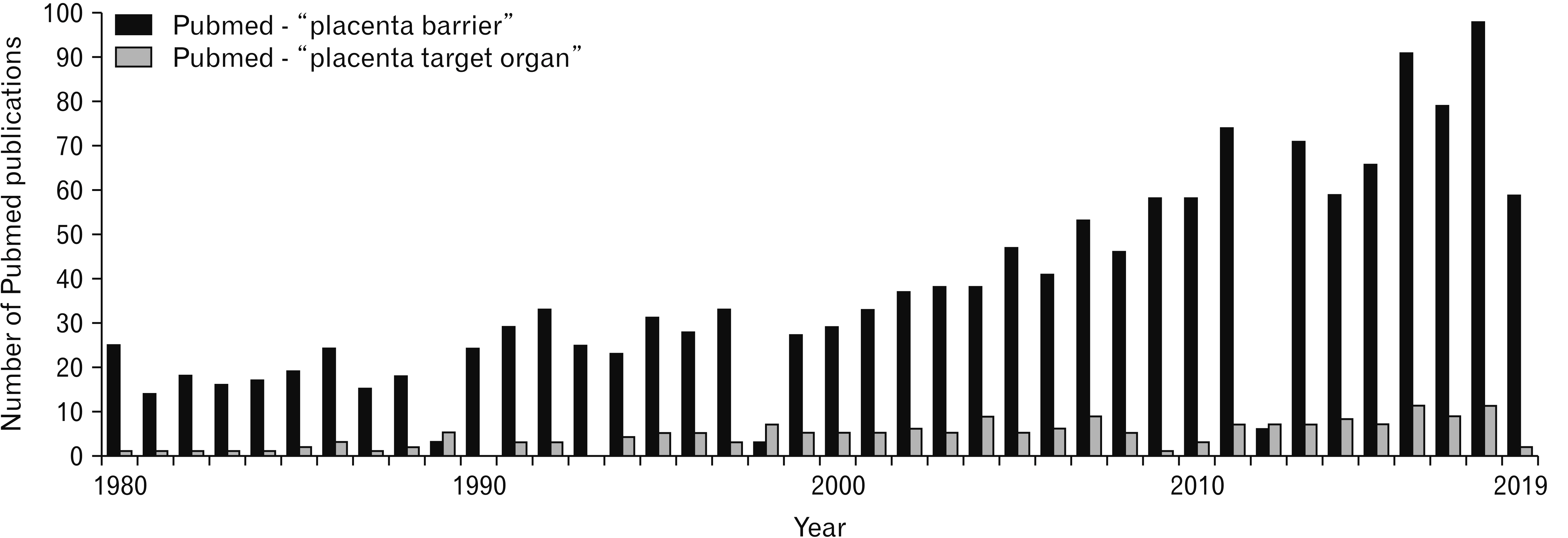
Fig. 1 Comparison of Pubmed publications using “placenta barrier” or “placenta target organ” as keywords in the research tool.

Fig. 2 Proliferation of JEG-3 cells in culture medium supplemented with different FBS concentrations. JEG-3 cells were incubated with three different concentrations of FBS for 24 or 72 hours, cell count was conducted to quantify the effect of FBS on JEG-3 cell proliferation. Black: 10% FBS, grey: 2.5% FBS, light grey: 0% FBS. FBS, fetal bovine serum; NS, not significant.
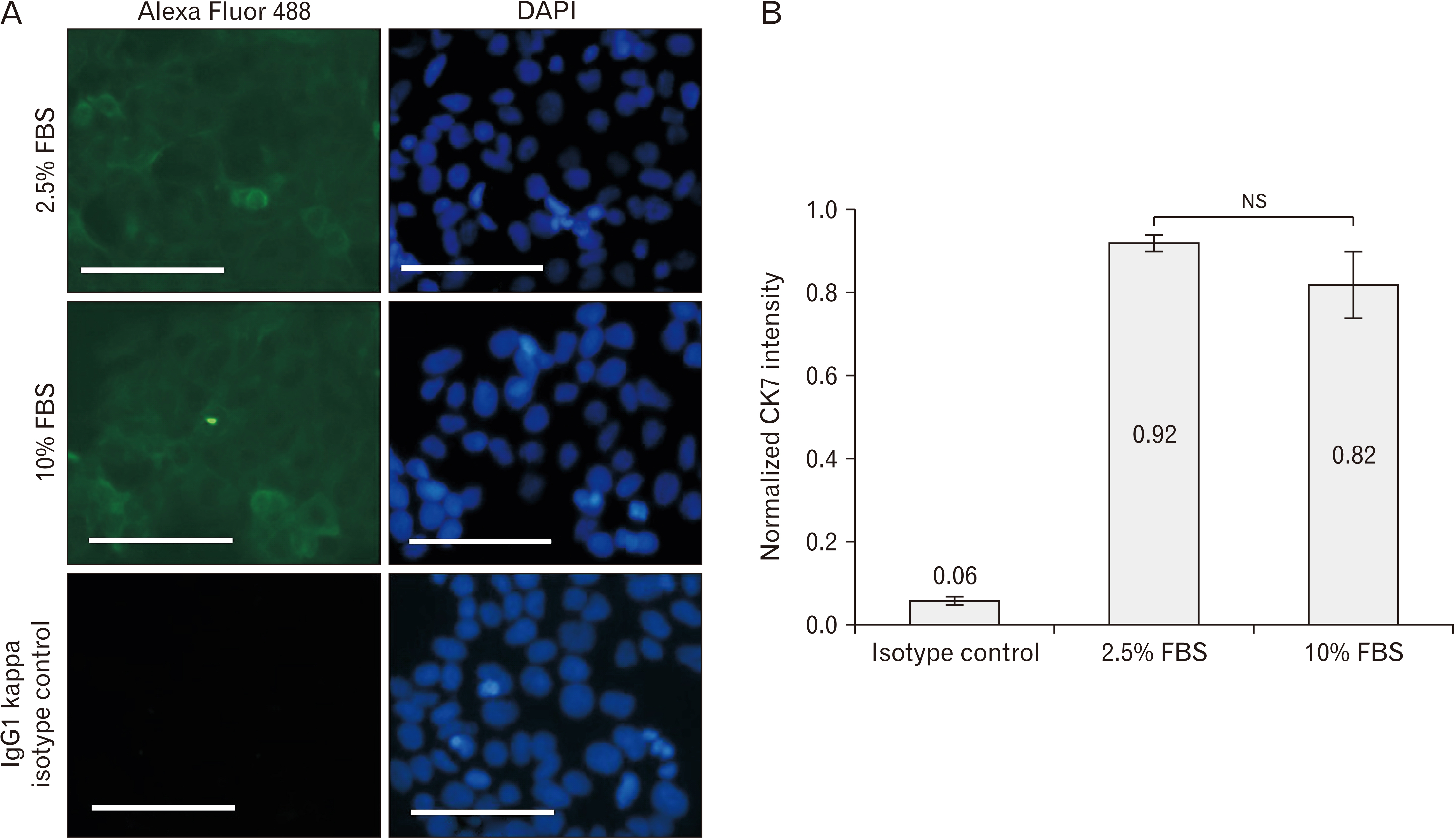
Fig. 3 Expression of CK7 in JEG-3 cells in 2.5% and 10% FBS. (A) Microscopic observation of JEG-3 cells stained with either anti-CK7 antibody or isotype control and then stained with Alexa Fluor 488 (green). DAPI was used to stain DNA (blue) (Magnification, ×200). Data are representative of at least 3 independent experiments. (B) Quantification of CK7 fluorescence using ImageJ software. Normalized CK7 fluorescence intensity was obtained by dividing green fluorescence intensity by blue fluorescence intensity to take into account the difference in cell numbers in the selected microscopic fields. CK7, cytokeratin-7; FBS, fetal bovine serum; NS, not significant. Scale bar=100 μm (A).
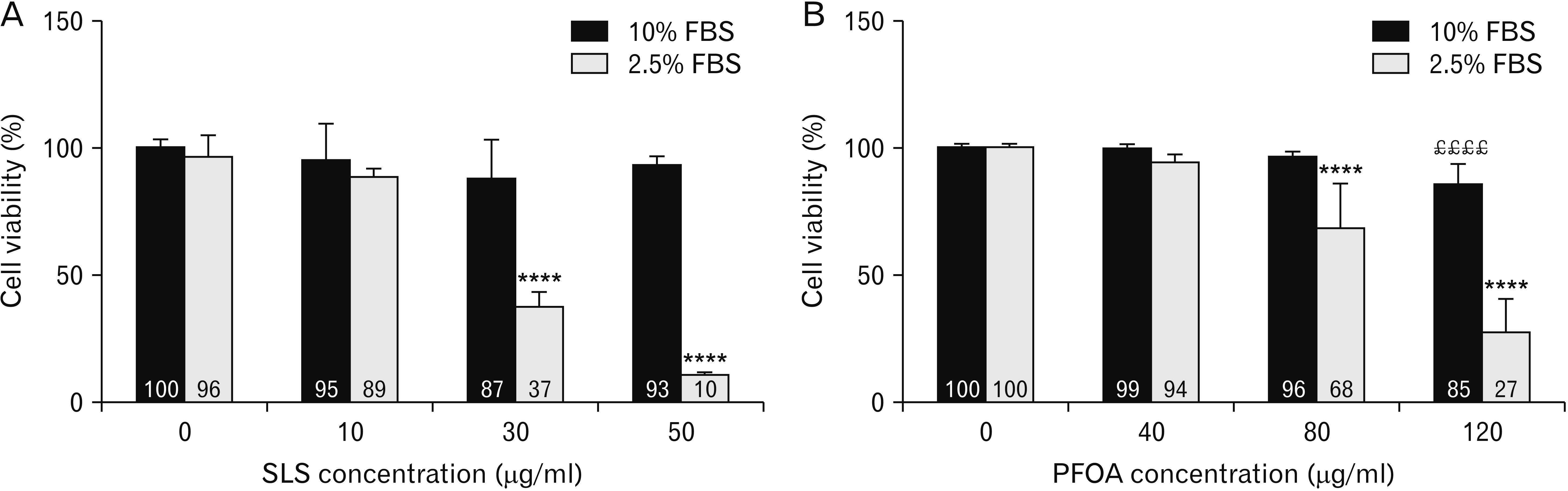
Fig. 4 Comparison of JEG-3 cell viability after incubation with SLS (A) or PFOA (B) in FBS 10% or FBS 2.5%. JEG-3 cells were incubated with SLS from 10 to 50 μg/ml or PFOA from 40 to 120 μM for 24 hours. Cell viability was determined using the neutral red assay. ££££P<0.0001 compared to negative control in 10% FBS, ****P<0.0001 compared to negative control in 2.5% FBS (n=3). FBS, fetal bovine serum; PFOA, perfluorooctanic acid; SLS, sodium lauryl sulfate.

Fig. 5 Evaluation of cell viability and chromatin condensation of JEG-Tox cells after incubation with apoptosis inducers for 24 hours. Cell viability and chromatin condensation were quantified using the Alamar blue and Hoechst 33342 assays, respectively. Dashed line: cell viability, solid line: chromatin condensation. *P<0.05, **P<0.01, ***P<0.001, and ****P<0.0001 compared to negative control (n=3). BAC, benzalkonium chloride; PFOA, perfluorooctanic acid; 4,4’DDT, 4,4’-dichlorodiphenyltrichloroethane.
Reference
-
References
1. Griffiths SK, Campbell JP. 2015; Placental structure, function and drug transfer. Contin Educ Anaesth Crit Care Pain. 15:84–9. DOI: 10.1093/bjaceaccp/mku013.
Article2. Murphy VE, Smith R, Giles WB, Clifton VL. 2006; Endocrine regulation of human fetal growth: the role of the mother, placenta, and fetus. Endocr Rev. 27:141–69. DOI: 10.1210/er.2005-0011. PMID: 16434511.
Article3. Chaddha V, Viero S, Huppertz B, Kingdom J. 2004; Developmental biology of the placenta and the origins of placental insufficiency. Semin Fetal Neonatal Med. 9:357–69. DOI: 10.1016/j.siny.2004.03.006. PMID: 15691771.
Article4. Fisher SJ. 2004; The placental problem: linking abnormal cytotrophoblast differentiation to the maternal symptoms of preeclampsia. Reprod Biol Endocrinol. 2:53. DOI: 10.1186/1477-7827-2-53. PMID: 15236649. PMCID: PMC493282.5. Ilekis JV, Tsilou E, Fisher S, Abrahams VM, Soares MJ, Cross JC, Zamudio S, Illsley NP, Myatt L, Colvis C, Costantine MM, Haas DM, Sadovsky Y, Weiner C, Rytting E, Bidwell G. 2016; Placental origins of adverse pregnancy outcomes: potential molecular targets: an Executive Workshop Summary of the Eunice Kennedy Shriver National Institute of Child Health and Human Development. Am J Obstet Gynecol. 215(1 Suppl):S1–46. DOI: 10.1016/j.ajog.2016.03.001. PMID: 26972897. PMCID: PMC4925329.
Article6. Innis SM. 2007; Fatty acids and early human development. Early Hum Dev. 83:761–6. DOI: 10.1016/j.earlhumdev.2007.09.004. PMID: 17920214.
Article7. Zhu Y, Mordaunt CE, Yasui DH, Marathe R, Coulson RL, Dunaway KW, Jianu JM, Walker CK, Ozonoff S, Hertz-Picciotto I, Schmidt RJ, LaSalle JM. 2019; Placental DNA methylation levels at CYP2E1 and IRS2 are associated with child outcome in a prospective autism study. Hum Mol Genet. 28:2659–74. DOI: 10.1093/hmg/ddz084. PMID: 31009952. PMCID: PMC6687952.
Article8. Smith SC, Baker PN, Symonds EM. 1997; Increased placental apoptosis in intrauterine growth restriction. Am J Obstet Gynecol. 177:1395–401. DOI: 10.1016/S0002-9378(97)70081-4. PMID: 9423741.
Article9. Erel CT, Dane B, Calay Z, Kaleli S, Aydinli K. 2001; Apoptosis in the placenta of pregnancies complicated with IUGR. Int J Gynaecol Obstet. 73:229–35. DOI: 10.1016/S0020-7292(01)00373-3. PMID: 11376669.
Article10. Enders AC, Blankenship TN. 1999; Comparative placental structure. Adv Drug Deliv Rev. 38:3–15. DOI: 10.1016/S0169-409X(99)00003-4. PMID: 10837743.
Article11. Malassiné A, Frendo JL, Evain-Brion D. 2003; A comparison of placental development and endocrine functions between the human and mouse model. Hum Reprod Update. 9:531–9. DOI: 10.1093/humupd/dmg043. PMID: 14714590.12. Schmidt A, Morales-Prieto DM, Pastuschek J, Fröhlich K, Markert UR. 2015; Only humans have human placentas: molecular differences between mice and humans. J Reprod Immunol. 108:65–71. DOI: 10.1016/j.jri.2015.03.001. PMID: 25817465.
Article13. Ceccaldi PF, Mandelbrot L, Farinotti R, Forestier F, Gil S. 2010; [Contributions of the ex vivo human perfused placenta in the study of placental transfer of drugs]. J Gynecol Obstet Biol Reprod (Paris). 39:601–5. French. DOI: 10.1016/j.jgyn.2010.06.010. PMID: 20692775.14. Abou-Kheir W, Barrak J, Hadadeh O, Daoud G. 2017; HTR-8/SVneo cell line contains a mixed population of cells. Placenta. 50:1–7. DOI: 10.1016/j.placenta.2016.12.007. PMID: 28161053.
Article15. Mitchell AM, Yap AS, Payne EJ, Manley SW, Mortimer RH. 1995; Characterization of cell polarity and epithelial junctions in the choriocarcinoma cell line, JAR. Placenta. 16:31–9. DOI: 10.1016/0143-4004(95)90079-9. PMID: 7716126.
Article16. Rothbauer M, Patel N, Gondola H, Siwetz M, Huppertz B, Ertl P. 2017; A comparative study of five physiological key parameters between four different human trophoblast-derived cell lines. Sci Rep. 7:5892. DOI: 10.1038/s41598-017-06364-z. PMID: 28724925. PMCID: PMC5517571.
Article17. Bohets HH, Nouwen EJ, De Broe ME, Dierickx PJ. 1994; Effects of foetal calf serum on cell viability, cytotoxicity and detoxification in the two kidney-derived cell lines LLC-PK1 and MDCK. Toxicol In Vitro. 8:559–61. DOI: 10.1016/0887-2333(94)90016-7. PMID: 20692960.
Article18. Lordan S, Higginbotham CL. 2012; Effect of serum concentration on the cytotoxicity of clay particles. Cell Biol Int. 36:57–61. DOI: 10.1042/CBI20100587. PMID: 21883092.
Article19. Pouzaud F, Dutot M, Martin C, Debray M, Warnet JM, Rat P. 2006; Age-dependent effects on redox status, oxidative stress, mitochondrial activity and toxicity induced by fluoroquinolones on primary cultures of rabbit tendon cells. Comp Biochem Physiol C Toxicol Pharmacol. 143:232–41. DOI: 10.1016/j.cbpc.2006.02.006. PMID: 16574493.
Article20. Dutot M, Fagon R, Hemon M, Rat P. 2012; Antioxidant, anti-inflammatory, and anti-senescence activities of a phlorotannin-rich natural extract from brown seaweed Ascophyllum nodosum. Appl Biochem Biotechnol. 167:2234–40. DOI: 10.1007/s12010-012-9761-1. PMID: 22692848.
Article21. Wakx A, Dutot M, Massicot F, Mascarelli F, Limb GA, Rat P. 2016; Amyloid β peptide induces apoptosis through P2X7 cell death receptor in retinal cells: modulation by marine omega-3 fatty acid DHA and EPA. Appl Biochem Biotechnol. 178:368–81. DOI: 10.1007/s12010-015-1878-6. PMID: 26467741. PMCID: PMC4718936.
Article22. Ghazi K, Deng-Pichon U, Warnet JM, Rat P. 2012; Hyaluronan fragments improve wound healing on in vitro cutaneous model through P2X7 purinoreceptor basal activation: role of molecular weight. PLoS One. 7:e48351. DOI: 10.1371/journal.pone.0048351. PMID: 23173033. PMCID: PMC3500239.23. Reid Y, Storts D, Riss T, Minor L. c2004. Authentication of human cell lines by STR DNA profiling analysis [Internet]. Eli Lilly & Company and the National Center for Advancing Translational Sciences;Bethesda, MD: Available from: http://www.ncbi.nlm.nih.gov/books/NBK144066/. cited 2019 Jul 19.24. Maldonado-Estrada J, Menu E, Roques P, Barré-Sinoussi F, Chaouat G. 2004; Evaluation of Cytokeratin 7 as an accurate intracellular marker with which to assess the purity of human placental villous trophoblast cells by flow cytometry. J Immunol Methods. 286:21–34. DOI: 10.1016/j.jim.2003.03.001. PMID: 15087219.
Article25. Szuster-Ciesielska A, Plewka K, Daniluk J, Kandefer-Szerszeń M. 2008; Zinc inhibits ethanol-induced HepG2 cell apoptosis. Toxicol Appl Pharmacol. 229:1–9. DOI: 10.1016/j.taap.2007.11.019. PMID: 18396304.
Article26. Zerin T, Song HY, Kim YS. 2015; Quinalphos induced intracellular ROS generation and apoptosis in human alveolar A549 cells. Mol Cell Toxicol. 11:61–9. DOI: 10.1007/s13273-015-0008-4.
Article27. Mokra K, Kocia M, Michałowicz J. 2015; Bisphenol A and its analogs exhibit different apoptotic potential in peripheral blood mononuclear cells (in vitro study). Food Chem Toxicol. 84:79–88. DOI: 10.1016/j.fct.2015.08.007. PMID: 26271707.28. Carvalho CM, Menezes PF, Letenski GC, Praes CE, Feferman IH, Lorencini M. 2012; In vitro induction of apoptosis, necrosis and genotoxicity by cosmetic preservatives: application of flow cytometry as a complementary analysis by NRU. Int J Cosmet Sci. 34:176–82. DOI: 10.1111/j.1468-2494.2011.00698.x. PMID: 22118339.29. Eggert A, Cisneros-Montalvo S, Anandan S, Musilli S, Stukenborg JB, Adamsson A, Nurmio M, Toppari J. 2019; The effects of perfluorooctanoic acid (PFOA) on fetal and adult rat testis. Reprod Toxicol. 90:68–76. DOI: 10.1016/j.reprotox.2019.08.005. PMID: 31412280.
Article30. Jin X, Song L, Liu X, Chen M, Li Z, Cheng L, Ren H. 2014; Protective efficacy of vitamins C and E on p,p'-DDT-induced cytotoxicity via the ROS-mediated mitochondrial pathway and NF-κB/FasL pathway. PLoS One. 9:e113257. DOI: 10.1371/journal.pone.0113257. PMID: 25464339. PMCID: PMC4252254.
Article31. Pozarowska D, Pozarowski P. 2011; Benzalkonium chloride (BAK) induces apoptosis or necrosis, but has no major influence on the cell cycle of Jurkat cells. Folia Histochem Cytobiol. 49:225–30. DOI: 10.5603/FHC.2011.0031. PMID: 21744321.
Article32. Gupta RK, Gupta RC. Gupta RC, editor. 2017. Placental toxicity. Reproductive and Developmental Toxicology. 2nd ed. Academic Press;London: p. 1301–25. DOI: 10.1016/B978-0-12-804239-7.00068-8.
Article33. Nykjaer C, Alwan NA, Greenwood DC, Simpson NA, Hay AW, White KL, Cade JE. 2014; Maternal alcohol intake prior to and during pregnancy and risk of adverse birth outcomes: evidence from a British cohort. J Epidemiol Community Health. 68:542–9. DOI: 10.1136/jech-2013-202934. PMID: 24616351. PMCID: PMC4033207.
Article34. Ferguson KK, Rosen EM, Rosario Z, Feric Z, Calafat AM, McElrath TF, Vélez Vega C, Cordero JF, Alshawabkeh A, Meeker JD. 2019; Environmental phthalate exposure and preterm birth in the PROTECT birth cohort. Environ Int. 132:105099. DOI: 10.1016/j.envint.2019.105099. PMID: 31430608. PMCID: PMC6754790.
Article35. Ferguson KK, McElrath TF, Meeker JD. 2014; Environmental phthalate exposure and preterm birth. JAMA Pediatr. 168:61–7. DOI: 10.1001/jamapediatrics.2013.3699. PMID: 24247736. PMCID: PMC4005250.
Article36. Barkoski JM, Busgang SA, Bixby M, Bennett D, Schmidt RJ, Barr DB, Panuwet P, Gennings C, Hertz-Picciotto I. 2019; Prenatal phenol and paraben exposures in relation to child neurodevelopment including autism spectrum disorders in the MARBLES study. Environ Res. 179(Pt A):108719. DOI: 10.1016/j.envres.2019.108719. PMID: 31627027. PMCID: PMC6948181.
Article37. Herzog E, Byrne HJ, Davoren M, Casey A, Duschl A, Oostingh GJ. 2009; Dispersion medium modulates oxidative stress response of human lung epithelial cells upon exposure to carbon nanomaterial samples. Toxicol Appl Pharmacol. 236:276–81. DOI: 10.1016/j.taap.2009.02.007. PMID: 19233222.
Article38. Charles I, Khalyfa A, Kumar DM, Krishnamoorthy RR, Roque RS, Cooper N, Agarwal N. 2005; Serum deprivation induces apoptotic cell death of transformed rat retinal ganglion cells via mitochondrial signaling pathways. Invest Ophthalmol Vis Sci. 46:1330–8. DOI: 10.1167/iovs.04-0363. PMID: 15790899.
Article39. Koyama Y, Kimura Y, Yoshioka Y, Wakamatsu D, Kozaki R, Hashimoto H, Matsuda T, Baba A. 2000; Serum-deprivation induces cell death of rat cultured microglia accompanied with expression of Bax protein. Jpn J Pharmacol. 83:351–4. DOI: 10.1016/S0021-5198(19)30572-4. PMID: 11001183.
Article40. Kulkarni GV, McCulloch CA. 1994; Serum deprivation induces apoptotic cell death in a subset of Balb/c 3T3 fibroblasts. J Cell Sci. 107(Pt 5):1169–79. PMID: 7929626.41. Sharp AN, Heazell AE, Crocker IP, Mor G. 2010; Placental apoptosis in health and disease. Am J Reprod Immunol. 64:159–69. DOI: 10.1111/j.1600-0897.2010.00837.x. PMID: 20367628. PMCID: PMC3025811.
Article
- Full Text Links
-
- Actions
-
Cited
- CITED
-
- Close
- Share
-
- Similar articles
-
- Comparative study between pregnancies with and without hypertensive disorders in placental abruption
- The effect of pregnancy induced hypertensive disorders on perinatal death in placental abruption
- A Case of Perforated Stomach Cancer with Placental Abruption in Pregnancy
- Insulin Resistance During Pregnancy
- Pure Red Cell Aplasia in Pregnancy
