Cadaveric anatomy of the lumbar triangular safe zone of Kambin’s in North West Indian population
- Affiliations
-
- 1Department of Anatomy, Postgraduate Institute of Medical Education and Research, Chandigarh, India
- 2Department of Orthopaedics, Postgraduate Institute of Medical Education and Research, Chandigarh, India
- KMID: 2514581
- DOI: http://doi.org/10.5115/acb.20.243
Abstract
- A three dimensional triangular space ‘the Kambin’s triangle (KT)’ present on the dorsolateral aspect of the intervertebral disc, is considered to be a safe area for transforaminal approaches. It allows access to the exiting and traversing nerve roots, the thecal sac and to the intervertebral disc spaces. Our aim was to calculate the area of the triangle by measuring the height and base at all the intervertebral spaces bilaterally in the lumbar region in North West Indian cadavers and to assess the diameter of circle inscribed within this triangle which will correspond to the size of cannula inserted for the minimally invasive transforaminal approaches in this population. Five randomly chosen adult cadavers were used for this study. After clearing the area, the exiting nerve was identified. The height and base of the bony KTs (n=40) were measured with the help of digital Vernier’s calliper (accuracy 0.02 mm) to calculate the area of the KT. There is a steady increase in the area of the bony KT reaching maximum at the level of L4-5 intervertebral space. Statistically there were no differences in the calculated areas between right and left side. The mean diameter of inscribed circle within the triangle also showed gradual increase from 5.82 mm at L1-2 level, reaching maximum value of 7.26 mm at L4-5 level on the right side while on the left side the values were 5.66 mm and 8.16 mm respectively. Careful anatomical consideration is of utmost importance in transforaminal approaches during surgical or interventional procedures in this region. Cannula having external diameter ranging 6–8 mm is recommended for any interventional approach through Kambin’s space.
Figure
-

Fig. 1 (A, B) Schematic diagram of the Kambin’s triangle. I-Inferior articular process of Cr, S-Superior articular process of Cd. A, point of intersection of the vertical plane with the exiting nerve root; AB, height; B, point of intersection of vertical and horizontal plane; BC, base, C, point of intersection of the horizontal plane with the exiting nerve root; CA, hypotenuse, Cd, caudal vertebra; Cr, cranial vertebra; r, radius of inscribed circle.

Fig. 2 Kambin’s triangle in a dissected cadaver (left lateral view) (black arrow). A, point of intersection of the vertical plane with the exiting nerve root; AB, plane along the superior articular process (star, black) of caudal lumbar vertebra (L3); B, point of intersection of vertical and horizontal plane; BC, Plane along superior surface of body of caudal lumbar vertebra (L3); C, point of intersection of the horizontal plane with the exiting nerve root; IVD, Intervertebral disc; L1,2,3,4,5, Lumbar vertebrae from cranial to caudal direction; T, Transverse process.
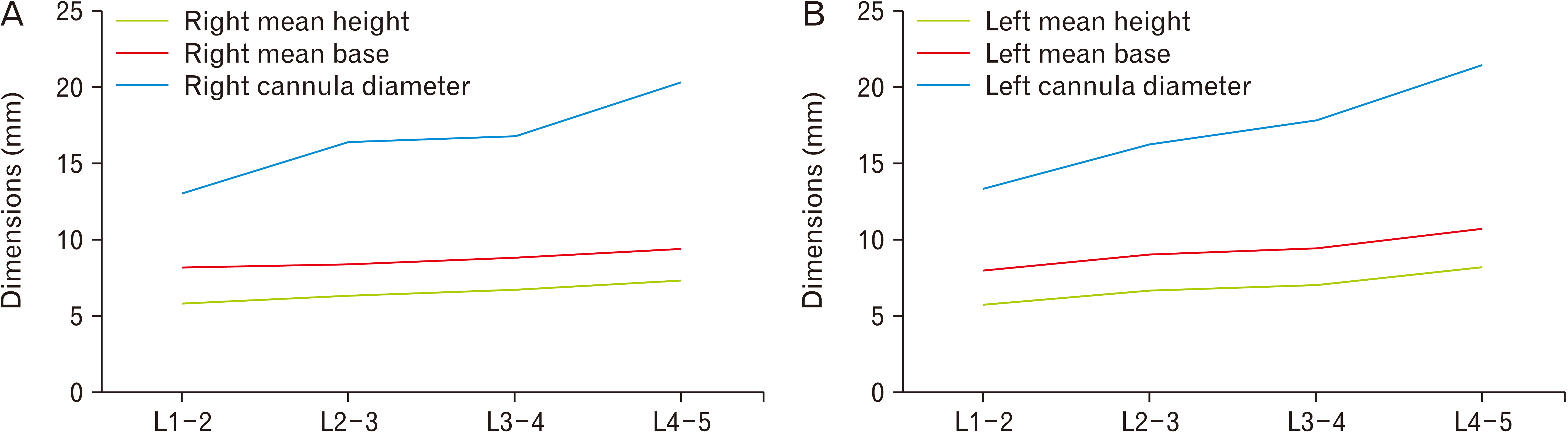
Fig. 3 (A, B) Line diagram showing the correlation of parameters between the lumbar levels on the right (A) and the left (B) side. L1-2, L2-3, L3-4, L4-5, lumbar intervertebral levels.
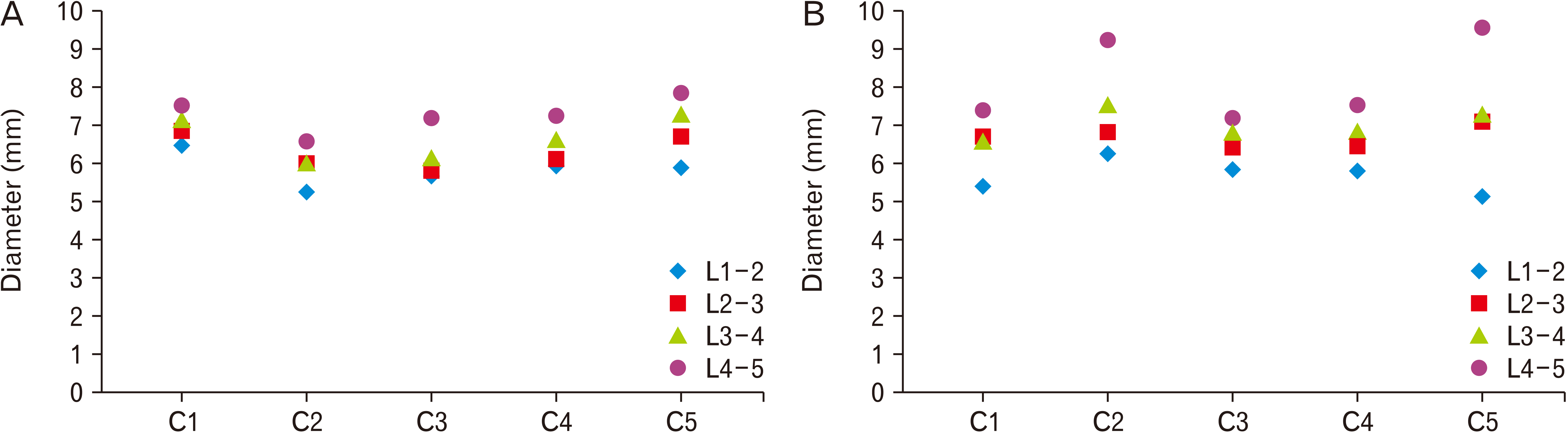
Fig. 4 (A, B) Scattered diagram showing the distribution of dimensions of diameter of inscribed circle within Kambin’s triangle at different levels on the right (A) and the left (B) side. C1-5, studied cadavers.
Reference
-
References
1. Glaser SE, Falco F. 2005; Paraplegia following a thoracolumbar transforaminal epidural steroid injection. Pain Physician. 8:309–14. DOI: 10.36076/ppj.2005/8/309. PMID: 16850088.2. Schaffer JL, Kambin P. 1991; Percutaneous posterolateral lumbar discectomy and decompression with a 6.9-millimeter cannula. Analysis of operative failures and complications. J Bone Joint Surg Am. 73:822–31. DOI: 10.2106/00004623-199173060-00005. PMID: 1830052.
Article3. Kambin P, Sampson S. 1986; Posterolateral percutaneous suction-excision of herniated lumbar intervertebral discs. Report of interim results. Clin Orthop Relat Res. (207):37–43. DOI: 10.1097/00003086-198606000-00008. PMID: 3720102.4. Botwin KP, Gruber RD, Bouchlas CG, Torres-Ramos FM, Sanelli JT, Freeman ED, Slaten WK, Rao S. 2002; Fluoroscopically guided lumbar transformational epidural steroid injections in degenerative lumbar stenosis: an outcome study. Am J Phys Med Rehabil. 81:898–905. DOI: 10.1097/00002060-200212000-00003. PMID: 12447088.5. Park JW, Nam HS, Cho SK, Jung HJ, Lee BJ, Park Y. 2011; Kambin's triangle approach of lumbar transforaminal epidural injection with spinal stenosis. Ann Rehabil Med. 35:833–43. DOI: 10.5535/arm.2011.35.6.833. PMID: 22506212. PMCID: PMC3309379.
Article6. Hoshide R, Feldman E, Taylor W. 2016; Cadaveric analysis of the Kambin's Triangle. Cureus. 8:e475. DOI: 10.7759/cureus.475. PMID: 27004152. PMCID: PMC4780690.
Article7. Fanous AA, Tumialán LM, Wang MY. 2019; Nov. 29. Kambin's triangle: definition and new classification schema. J Neurosurg Spine. [Epub]. https://doi.org/10.3171/2019.8.SPINE181475. DOI: 10.3171/2019.8.SPINE181475. PMID: 31783346.
Article8. Pairaiturkar PP, Sudame OS, Pophale CS. 2019; Evaluation of dimensions of Kambin's triangle to calculate maximum permissible cannula diameter for percutaneous endoscopic lumbar discectomy: a 3-dimensional magnetic resonance imaging based study. J Korean Neurosurg Soc. 62:414–21. DOI: 10.3340/jkns.2018.0091. PMID: 31079448. PMCID: PMC6616981.9. Lertudomphonwanit T, Keorochana G, Kraiwattanapong C, Chanplakorn P, Leelapattana P, Wajanavisit W. 2016; Anatomic considerations of intervertebral disc perspective in lumbar posterolateral approach via Kambin's triangle: cadaveric study. Asian Spine J. 10:821–7. DOI: 10.4184/asj.2016.10.5.821. PMID: 27790308. PMCID: PMC5081315.
Article10. Mirkovic SR, Schwartz DG, Glazier KD. 1995; Anatomic considerations in lumbar posterolateral percutaneous procedures. Spine (Phila Pa 1976). 20:1965–71. DOI: 10.1097/00007632-199509150-00001. PMID: 8578369.
Article11. Datar GP, Shinde A, Bommakanti K. 2017; Technical consideration of transforaminal endoscopic spine surgery for central herniation. Indian J Pain. 31:86–93. DOI: 10.4103/ijpn.ijpn_37_17.
Article12. Kapetanakis S, Gkasdaris G, Angoules AG, Givissis P. 2017; Transforaminal Percutaneous Endoscopic Discectomy using Transforaminal Endoscopic Spine System technique: pitfalls that a beginner should avoid. World J Orthop. 8:874–80. DOI: 10.5312/wjo.v8.i12.874. PMID: 29312845. PMCID: PMC5745429.
Article13. Kim M, Kim HS, Oh SW, Adsul NM, Singh R, Kashlan ON, Noh JH, Jang IT, Oh SH. 2019; Evolution of spinal endoscopic surgery. Neurospine. 16:6–14. DOI: 10.14245/ns.1836322.161. PMID: 31618807. PMCID: PMC6449828.
Article14. Zhang KH, Zhang WH, Xu BS, Dong XM, Guo L, Du LL, Xu HW. 2019; CT-based morphometric analysis of approach of percutaneous transforaminal endoscopic lumbar interbody fusion. Orthop Surg. 11:212–20. DOI: 10.1111/os.12434. PMID: 30895721. PMCID: PMC6594482.
Article15. Ozer AF, Suzer T, Can H, Falsafi M, Aydin M, Sasani M, Oktenoglu T. 2017; Anatomic assessment of variations in Kambin's triangle: a surgical and cadaver study. World Neurosurg. 100:498–503. DOI: 10.1016/j.wneu.2017.01.057. PMID: 28132923.
Article16. Wimmer C, Maurer H. 2000; Anatomic consideration for lumbar percutaneous interbody fusion. Clin Orthop Relat Res. (379):236–41. DOI: 10.1097/00003086-200010000-00028. PMID: 11039812.
Article17. Boonstra H, Oosterhuis JW, Oosterhuis AM, Fleuren GJ. 1983; Cervical tissue shrinkage by formaldehyde fixation, paraffin wax embedding, section cutting and mounting. Virchows Arch A Pathol Anat Histopathol. 402:195–201. DOI: 10.1007/BF00695061. PMID: 6420986.
Article18. Siu KF, Cheung HC, Wong J. 1986; Shrinkage of the esophagus after resection for carcinoma. Ann Surg. 203:173–6. DOI: 10.1097/00000658-198602000-00011. PMID: 3947154. PMCID: PMC1251066.
Article19. Pritt B, Tessitore JJ, Weaver DL, Blaszyk H. 2005; The effect of tissue fixation and processing on breast cancer size. Hum Pathol. 36:756–60. DOI: 10.1016/j.humpath.2005.04.018. PMID: 16084944.
Article20. Docquier PL, Paul L, Cartiaux O, Lecouvet F, Dufrane D, Delloye C, Galant C. 2010; Formalin fixation could interfere with the clinical assessment of the tumor-free margin in tumor surgery: magnetic resonance imaging-based study. Oncology. 78:115–24. DOI: 10.1159/000306140. PMID: 20357519.
Article
- Full Text Links
-
- Actions
-
Cited
- CITED
-
- Close
- Share
-
- Similar articles
-
- Anatomic Considerations of Intervertebral Disc Perspective in Lumbar Posterolateral Approach via Kambin's Triangle: Cadaveric Study
- Anatomy acts concerning body and organ donations across the globe: past, present and future with a special emphasis on the indian scenario
- Exploring the atlantic part of the vertebral artery in the South Indian population and its implications in spine surgery
- Morphometric Measurements of Cadaveric Thoracic Spine in Indian Population and Its Clinical Applications
- Surgical Morphometry of C1 and C2 Vertebrae: A Three-Dimensional Computed Tomography Analysis of 180 Chinese, Indian, and Malay Patients
