Pediatric split liver transplantation for congenital factor X deficiency: first 10-year follow-up of a case with portal vein stenting
- Affiliations
-
- 1Department of Surgery, Asan Medical Center, University of Ulsan College of Medicine, Seoul, Korea
- 2Department of Pediatrics, Asan Medical Center, University of Ulsan College of Medicine, Seoul, Korea
- KMID: 2514411
- DOI: http://doi.org/10.4285/kjt.20.0039
Abstract
- Congenital factor X (FX) deficiency is a rare autosomal-recessive disease that induces bleeding disorder. Herein, we present the 10-year posttransplant course of a pediatric patient who underwent liver transplantation (LT) with portal vein (PV) stenting for correction of severe congenital FX deficiency, with focus on long-term maintenance of coagulation function and patency of PV stenting. A 17-month-old infant with recurrent hemorrhagic episodes due to FX deficiency underwent split LT using a left lateral sec- tion graft. The graft-recipient weight ratio was 2.2%. The graft implantation procedures were performed by following the standard pediatric split LT procedure. Nevertheless, a wall stent was inserted due to PV anastomotic stenosis on posttransplant day 1. Graft function recovered slowly because of partial parenchyma infarct, and the patient was discharged at 46 days after LT operation. The FX activity started to increase soon after LT and gradually normalized; the coagulation profiles have been maintained well for the past 10 years. The patient has been doing well for the past 10 years after LT without any episodes of abnormal bleeding. Due to the risk of vascular complications owed to PV stenting, life-long follow-up is mandatory with special attention until attainment of complete physical growth to adolescent and adulthood.
Keyword
Figure
-
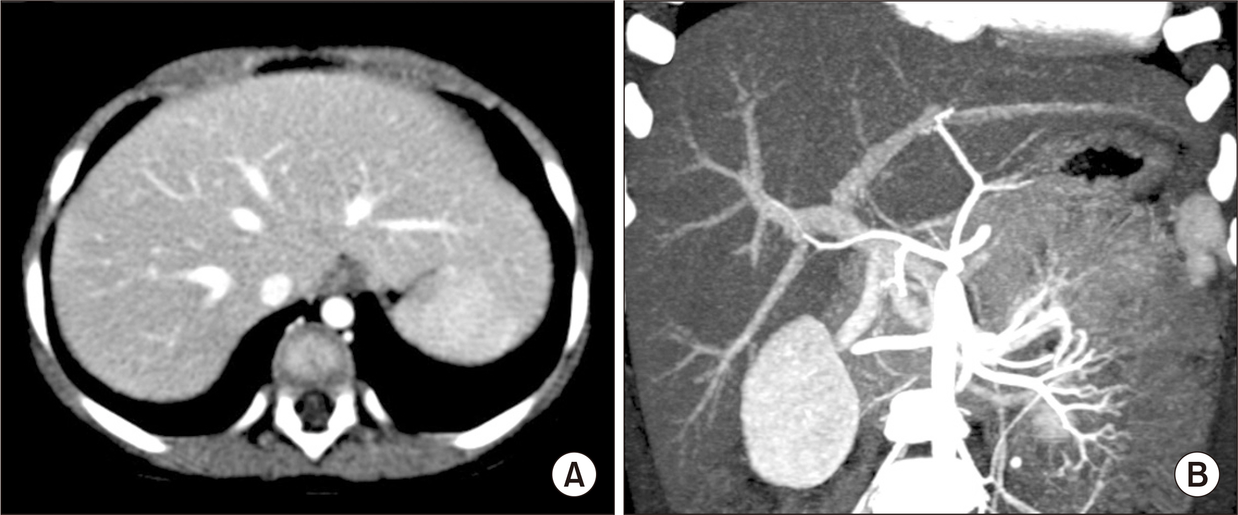
Fig. 1 Pretransplant computed tomography scan showing normal looking hepatic parenchyma (A) and perihilar vascular anatomy (B).
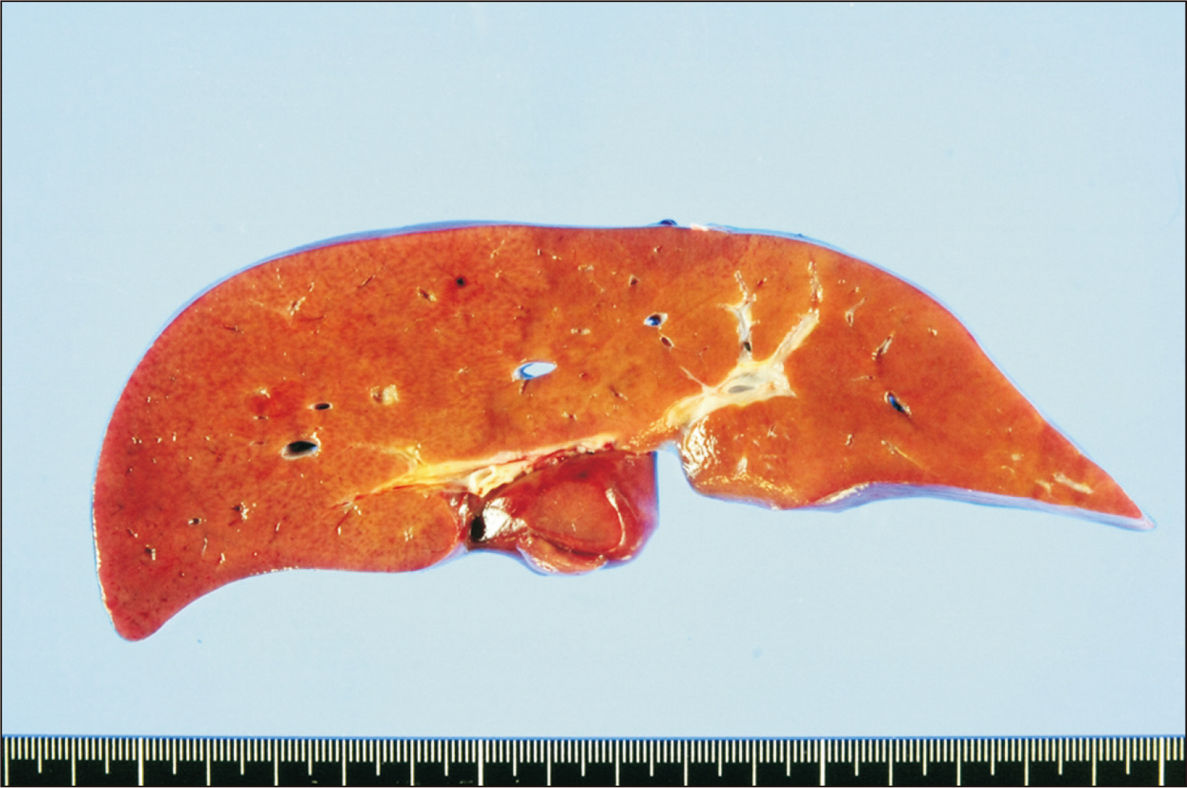
Fig. 2 Gross photographs of the explant liver showing normal-looking hepatic parenchyma.

Fig. 3 Posttransplant computed tomography scan taken at 1 day after transplantation. Heterogeneous low attenuation of the hepatic parenchyma (A) and segmental stenosis of the portal vein anastomosis site (B) are identified. Arrows indicate the site of anastomotic stenosis.
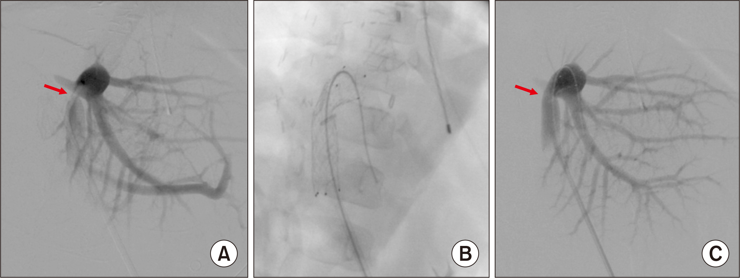
Fig. 4 Posttransplant portal vein stenting performed at 1 day after transplantation. (A) A significant stenosis portal vein anastomosis site is identified. (B) A 10 mm×40 mm-sized wall stent is inserted across the stenosis. (C) The stenotic site is expanded after stenting. Arrows indicate the site of anastomotic stenosis.

Fig. 5 Posttransplant computed tomography scan taken at 2 days after transplantation. (A) Heterogeneous low attenuation of the hepatic parenchyma was slightly improved. (B) The configuration of the reconstructed portal vein with an internal stent implicates the presence of tension and acute angle bending at the anastomosis site before stenting.
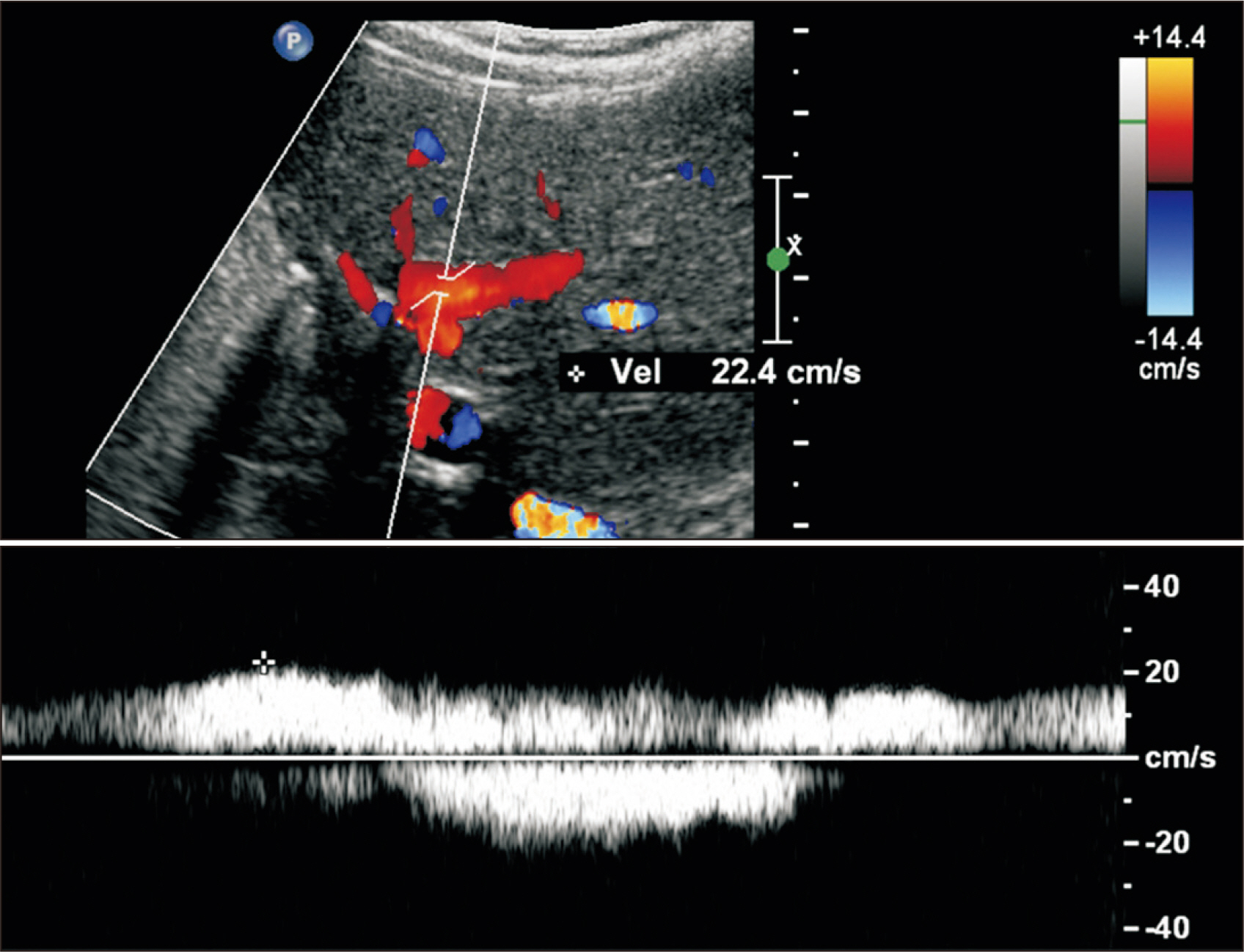
Fig. 6 Follow-up Doppler ultrasonography taken at 9 years after transplantation. The intrahepatic portal blood flow is well maintained (up to 20 cm/sec) with slight narrowing of the intrastent diameter. The intrastent diameter of the portal vein was about 6 mm, which remained unchanged during last 5 years despite gradual physical growth of the patient.
Reference
-
1. Brown DL, Kouides PA. 2008; Diagnosis and treatment of inherited factor X deficiency. Haemophilia. 14:1176–82. DOI: 10.1111/j.1365-2516.2008.01856.x. PMID: 19141158.
Article2. Funk DM, Casciato D. 2010; Factor X deficiency. Clin Lab Sci. 23:131–3. DOI: 10.29074/ascls.23.3.131. PMID: 20734884.
Article3. Bang SH, Oh SH, Kim KM, Song SM, Seo JJ, Yoo HW, et al. 2012; Successful liver transplantation for a child with life-threatening recurrent bleeding episodes due to congenital factor X deficiency: a case report. Transplant Proc. 44:583–4. DOI: 10.1016/j.transproceed.2012.01.075. PMID: 22506295.
Article4. Girolami A, Scandellari R, Scapin M, Vettore S. 2008; Congenital bleeding disorders of the vitamin K-dependent clotting factors. Vitam Horm. 78:281–374. DOI: 10.1016/S0083-6729(07)00014-3. PMID: 18374200.
Article5. Senturk S, Guzel E, Bayrak AH, Bukte Y, Guzel A. 2010; Factor X deficiency presenting with bilateral chronic subdural hematoma. Pediatr Neurosurg. 46:54–7. DOI: 10.1159/000315004. PMID: 20516741.
Article6. Gordon FH, Mistry PK, Sabin CA, Lee CA. 1998; Outcome of orthotopic liver transplantation in patients with haemophilia. Gut. 42:744–9. DOI: 10.1136/gut.42.5.744. PMID: 9659174. PMCID: PMC1727096.
Article7. Kang SH, Hwang S, Jung DH, Ahn CS, Moon DB, Ha TY, et al. 2013; Unification venoplasty to cope with recipient portal vein anomaly during living donor liver transplantation. Transplant Proc. 45:3000–4. DOI: 10.1016/j.transproceed.2013.08.073. PMID: 24157023.
Article8. Hwang S, Kim DY, Ahn CS, Moon DB, Kim KM, Park GC, et al. 2013; Computational simulation-based vessel interposition reconstruction technique for portal vein hypoplasia in pediatric liver transplantation. Transplant Proc. 45:255–8. DOI: 10.1016/j.transproceed.2012.05.090. PMID: 23375311.
Article9. Namgoong JM, Hwang S, Oh SH, Kim KM, Park GC, Ahn CS, et al. 2020; Living-donor liver transplantation with inferior vena cava replacement in an infant recipient with advanced hepatoblastoma. Ann Hepatobiliary Pancreat Surg. 24:72–7. DOI: 10.14701/ahbps.2020.24.1.72. PMID: 32181433. PMCID: PMC7061035.
Article10. Cho YP, Kim KM, Ha TY, Ko GY, Hwang JY, Park H, et al. 2014; Management of late-onset portal vein complications in pediatric living-donor liver transplantation. Pediatr Transplant. 18:64–71. DOI: 10.1111/petr.12204. PMID: 24341631.
Article11. Shim DJ, Ko GY, Sung KB, Gwon DI, Ko HK. 2018; Long-term outcome of portal vein stent placement in pediatric liver transplant recipients: a comparison with balloon angioplasty. J Vasc Interv Radiol. 29:800–8. DOI: 10.1016/j.jvir.2017.11.019. PMID: 29545104.
Article12. Yeh YT, Chen CY, Tseng HS, Wang HK, Tsai HL, Lin NC, et al. 2017; Enlarging vascular stents after pediatric liver transplantation. J Pediatr Surg. 52:1934–9. DOI: 10.1016/j.jpedsurg.2017.08.060. PMID: 28927979.
Article
- Full Text Links
-
- Actions
-
Cited
- CITED
-
- Close
- Share
-
- Similar articles
-
- Third retransplantation using a whole liver graft for late graft failure from hepatic vein stent stenosis in a pediatric patient who underwent split liver retransplantation
- Tailored standardization of portal vein reconstruction for pediatric liver transplantation at Asan Medical Center
- Portal Vein Anastomosis with using Growth Factor for Preventing Stricture and Thrombosis following Auxiliary Partial Liver Transplantation in Rat.
- Living donor liver transplantation in a pediatric patient having intrahepatic portocaval shunt with congenital absence of the intrahepatic portal vein
- Living donor liver transplantation with direct collateral portal vein anastomosis in a pediatric patient with congenital absence of the portal vein
